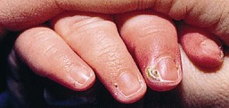
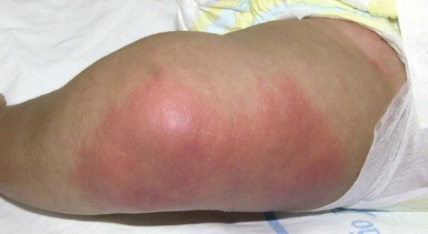
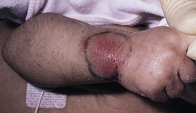
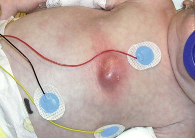
James Treat, Brian T. Fisher Bacterial skin infections in neonates and infants can present differently from those in older children, based on several factors: (1) the nature of the pathogen; (2) the developmental stage of the infant when infection is acquired: early (first or second trimester) versus late (third trimester) in gestation and early (first few days) versus late (2–8 weeks) postnatal life; and (3) the manner in which inoculation occurs (i.e. vertical transmission in utero, vertical transmission at the time of birth, or postnatally). Infection from hematogenous penetration of the placental barrier or through ruptured amniotic membranes can be more severe and widespread, involving multiple organ systems in addition to the skin, because of the vulnerable state of the developing neonate. Importantly, neonatal cutaneous infections, even those acquired postnatally, are potentially more serious compared with similar cutaneous infections in older children. The reason for these differences in severity is multifactorial, involving a complex interaction between host, pathogen, and environmental factors (Box 12.1). Furthermore, the potential impact of a localized cutaneous infection is of greater concern in neonates, where alterations of the normal skin barrier are more common and can serve as a conduit for systemic infection. This is especially true for very low-birthweight infants where organisms that normally colonize the skin are also implicated as etiologic agents of sepsis. This suggests that sepsis may be a consequence of bacterial penetration at sites of skin injury or through the immature epidermal barrier.1 Edwards and colleagues highlighted this concern when they revealed the association of simple bland emollients in very low-birthweight infants with increased risk of neonatal bacterial sepsis caused by normal skin flora.2 Given the risk for severe consequences, neonatal cutaneous infections mandate a prompt and thorough evaluation and aggressive treatment to minimize morbidity and mortality. The clinical presentation, pathogenesis, diagnostic modalities and suggested therapeutic interventions are reviewed for various common and less common neonatal cutaneous infections. The continued emergence of antibiotic resistance has complicated the empiric therapeutic options for some bacterial infections. This emergence of resistance has highlighted the importance of obtaining cultures when possible to identify the pathogen and its sensitivity profile. Recommendations regarding antibiotic choices and duration of therapy specific to the infection are provided for each clinical scenario noting the potential impact of resistance on antibiotic choices. Impetigo is the single most common primary skin infection in children,3 and impetiginization of atopic dermatitis is the most important secondary skin infection.4 Nonbullous impetigo is a superficial infection localized to the subcorneal portion of the epidermis typically caused Staphylococcus aureus and less commonly Streptococcus pyogenes (Group A beta-hemolytic streptococci). Bullous impetigo is almost exclusively caused by Staphylococcus aureus, most commonly phage group 2 (types 71 and 55) that elaborates toxins.5 These toxins cause epidermolysis and subsequent blister formation. Bullous impetigo is thought to be a localized form of staphylococcal scalded skin syndrome (SSSS).6 Nonbullous impetigo is characterized by erythematous, honey-colored crusted plaques (Fig. 12.1). Lesions tend to be localized in primary impetigo, but may become more widespread when superimposed on diseased skin (e.g., atopic dermatitis). Moist intertriginous, periorificial and periumbilical areas are commonly involved in both nonbullous and bullous impetigo. Bullous impetigo often presents during the first 2 weeks of life with flaccid, transparent, subcorneal bullae, which may be single or clustered, and often lack underlying cutaneous erythema. The bullae may contain pus that layers out in the dependent portion. The lesions rupture easily, leaving a shallow erosion surrounded by a narrow rim of peeling that heals without scarring (Fig. 12.2 Most cases of impetigo, including neonatal bullous impetigo, are unaccompanied by constitutional signs of illness. Occasionally, hematogenous spread of bacteria can result in osteomyelitis, septic arthritis, pneumonia, or septicemia, particularly in neonates with bullous impetigo.3,7 S. aureus is isolated from approximately 85% of impetigo lesions and in 50–60% of cases is the sole pathogen. Streptococcus pyogenes causes the remainder of cases of nonbullous impetigo. The staphylococcal organisms that cause nonbullous impetigo are variable, but are generally not from phage group 2, whereas bullous impetigo is most commonly (80%) the result of S. aureus from phage group 2.8 Bullae production results from locally produced exfoliative or epidermolytic toxins A or B (ETA and ETB) which cleave desmoglein 1 (DSG1) within desmosomes, via serine proteases.9 If there is enough toxin produced, it can disseminate and cleave DSG1 in broader areas, leading to staphylococcal scalded skin syndrome (SSSS, see below). ETA is more commonly associated with bullous impetigo than is ETB. High titers of neutralizing antibodies against ETA and/or the inability of ETA to penetrate through the epidermis may explain why ETA does not commonly cause SSSS.10 Methicillin-resistant Staphylococcus aureus (MRSA) can cause impetigo, bullous impetigo, orbital cellulitis,11 abscesses, SSSS, necrotizing fasciitis, and toxic shock syndrome that are clinically indistinguishable from similar infections caused by methicillin-sensitive Staphylococcus aureus (MSSA). As the clinical presentation of MRSA and MSSA bullous impetigo and S. aureus and S. pyogenes nonbullous impetigo are indistinguishable, obtaining appropriate cultures is imperative to guide therapeutic choices. Culture of fluid from a vesicle, pustule or from beneath the lifted edges of a crusted plaque of impetigo, is usually sufficient to establish a diagnosis. Gram stains can be useful in establishing a rapid presumptive diagnosis but are less sensitive and less specific than a culture. When the diagnosis is in question, and Gram stain and culture are negative, a skin biopsy can be useful, although this is seldom necessary. When a biopsy is performed, the histopathology often reveals a vesicle or pustule in the subcorneal or granular region of the epidermis with marked dermal inflammation; the cavity is larger in the bullous form. Staphylococcal impetigo is clinically indistinguishable from streptococcal impetigo unless it is bullous, which is only caused by Staphylococcus aureus.3 Impetigo may be confused with several other infectious vesiculobullous or pustular disorders, such as herpes simplex virus (HSV), varicella, enterovirus, congenital cutaneous candidiasis, listeriosis, and scabies, as well as noninfectious disorders such as erythema toxicum neonatorum, transient neonatal pustular melanosis, eosinophilic pustulosis, chronic bullous disease of childhood, incontinentia pigmenti, epidermolysis bullosa, pemphigus vulgaris, pemphigus foliaceus, and bullous pemphigoid. For superficial skin infections such as impetigo, the course is often self-limiting. Recently, there has been a trend towards using topical mupirocin, retapamulin (in children over 9 months of age) or fusidic acid (not available in the USA) for localized forms of impetigo and reserving oral antibiotics for those patients with more diffuse involvement or resistant organisms.12 When systemic antibiotic therapy is deemed necessary, antibiotic coverage should be inclusive of S. aureus. Oral antibiotics that are not cleaved by pencillinases present in most S. aureus isolates can be effective in this setting. This includes options such as cephalexin, cloxacillin, or dicloxacillin. Each of these options also has the benefit of covering S. pyogenes in the setting of nonbullous impetigo. Knowledge of the local epidemiology of MRSA infections is imperative, as a high rate of MRSA would prompt empiric coverage for MRSA in the setting of impetigo. Clindamycin is a reasonable empiric option; however, clindamycin resistance among MSSA and MRSA isolates is also on the rise.13 Strains of MRSA have been grouped primarily by the type of resistance genes that they carry in a genetic element referred to as the cassette chromosome element, called SCCMEC (SCCmec). Nosocomial MRSA contains types 1, 2, and 3 SCCmec, conferring multidrug resistance to non-β-lactam antibiotics. Community-acquired MRSA contains types 4 or 5 SCCmec, making this strain more susceptible to antimicrobial agents.14,15 There are a number of additional second-line oral agents that may be considered for the treatment of uncomplicated impetigo including cefadroxil, cefprozil, loracarbef, and amoxicillin/clavulanate. It should be noted that none of these second-line agents is effective against MRSA. The changing landscape of resistance necessitates that cultures be performed in order to guide appropriate therapeutic decisions.16 For isolates that are both MRSA and clindamycin resistant, trimethoprim-sulfamethoxazole may be an effective oral option but should not be used in children under 2 months of age, due to risk of hyperbilirubinemia and kernicterus. Additionally, the clinician should note that trimethoprim-sulfamethoxazole may not adequately cover Streptococcus pyogenes. Various clinical scenarios of skin bacterial infection warrant the use of parenteral therapy. These include: infants presenting with deeper infections such as furuncles, abscesses, cellulitis, osteomyelitis, endocarditis, and pneumonia. Additionally, as bullous impetigo in neonates may advance rapidly, systemic enteral or parenteral therapy (instead of topical) with close clinical follow-up is encouraged.17 As with oral therapy, initial empiric parenteral therapy should be guided by local resistance patterns. Regions with low rates of MRSA can employ oxacillin or nafcillin as a first line agent. Those areas with higher rates of MRSA should use clindamycin or vancomycin empirically. Clindamycin, a protein synthesis inhibitor, may be advantageous for decreasing epidermolytic toxin production and is often used as an adjunctive agent in SSSS caused by MSSA or MRSA. Linezolid also shows excellent activity against MRSA and can be considered as an alternative parenteral option. The mortality rate in NICUs due to MRSA infection has been as high as 38% in some cohorts.18 Mortality rates are highest in infants developing bacteremia and septic shock.14,15,18 Late-onset neonatal sepsis, occurring 2–3 weeks after birth, has been seen with nosocomial MRSA infections. Thus, patients should be followed closely, even if they appear healthy. Once signs of infection have begun to subside, and if constitutional signs are absent, therapy may be completed orally. Duration of therapy should be dictated by the clinical scenario as well as any associated complications. For uncomplicated impetigo, a 7-day course of therapy is typically sufficient. Secondary complications such as bacteremia, osteomyelitis and endocarditis warrant longer parenteral therapeutic courses, which should be managed with consultation from an infectious disease specialist. Intertrigo is an acute inflammation in a skin fold that can be irritant or infectious in etiology. Neonates and infants have abundant subcutaneous tissue and are often not mobile enough to allow the folds to air out. This occluded skin (neck, axillae and inguinal folds) is prone to overgrowth of yeast and bacteria. Clinicians often overlook staphylococcal and streptococcal infections as important and potentially dangerous causes of intertrigo. Bacterial intertrigo typically presents with red, slightly eroded, tender, shiny exudative patches centered on a skin fold (Figs 12.4, 12.5). Although the inguinal areas are most common, infants and neonates have many occluded skin folds due to their body habitus (including neck, axillae, antecubital and popliteal fossae) and any of these can become infected. The perianal area can be the initial source of infection and will also have a bright red, eroded appearance when infected with Strep. pyogenes or S. aureus. Intertrigo can also be present in several skin folds.19,20 Intertrigo is typically not associated with fever or extracutaneous spread. Untreated streptococcal disease can lead to post-streptococcal glomerulonephritis. Cutaneous streptococcal infections have also been associated with the subsequent development of psoriasis. Specifically, guttate psoriasis or napkin/perineal psoriasis should prompt an evaluation for perianal streptococcal infection in infants.21 Bacterial intertrigo and perianal infection is most commonly due to Strep. pyogenes but S. aureus (including MRSA) is becoming increasingly recognized as an etiologic pathogen for intertrigo.22,23 Pseudomonas species are less common causes of intertrigo and are more commonly indicative of a polymicrobial infection. Intertrigo can be due to irritation from over-washing or use of baby wipes. Seborrheic dermatitis, atopic dermatitis and contact dermatitis can also present with red patches in skin folds and each can become superinfected with bacteria or yeast. Candidal yeast infections often infect skin folds and present with peeling and satellite pustules. Although the clinical presentation is often enough to make the diagnosis, a Gram stain and culture from the most eroded or purulent area can be helpful in defining the causative pathogen. However, the results of these cultures need to be interpreted with the understanding that typical colonizing organisms, especially S. aureus, may not represent true infection. Although a rapid Strep test may be positive in streptococcal perianal dermatitis, this test has not been approved for use on non-posterior pharyngeal specimens. Potassium hydroxide scrapings and fungal cultures can help diagnose yeast infections. Intertrigo is worsened by skin occlusion, so allowing the area to aerate is important. Therapy with topical antibiotics such as mupirocin for S. aureus or Strep. pyogenes or bacitracin-polymyxin B for Pseudomonas spp. is often effective. For premature or immunosuppressed infants, or for multifocal and/or complicated infections, systemic therapy may be necessary. A similar approach, as described above for impetigo, should be employed when choosing an oral or parenteral therapeutic agent for intertrigo. A Gram stain and culture of the affected area should be taken before initiating empiric systemic antibiotics. If the culture results suggest a polymicrobial infection or a Pseudomonas spp. infection and the clinical scenario requires systemic therapy, then additional input from an infectious disease specialist should be considered. Pustules in the neonate or infant can be benign but must be differentiated from infectious pustulosis (bacterial, yeast, herpetic). Bacterial folliculitis is a superficial infection of the hair follicle ostium. Noninfectious pustulosis such as erythema toxicum neonatorum and transient neonatal pustular melanosis are addressed elsewhere in this book but typically are differentiated by their clinical presentation, including time course and superficial nature. Folliculitis presents as discrete, dome-shaped pustules with an erythematous base located at the ostia of the pilosebaceous canals. The lesions are usually asymptomatic, but may be pruritic or painful. Occasionally, a lesion may extend to involve deeper tissues and form an abscess (e.g., furuncle, carbuncle). Extracutaneous findings are not typically present. Rarely, the local infection can progress to a systemic illness that is evident by fever onset and an ill-appearing child. Folliculitis is caused predominantly by S. aureus, although coagulase-negative staphylococci are occasionally involved. A moist environment, maceration, poor hygiene, application of an occlusive emollient, and drainage from adjacent wounds and abscesses can be provoking factors. Pseudomonas aeruginosa is a rare cause of folliculitis in infants. It is found in circulating water, and the bacteria become trapped under the clothing, leading to infection. If pustulosis is due to P. aeruginosa it is often concentrated in areas that are covered by swimming suits or occluded such as axillae, inguinal folds and trunk.24 The causative organism of folliculitis can be identified by Gram stain and culture of purulent material from the follicular orifice. Benign pustulosis of infancy is often on the differential of staphylococcal pustulosis in the neonate. Erythema toxicum neonatorum tends to be more transient and have a splotchy patch of erythema around fragile superficial pustules and the pustule contents contain eosinophils. Transient neonatal pustular melanosis is typically present at birth and the pustules rupture easily and are gone within hours to days; the pustules contain neutrophils similar to bacterial folliculitis but a Gram stain should be negative (although Gram stains have a significant false-negative rate in bacterial infection). Candida species, Pityrosporum ovale, and Malassezia furfur are all yeast pathogens capable of causing follicular papules and/or pustules. Identification of these fungal organisms can be made by potassium hydroxide examination of lesion scrapings or by skin biopsy. Several other conditions may mimic folliculitis, including miliaria, eosinophilic pustular folliculitis, acne neonatorum, tinea corporis, congenital cutaneous candidiasis, scabies, and erythema toxicum neonatorum. An attempt should be made to identify and eliminate predisposing factors. Localized uncomplicated folliculitis may be managed by removing causative factors, antiseptic cleansing (e.g., chlorhexidine in children over 2 months), and topical antibiotics. Severe or refractory folliculitis should be managed with oral therapy. Similar to impetigo and intertrigo, bacterial Gram stain and culture can be extremely useful to guide appropriate therapy. Initial empiric therapy should be directed at S. aureus. In areas with limited MRSA rates, cephalexin, cloxacillin or dicloxacillin are reasonable first-line agents. For regions where MRSA prevalence is higher, initial empiric therapy with clindamycin should be considered. Neonates and immunosuppressed infants may require intravenous therapy. Recurrent episodes should warrant investigation into asymptomatic nasal carriage of S. aureus by the patient, immediate caregivers, pets or other contaminated objects in the child’s environment.25 Decolonizing efforts including intranasal mupirocin and antibacterial washes should be considered for direct caretakers of infected infants, especially if the infection is recurrent. Open wounds that are not initially caused by a bacterial infection, such as those caused by epidermolysis bullosa (EB), autoimmune blistering diseases, ulcerated hemangiomas or vesicular infections (herpes simplex or varicella) often become colonized with bacteria but can also become secondarily infected. It is important for the clinician to be able to differentiate colonization from infection to determine if antibiotics are necessary. Erosions and ulcerations that are secondarily infected can present with a myriad of symptoms that include purulent drainage, crusting, erythema expanding from the wound, pain, and acute worsening that cannot be explained by the primary process. Green discharge or discoloration on dressings may be a marker of Pseudomonas infection. Sepsis can occur secondary to bacterial infection of pre-existing erosions or ulcerations leading to fever, hypotension and potentially death. Sepsis complicating varicella-related skin ulcerations has been reported secondary to Gram-positive organisms including S. aureus and Strep. pyogenes, as well as Gram-negative pathogens such as Pseudomonas spp. and Escherichia coli.26 Cutaneous ulcerations are most typically colonized with normal skin flora such as diphtheroids, coagulase-negative staphylococci or anaerobes (especially in the perineum). S. aureus, streptococci (especially S. pyogenes) and Pseudomonas can also flourish in open wounds and are more pathogenic. The open wound lacks the physical protection of the epidermis and creates a warm, moist environment for bacterial overgrowth. Clinical interpretation of the likelihood of infection versus colonization is important but if infection is suspected, the wound should be cultured. The amount of bacteria, rapidity of bacterial growth in culture and the presence of just one pathogenic bacteria that has outcompeted the normal skin flora all suggest a true infection versus colonization. Ulcers can acutely worsen and drain due to the primary disease process, such as in pyoderma gangrenosum. Herpetic infections as well as yeast infections such as those caused by Candida spp. can also superinfect open wounds. Infected ulcers and erosions will often fail to heal at the same rate as the non-infected ulcers or erosions. Untreated, the areas may never heal, and may progress to cellulitis or systemic infection. Some erosions or ulceration in patients with EB are chronically colonized and so therapy should only be started if there is a clinical change or an area that is determined not to heal because of secondary infection. Topical therapy (such as mupirocin or bacitracin-polymyxin B) is often enough for localized areas. Metronidazole is often used for ulceration of perineal hemangiomas in order to cover anaerobic gut flora.27 Anecdotally, patients with chronic wounds such as those with EB will often rotate the type of topical antibiotic they use so as not to induce resistance. Systemic therapy should be reserved for patients with widespread infection, cellulitis or worsening infection, despite adequate topical therapy. As has been noted previously, therapy should be guided by the results of bacterial cultures. Initial empiric therapy should be directed at Gram-positive organisms with cephalexin, cloxacillin, dicloxacillin, or clindamycin. Infection secondary to S. pyogenes should always dictate systemic antibiotic therapy because of its potential for causing glomerulonephritis. Although less frequent, Gram-negative pathogens are possible and thus expanding antibiotic coverage for such pathogens should be considered if the clinical scenario is worsening, despite Gram-positive coverage, or if culture results suggest a Gram-negative pathogen. Paronychia is a localized inflammation of the nail fold, which is usually acquired, rather than congenital and can be either acute or chronic.28 Bacterial paronychia typically presents with the lateral nail fold becoming warm, erythematous, edematous, and painful (Fig. 12.6). A purulent exudate may develop. Dermatitis and loss of the cuticle in the affected area may contribute to initiation and/or perpetuation of the problem. Although the primary disorder is separation of the eponychium from the nail plate, secondary infection is common. S. aureus and Strep. pyogenes are the most common aerobic organisms. Occasionally, Gram-negative organisms such as Pseudomonas spp., Proteus spp., and E. coli are involved.29 Paronychia is often associated with thumb-sucking. The diagnosis is usually made clinically; in selected cases both aerobic and anaerobic cultures of purulent material may be useful in directing appropriate therapy. The differential diagnosis includes Candida spp. infection, a frequent cause of acute and chronic paronychia in neonates, herpetic whitlow, and atopic dermatitis. Treatment includes measures directed at eliminating or reducing predisposing factors of nail-fold maceration and trauma (such as thumb-sucking). Warm compresses generally are curative for superficial lesions. Drainage of the abscess may be facilitated by gently pushing the nail fold away from the nail plate. In addition to incision and drainage, antibiotics are needed for treatment of deeper lesions. Dicloxacillin, cloxacillin, cephalexin or clindamycin (if MRSA is prevalent in the locality) are the antibiotics of choice for treatment of infections caused by S. aureus, whereas amoxicillin plus clavulanic acid is preferred for empiric treatment when additional anti-anaerobic therapy is desired. Concomitant Candida albicans can be present requiring topical or systemic anti-fungal therapy. Funisitis is inflammation of the umbilical cord characterized by increased secretions and a foul odor. Funisitis may accompany chorioamnionitis. Omphalitis is an infection of the umbilical stump. Low-birthweight infants and those with complicated deliveries are at increased risk for omphalitis.30 Excessive exudate from the umbilical stump, as seen in funisitis, may be a harbinger of subsequent infection. The exudate may be accompanied by bleeding from the umbilical vessels caused by a delay in closure. Omphalitis shows periumbilical erythema, edema, and tenderness, with or without discharge. Typically, signs of infection occur on the third day of life. The infection may extend subcutaneously to cause cellulitis, or along abdominal wall fascial planes leading to necrotizing fasciitis. Black discoloration or crepitus of the periumbilical tissues suggests more advanced infection. The umbilical cord of the newborn infant is a particularly common portal of entry for invasive bacterial pathogens. Invasion may occur directly into the peritoneal cavity, with resultant peritonitis. Ascending infection along the umbilical vein is a particularly serious complication. Septic umbilical arteritis or suppurative thrombophlebitis of umbilical or portal veins, portal vein thrombosis, and liver abscesses may occur. Septic embolization from infected umbilical vessels (arteries or the vein) is uncommon, but may seed various organs, including the lungs, pancreas, kidneys, liver, brain, heart (i.e., endocarditis), or skin. The umbilical cord stump may become highly colonized with pathogenic bacteria shortly after birth, including the mother’s vaginal and skin flora and bacteria from the hands of caregivers, including healthcare workers. Omphalitis is caused by a variety of these colonizing organisms, but S. aureus and Gram-negative organisms are most common, while anaerobes are an infrequent cause.31,32 Candidal funisitis has also been reported.33 Gram stain and culture of moist umbilical cord or stump material may show organisms and be helpful in the early diagnosis and therapeutic decision process for funisitis or omphalitis. As with all cutaneous infections, the results of Gram stain and culture must be interpreted along with clinical signs to determine true infection as cultures will likely yield colonizing organisms, even in the absence of infection. The serous secretions of funisitis/omphalitis must be distinguished from those of a vitelline duct remnant, umbilical papilloma, or urachal remnant. Inflammatory disorders, such as seborrheic dermatitis and psoriasis, can affect the umbilical region. As progression to severe sequelae is a major concern, patients should be initiated on parenteral broad-spectrum antibiotics directed against Gram-positive and Gram-negative organisms.34 Definitive recommendations on empiric therapeutic choices are difficult owing to the variation in regional resistance rates. Empiric therapy should be dictated by each institution’s local resistance profile for S. aureus and Gram-negative organisms. Ampicillin-sulbactam is a reasonable empiric therapeutic option but if MRSA is a concern, the combination of clindamycin or vancomycin and gentamicin would be reasonable. In an infant who is ill-appearing, broadening coverage to include vancomycin, a higher generation cephalosporin and metronidazole may be warranted. Intravenous antibiotics should be continued until the erythema and drainage subside. Complications include evisceration, umbilical hernia, necrotizing fasciitis, cellulitis, peritonitis, superficial abscess, liver abscess and peritoneal adhesions.35 Predictors of poor outcome include early onset of the infection, unplanned home delivery, and temperature instability. Patients should be monitored closely for signs of necrotizing fasciitis, which has a high mortality rate. Cellulitis is characterized by infection of the dermis and subcutaneous tissues with relative sparing of the epidermis. A break in the skin resulting from trauma, surgery, or an underlying skin lesion predisposes to cellulitis. Cellulitis may be seen more commonly in patients predisposed to abnormal lymphatic drainage (e.g., Klippel–Trenaunay syndrome and lymphatic malformations) as well as in immunodeficiency disorders, but also can occur in otherwise healthy infants. Cellulitis presents as an area of edema, warmth, erythema, and tenderness (Fig. 12.7). The lateral margins tend to be indistinct because the process is deep in the skin. Application of pressure may produce pitting and pain. Cellulitis caused by S. aureus tends to be more localized and may be suppurative. Erysipelas is a superficial infection of the lymphatics typically caused by S. pyogenes that presents with bright red patches that tend to spread more rapidly and is often quite tender to palpation. There is often some break in the skin that is a portal of entry for the bacteria, leading to infection. Regional adenopathy and constitutional signs and symptoms of fever, chills, and malaise are common. Complications of cellulitis include bacteremia with additional sequelae such as osteomyelitis, septic arthritis, and thrombophlebitis. Glomerulonephritis also can follow infection with S. pyogenes. Besides S. aureus and Strep. pyogenes, group B streptococci are of major concern in the neonatal period. Additionally, Streptococcus pneumoniae, group G or C streptococci and Gram-negative agents, such as E. coli, have been implicated in cellulitis. In premature newborns, or newborns with immunologic defects, a number of other bacterial or fungal organisms need to be considered. Pasteurella multocida is implicated in cellulitis that follows dog or cat bites, whereas human bites may become infected with Eikenella corrodens. Immunization against Haemophilus influenzae type B has significantly reduced the incidence of cellulitis due to this organism.36 Although not routinely necessary, aspirates from the leading edge of inflammation, skin biopsy, and blood cultures collectively allow for identification of the causal organism in approximately 25% of cellulitis cases. Importantly, an aspirate taken from the point of maximum inflammation yields the causal organism more often than does a leading-edge aspirate, and thus this approach may be favored.37 Lack of success in isolating an organism stems primarily from the low number of organisms present within the lesion. Wells syndrome, insect bites, drug reactions, contact dermatitis, subcutaneous fat necrosis, and cold panniculitis may resemble cellulitis. Empiric therapy for cellulitis should be directed by the history of the illness, the location and character of the cellulitis, and the age and immune status of the patient. Cellulitis in the neonate should prompt a sepsis workup, followed by initiation of empiric therapy intravenously with a penicillinase-stable antistaphylococcal antibiotic such as cefazolin, oxacillin, or nafcillin, and an aminoglycoside such as gentamicin or a cephalosporin such as cefotaxime. In areas with a high prevalence of MRSA, clindamycin or, if the child is severely ill, vancomycin should be considered. Once the regional erythema, warmth, edema, and fever have decreased significantly, transition to an oral regimen for completion in the outpatient setting is reasonable, provided that other sites of infection (e.g., the CSF) have been excluded. Infection of the soft tissues surrounding the eye is classified as periorbital (preseptal) cellulitis or orbital (postseptal) cellulitis. The orbital septum connects the orbital periosteum to the upper and lower eyelid structures. This tough fibrous band acts to prevent superficial infections from extending into the orbit and threatening vision. Periorbital cellulitis is a superficial infection that presents with redness, swelling, and tenderness of soft tissues anterior to the orbital septum. Orbital cellulitis involves deeper structures behind the orbital septum. Orbital cellulitis can be associated with sinusitis or direct penetrating trauma to the orbital septum. Orbital cellulitis presents with eyelid erythema, edema, and conjunctival hyperemia, and is distinguished clinically from periorbital cellulitis by ocular involvement. Periorbital cellulitis can be associated with bacteremia, particularly when Streptococcus pneumoniae is involved. Orbital cellulitis is associated with proptosis, limited painful eye movement, decreased vision, and loss of corneal sensation. Constitutional signs such as fever can be seen with both periorbital and orbital cellulitis. As noted above, periorbital cellulitis is a superficial infection and as such is more frequently caused by skin colonizers such as Staphylococcus spp. and Streptococcus spp.38 Orbital cellulitis is most commonly caused by an extension of ethmoiditis into the surrounding soft tissue. Although other sinus pathogens are linked to orbital cellulitis in children, S. aureus and, less commonly, Strep. pneumoniae are the more common pathogens described in neonatal orbital cellulitis.22,39 MRSA has less commonly been associated with orbital cellulitis in newborns23,24; however, with the recent increase in MRSA, it is likely that the frequency of neonatal orbital cellulitis caused by MRSA will also continue to increase.40 As the landscape of antibiotic resistance changes, surgical intervention for drainage and attainment of cultures, if possible, can be extremely helpful in guiding antibiotic therapy choices. Positive blood cultures can be helpful in directing therapy but they are positive in a minority of cases. Ultrasound cannot reliably detect deep soft tissue infection; therefore, computed tomography (CT) is the most appropriate imaging study to define the extent of infection. Prompt imaging should be undertaken in any infant with proptosis and limited eye movement, as a delay in diagnosing orbital cellulitis can threaten vision and result in death.41 Adenoviral conjunctivitis may mimic preseptal and septal cellulitis. Orbital pseudotumor, rhabdomyosarcoma, and Aspergillus fungal sinusitis must be excluded in the patient with periorbital swelling. A stye, or insect bite, can cause localized redness and edema in the eyelid, mimicking preseptal cellulitis but typically, there is a lack of fever, pain and constitutional symptoms. Deep soft tissue infection in the orbital region is a medical emergency. Making definitive recommendations on empiric antibiotic therapy for neonatal periorbital and orbital cellulitis is challenging. Clinicians need to be aware that the epidemiology of pathogens causing neonatal orbital cellulitis differs from that of older children.42 Generally speaking, antibiotic therapy should be directed at S. aureus and Strep. pneumoniae with less concern for anaerobes and other sinus commensals. In regions where MRSA rates are high, initial Gram-positive coverage should be broadened to include MRSA. Most pain can be managed adequately with decongestants and analgesics. Prompt consultation with ophthalmology and otorhinolaryngology should be obtained. Signs of optic nerve compression, such as decreased visual acuity, require urgent surgical decompression of the orbital space and infected sinuses.24 Transition to oral therapy can be considered once source control is established and clinical improvement has been shown to be robust. Although the most appropriate duration of therapy is not well studied, most experts continue therapy for 2–3 weeks.43 An abscess is a localized collection of pus in a cavity formed by the disintegration or necrosis of tissue, resulting in a firm, tender, erythematous nodule that becomes fluctuant (Figs 12.8, 12.9). In the neonatal period, an abscess is a potentially serious infection because of the higher risk for bacterial dissemination. Specific scenarios of breast and scalp abscesses are discussed below. Abscesses present with erythema, induration and pain to palpation. Depending on their depth and amount of time present, they may have fluctuance, or a pustule on top or drain to the surface. Most localized furuncles are not associated with systemic findings. However, infants with an abscess should be closely monitored for signs and symptoms of bacterial sepsis, including temperature instability, irritability, poor oral intake, and leukocytosis (>15 000/mm3). Bacteremia can lead to hematogenous spread of the pathogen to other viscera and bones. S. aureus is the single most common pathogen causing abscesses but Strep. pyogenes is found commonly as well. MRSA sometimes has virulence factors making infections caused by MRSA more likely to lead to abscess or furuncle formation. Abscesses can be seen in normal newborns, but multiple abscesses are characteristic of immunodeficiency syndromes, such as hyper-IgE syndrome and leukocyte adhesion deficiency. Although S. aureus dominates as the causative pathogen of skin abscesses, other pathogens are possible. Prior to routine vaccination, Haemophilus influenzae type B was a well-documented cause of head and neck abscesses. In the setting of perianal and perineal abscesses, aerobic and anaerobic flora from the gastrointestinal and urinary tract should be considered. Neisseria gonorrhoeae has even been identified as the source of an abdominal abscess in a child.44 Identification of organisms not typically associated with abscess formation, especially organisms of low virulence, should raise concerns about underlying immunodeficiency. An abscess is typically a clinical diagnosis based on warmth, redness, induration, as well as pain and fluctuance on palpation. If the diagnosis is in question, an ultrasound can be useful to establish the presence of, size, and extent of any pocket of purulence. The therapy is to incise and drain amenable lesions; and the process of draining fluid also confirms the diagnosis of an abscess. Additional diagnostic testing such as blood cultures should be performed on a case-by-case basis. Recent data suggest that blood cultures in afebrile neonates with skin and soft tissue infections, including abscesses, are unnecessary.45 However, in febrile patients, blood cultures should be performed routinely. Additionally, blood cultures should be considered for premature and very low-birthweight infants as well as any child who is immunosuppressed. Incision and drainage is the treatment of choice for localized, uncomplicated abscesses in full-term neonates and infants who are otherwise well. Appropriate cultures should always be ordered for bacterial identification and sensitivities. The need for additional antibiotic coverage in the well-appearing neonate with adequate source control is not clear. Studies in children 6 months of age and older have shown that adequate drainage of an uncomplicated skin and soft tissue infection without subsequent administration of an antibiotic active against the identified pathogen is an effective approach.46 Although this study did not include those less than 6 months of age, it is reasonable to consider a similar approach assuming that close follow-up can be established. However, for premature, very low-birthweight or immunosuppressed neonates, intravenous antibiotics with MRSA coverage should be initiated (vancomycin or clindamycin typically); and an appropriate systemic workup should be done, including blood cultures.47 There are a few locations of abscesses in neonates and infants that have unique clinical diagnostic and therapeutic implications and are presented separately. Breast abscesses develop in full-term neonates during the first 1–6 weeks of life, most commonly during the 4th and 5th weeks.48 The incidence is approximately equal in males and females during the first 2 weeks of life, but thereafter, the incidence in girls is nearly twice that in boys. Breast abscesses present initially with breast enlargement, accompanied by varying degrees of erythema, induration, and tenderness. Fluctuance may or may not occur, depending in part on how early antibiotic therapy is initiated. Bilateral infection occurs in less than 5% of cases. Breast abscesses caused by S. aureus are accompanied by cutaneous pustules or bullae on the trunk, particularly in the perineal region, in 25–50% of patients. The symptoms, age at presentation, and clinical findings of infants with breast abscess caused by Gram-negative bacilli or anaerobes, are similar to those of infants infected with S. aureus. A noted exception is for infants infected with Salmonella species; they generally also have gastrointestinal illness. A minority of patients develop fever (approx. one-third) and constitutional symptoms, such as irritability or toxicity, are uncommon. Leukocytosis (>15 000/mm3) is found in approximately half to two-thirds of patients. Concomitant bacteremia, pneumonia, osteomyelitis, or sepsis is unusual. Breast abscess is usually due to S. aureus, but occasionally is caused by group B streptococci, E. coli, Salmonella spp., Proteus mirabilis, Pseudomonas aeruginosa, or Ureaplasma urealyticum.49 Although anaerobic organisms can be isolated from up to 40% of infections, their pathogenic role in neonates is questionable, and therapy directed specifically against them is generally unnecessary. The increased incidence in girls after 2 weeks of age (when breast gland development is more pronounced in girls than boys) and the lack of the disorder in the underdeveloped breast of premature infants, suggest that increased ductal tissue may be a factor in pathogenesis. Breast manipulation has also been suggested as a predisposing factor. Infants with S. aureus breast abscesses are typically colonized with the same organism in the nose or pharynx. It seems likely that S. aureus colonizing the skin of the nipple subsequently tracks up the ducts of the physiologically enlarged, predisposed breast, perhaps facilitated by breast manipulation, to infect deeper tissues. Gram stain of material expressed from the nipple or obtained by needle aspiration or incision and drainage can help identify the organism and guide initial antibiotic therapy. The presence of accompanying cutaneous vesicles or bullae may help in identifying S. aureus as the causal agent. Typically, breast abscesses remain localized and resolve with appropriate therapy. If fluctuance is absent, systemic antibiotic therapy may be curative and prevent abscess development. If fluctuance is present, the abscess must be drained by needle aspiration, by gently expressing pus from the nipple, or surgically, and Gram stain and culture obtained. Data in older children suggest that breast abscesses often result in the need for hospital admission.50 Although fewer data exist relative to neonatal breast abscesses, it is prudent to admit patients for parenteral antibiotic therapy. Antibiotics directed against S. aureus should be initiated as primary therapy. If local resistant patterns denote high rates of MRSA, then an agent such as vancomycin or clindamycin should be started. Blood cultures have been recommended in this clinical scenario, however the clinical impact of such testing has not been established.51
Bacterial Infections
Introduction
Superficial infections
Impetigo
Cutaneous findings
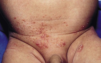
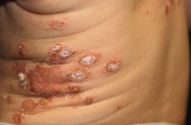
 ). However, postinflammatory pigmentary changes may persist for weeks to months. Multiple small pustules on the abdomen and diaper area are characteristic of staphylococcal pustulosis (Fig. 12.3).7 The healing sites from circumcision and the healing umbilical cord can be important niduses of infection in neonates.
Extracutaneous findings
Etiology and pathogenesis
Diagnosis
Differential diagnosis
Course, management, treatment, and prognosis
Intertrigo
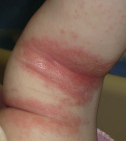
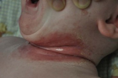
Cutaneous findings
Extracutaneous findings
Etiology and pathogenesis
Differential diagnosis
Diagnosis
Course, management, treatment, and prognosis
Folliculitis/pustulosis/furunculosis
Cutaneous findings
Extracutaneous findings
Etiology and pathogenesis
Diagnosis
Differential diagnosis
Course, management, treatment, and prognosis
Infection of pre-existing erosions and ulcerations
Cutaneous findings
Extracutaneous findings
Etiology and pathogenesis
Diagnosis
Differential diagnosis
Course, management, treatment, and prognosis
Paronychia
Funisitis/omphalitis
Cutaneous findings
Extracutaneous findings
Etiology and pathogenesis
Diagnosis
Differential diagnosis
Course, management, treatment, and prognosis
Cellulitis
Cutaneous findings
Extracutaneous findings
Etiology and pathogenesis
Diagnosis
Differential diagnosis
Course, management, treatment, and prognosis
Periorbital/orbital cellulitis
Cutaneous findings
Extracutaneous findings
Etiology and pathogenesis
Diagnosis
Differential diagnosis
Course, management, treatment, and prognosis
Subcutaneous and systemic infections
Abscesses
Cutaneous findings
Extracutaneous findings
Etiology and pathogenesis
Diagnosis
Course, management, treatment, and prognosis
Breast abscess
Cutaneous findings
Extracutaneous findings
Etiology and pathogenesis
Diagnosis
Course, management, treatment, and prognosis
Related posts:
![]()
Stay updated, free articles. Join our Telegram channel

Full access? Get Clinical Tree


Bacterial Infections
12