Facial volume loss is an important component of facial aging, especially in the periocular region. The authors evaluate the normal and aging anatomy of the periocular region and then discuss volume restoration of this region using hyaluronic acid, calcium hydroxylapatite, and autologous fat transfer. Preoperative assessment, operative technique, postoperative care, and complications are addressed.
Volume loss has increasingly been recognized as an important aspect of facial aging. This is especially true of the periocular region. Restoration of this lost volume can be achieved through placement of syringe-based fillers, autologous fat, or implants. This article discusses the use of syringe-based fillers (hyaluronic acid, calcium hydroxylapatite) and autologous fat to rejuvenate the periorbital region.
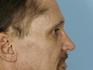
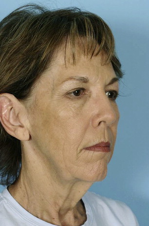
The periorbital complex consists of the brow, superior orbital rim, upper eyelid, lateral canthus, lower eyelid, inferior orbital rim, and upper cheek. The most important of these in the aging process of volume loss is the interface between the lower eyelid and upper cheek or midface. Systematic aging begins throughout the periorbital complex beginning in the patient’s mid-to-late 30s. The extent and rapidity of periorbital aging varies between individuals and is strongly dependent on the relationships between the bony orbit, globe, and malar complex. The periorbital area ages at a faster pace and earlier in life with a negative vector midface, much as the jaw line and neck age earlier in people with microgenia and a short thyromental distance ( Fig. 1 ). Some individuals even display “lower eyelid bags” in youth; these bags appear early because of the negative vector produced by the deficient anterior projection of the inferior orbital rim in relation to the globe.
Youth
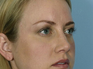
The youthful upper periorbital complex consists of a brow that is full over its entire height, being propped up by the volume of the brow fat pad. Entire articles have been written and rewritten about the normal aesthetic height of the brow. The authors are well aware of these aesthetic norms but maintain that patients differ tremendously regarding their natural brow height. They ask their patients routinely about their brow position in youth and strive to restore this relationship, only changing natural brow position after careful consideration. Comparing photos in the latest fashion magazine shows many examples of models, all of whom are exquisitely attractive, with significantly differing relationships between the brow and superior orbital rim ( Fig. 2 ). The upper eyelid also shows a variable fullness between patients; all may be considered youthful and attractive. Some individuals have significant tarsal show with a deep superior orbital sulcus, a high lid crease, and very little dermatochalasia. Others have very little tarsal show with a more prominent orbital fat component and therefore, a much fuller-appearing upper eyelid and a tendency toward greater dermatochalasia. In the authors’ opinion, the restoration of the youthful upper eyelid and brow complex must be tailored to each patient’s unique characteristics and must strive toward the restoration of youthfulness and not an ideal appearance based on the biases of the surgeon. Some rejuvenation of the upper eyelid complex relies on surgical lifting procedures that are beyond the scope of this article. However, restoration of the upper periorbital volume is addressed.
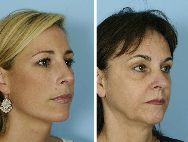
The youthful lower eyelid complex revolves around the appearance of a short lower eyelid, or rather, a superiorly placed and full upper cheek. The lower eyelid cheek interface should be at the lower tarsal border and flow into a full convex upper anterior cheek. This natural convex fullness results from the quantity of the lower eyelid suborbicularis oculi fat or SOOF and also depends heavily on inferior orbital rim projection ( Fig. 3 ). The youthful lower eyelid must also lack pseudoherniation of orbital fat. The cheek skin should be smooth over the underlying fat and the malar cheek fat pad should be shaped as a teardrop, with the rounded leading edge of the tear inferior-medial and tapering laterally over the anterior aspect of the zygomatic arch. The inferior aspect of this teardrop should create a subtle shadow in its interface with the buccal region that parallels that of the jaw line ( Fig. 4 ). These features lead to the overall appearance of the heart-shaped face of youth.
Aging
Aging is the culmination of a multifactorial process that includes the actions of gravity, volume loss, and skin changes due to intrinsic and extrinsic factors. Volume loss in the periorbital area leads to exposure of harsh bony contours and the creation of shadows indicating aging. In the superior rim, this also leads to an apparent descent of the brow. As volume is lost over the bony orbital rim, the support for the soft tissue brow is lost. This effectively raises the position of the bony rim, which is now harsh and skeletonized, and leads to an apparent drop in brow height because the hair-bearing eyebrow now rests in a lower position relative to the superior orbital rim. This volumetric contribution to brow aging is not the only component. Forehead, brow, and upper eyelid aging is a complex process with a gravitational contribution that may benefit from surgical lifting procedures; however, volume is an important consideration. Photos of the patient when young are a valuable tool in assessing the relative contributions of different factors in periorbital aging, thus assisting the surgeon in the appropriate selection of rejuvenation techniques.
Lower periorbital/cheek aging is most significantly influenced by volume-related changes. With time, the heart-shaped youthful face gives way to the more rectangular face of age. Some of this is due to the development of jowling and lower facial aging, which is beyond the scope of this discussion. The loss of volume in the lower eyelid/cheek complex, however, is a key contributor to the rectangular face. Additionally, volume loss allows the appearance of shadows, as tissues fall and become tethered by various retaining ligaments. Double contours arise in the lower eyelid and cheek, with exposed orbital fat creating a bulge, the exposed bony orbital rim creating a hollow, malar mound bulge, and malar septum hollow ( Fig. 5 ). The lower eyelid gains apparent length in this process. Pseudoherniation of lower eyelid orbital fat combined with loss of orbital rim volume leads to lengthening of the lower lid height and inferior placement of the lower eyelid cheek junction. This is the orbital groove/tear trough deformity. Below this, the malar mound and malar septum create a second double contour in the cheek region. Restoration of youth combines removal of orbital fat pseudoherniation, if indicated, and placement of volume into the orbital groove/tear trough and cheek region to restore the single convexity of the cheek and raise the cheek eyelid junction, thereby shortening the apparent lower lid height.
Patient assessment
Although people are becoming more educated about volume loss as a cause of their facial aging, patients rarely present for consultation requesting a fat transfer. Patients express concern that they appear tired or that they have persistent dark circles under their eyes. A detailed assessment is necessary to determine the cause of their concerns and what part of this is due to volume loss. Review of patient photographs from their youth is helpful, but if these are not available, pointed questions about eyelid appearance and brow height can help differentiate between age-related changes and normal anatomic variation. A detailed history and physical examination is indicated; however, the authors limit discussion to the key factors related to volume replacement. Much of forehead and upper eyelid rejuvenation relies on incisional techniques. However, brow position and brow fat quantity should be assessed. In many instances, the brow height is adequate, and adding volume alone to the superior orbital rim will restore a youthful appearance. A hollow superior orbital sulcus may also be restored with volume, although this is an advanced technique and careful patient consultation should take place regarding the likelihood of contour irregularities. The authors address the hollow superior orbital sulcus with a patient only if it is a specific concern and usually offer intervention only if it is due to previous surgical misadventures and not a natural occurrence. The mainstay of patient assessment for volume replacement involves the lower eyelid and cheek.
Assessment of the lower eyelid and cheek begins with determining if orbital fat pseudoherniation requires addressing. This determination is made by evaluating the patient in oblique and lateral views. If the orbital fat protrudes anterior, beyond the surgeon’s perception of a natural convex cheek eyelid interface, a blepharoplasty should be considered ( Fig. 6 ). This usually involves only the medial and middle fat compartments because the lateral fat compartment less commonly protrudes beyond the desired convex line. A transconjunctival technique is used with minimal exposure because the tissue planes need to be preserved over the orbital rim for concurrent fat grafting. An assessment of overall midface volume and position is also performed, noting a negative vector midface (prominent eyes), the presence of a malar groove, malar septum, and malar mound, especially if early festooning is present. Assessment of the lower cheek is also made, including buccal hollowing and perioral volume loss. Once assessments of the relative contributions of volume loss have been made, a conversation with the patient may begin regarding intervention.
Related posts:


Stay updated, free articles. Join our Telegram channel

Full access? Get Clinical Tree


