© Springer-Verlag Berlin Heidelberg 2015
Miranda A. Farage, Kenneth W. Miller, Nancy Fugate Woods and Howard I. Maibach (eds.)Skin, Mucosa and Menopause10.1007/978-3-662-44080-3_1414. Atrophic Vaginitis in the Menopause
(1)
Department of Obstetrics and Gynecology, Jefferson Medical College, Jefferson University Hospitals, 834 Chestnut St., Suite 400, Philadelphia, PA 19107, USA
(2)
Division of Infectious Diseases, Department of Internal Medicine, Detroit Medical Center, Wayne State University School of Medicine, Detroit, MI, USA
Vulvovaginal atrophy (VVA) is a chronic medical entity in both postmenopausal women and women during menopause transition secondary to decreased levels of circulating and consequent local vulvovaginal estrogen [1–3]. Atrophic vaginitis is considered by some to represent a more advanced form of vaginal atrophy in which evidence of vaginal inflammation supervenes with accompanying additional signs and symptoms. Traditionally, however, many authors have used the terms vaginal atrophy and atrophic vaginitis interchangeably.
It is estimated that up to 20–60 % of postmenopausal women experience symptoms of vaginal atrophy with 20–25 % of symptomatic women seeking medical treatment [2]. VVA and atrophic vaginitis are significantly increased in women taking aromatase inhibitors and some selective estrogen receptor modulators (SERMs, e.g., tamoxifen, raloxifene) [4].
The results of Women’s Health Initiative (WHI) led to a dramatic decline in routine use of estrogen as preventative therapy for a variety of host maladies in postmenopausal women. With estrogen elimination the incidence of VVA has markedly increased.
14.1 Pathophysiology
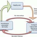
Estrogen components influence vulvovaginal physiology at several levels. The progressive decline in levels of endogenous estrogens is accompanied by a decrease in vaginal epithelial glycogen which serves as a major nutritional substrate both for epithelial cells per se and for vaginal microorganisms or flora (microbiota). Reduced glycogen substrate discourages the presence and population numbers of commensal protective bacteria, predominantly dominant Lactobacillus species which profoundly influence vaginal microbial communities. Accordingly with time, Lactobacillus numbers decline progressively and with significant consequences. Firstly decreased bacterial glycogenolysis results in decreased production of several organic acids but predominantly lactic acid, and consequently vaginal pH is altered with a progressive increase in pH above the normal acidic range (3.8–4.5). The normal acidic environment which fosters the presence of protective acidophilic Lactobacillus species is also an adversarial barrier to organisms originating from the neighboring gastrointestinal tract, specifically coliform bacteria. Accordingly, declining serum and local estrogen concentrations directly influence the healthy normal bacterial community or microbiome present in the vagina. The profoundly altered vaginal microbiome does not directly contribute to vaginal symptoms associated with vulvovaginal atrophy; hence, simply artificially reducing vaginal pH per se may not be an effective treatment modality for VVA. The role of estrogen in directly influencing microbial growth is poorly studied. Details of the vaginal microbiome associated with VVA are outside the scope of this review and are found elsewhere [5]. The microbiome of the atrophic vagina is mixed or heterogeneously devoid of Lactobacillus species, instead consisting of streptococci, staphylococci, and anaerobes such as Prevotella spp. and finally diphtheroids [6].
The second pathophysiologic consequence of declining estrogen relates to the stratified squamous epithelium lining of the vagina. Estrogen enhances maturation of basal epithelial cells, facilitating transition through parabasal cells to reach the mature larger squamous cells. In doing so the vaginal lining becomes considerably thickened and allows enhanced transudation of serum-derived fluid into the vaginal lumen. Accordingly, with decline in estrogenic effect, vaginal epithelium becomes progressively thinned, atrophic, more fragile, and accompanied by vaginal dryness reflecting the reduced transudation.
Declining estrogen also reduces function of both striated and smooth muscle, including bladder and pelvic floor. This contributes to bladder, rectum, uterus, and vault prolapse, as well as loss of muscle tone resulting in a patulous or dilated introitus. Bladder muscle and sphincter function progressively decline contributing to incontinence, post-voiding urine retention, and susceptibility to bladder bacterial infection.
There are no established definitions that separate atrophic vaginitis from VVA. The most prominent feature in addition to severity of symptoms and signs relates to presence of large numbers of polymorphonuclear leukocytes (PMNs) evident on saline microscopy suggesting an inflammatory process that is superimposed upon atrophy. The causes of PMN influx are unknown. No microorganisms are currently incriminated in their arrival, and some experts attribute the inflammatory component as secondary to minor/major trauma or friction that occurs with coitus especially in the presence of vaginal dryness.
14.2 Clinical Manifestation
The North American Menopause Society and International Menopause Society estimate that 10–50 % of postmenopausal women experience symptoms of VVA [7]. The earliest manifestation of estrogen deficiency, long before changes in pH, microbiota, and epithelial lining thinning, is vaginal dryness first recognized only during intercourse. Over time, dryness is progressive after making intercourse difficult, uncomfortable, painful, and even impossible [8].
Even in the absence of intercourse, VVA may be associated with irritation and itching. With worsening atrophy and thinning of the vulvovaginal lining, increased tenderness supervenes, thereafter the vaginal mucosa becomes friable, with petechiae, occasional ulceration, and postcoital bleeding. Vaginal elasticity is also lost. In spite of dryness, occasionally women are troubled by a thin watery discharge often necessitating use of pad. Infrequently, women will report new onset of genital malodor, not fishy but unpleasant in the presence or absence of discharge. Other symptoms include vulvovaginal pruritus and paresthesia. Urinary symptoms including burning on micturition, frequent urinary tract infections, and incontinence are also commonly present.
Physical findings include a dilated or later narrowed introitus, vestibular thinning, and prominent urethral caruncle often with petechiae. Diffuse vaginal epithelial surface thinning, pallor, and loss of rugae reflect vaginal atrophy with petechiae and ecchymoses, e.g., indicating advanced estrogen deficiency. Dyspareunia may also be the result of fissuring, ulceration, or stretching of the deep tissue surrounding a stenotic introitus or narrowed or shortened vagina. Vulvar findings include pubic hair loss, pendulous labia majora, less distinct labia minora, and less labial fat pad.
Unlike vasomotor manifestations such as hot flashes or night sweats, signs and symptoms of vaginal atrophy tend to increase over time [3]. Bachman et al. have emphasized that the aforementioned symptoms of vulvovaginal atrophy frequently overlap with those of female sexual dysfunction, i.e., reduced libido, arousal disorder, orgasmic disorders, and sexual pain disorder common in up to 50 % of postmenopausal women [3]. The role of reduced estrogen activity in the pathogenesis of these symptoms is suggested, given their higher frequency in women with concomitant vaginal atrophy. Postmenopausal women with sexual difficulties are significantly more likely to have VVA than women without.
14.3 Laboratory Findings
In the early phases, even in the presence of vaginal dryness, all commonly performed bedside laboratory tests performed on vaginal samples are normal. Established estrogen deficiency is associated with an elevated vaginal pH, in excess of 4.5, often 5–6. The amine or whiff test is always negative. Saline microscopy reveals absence of clue cells and typical Lactobacillus morphotype organisms reflecting altered vaginal flora (AVF). Most importantly, diagnosis requires the presence of parabasal cells, an essential marker of impaired epithelial cell maturation. Atrophic vaginitis is characterized by the further addition of increased numbers of PMNs.
Many clinicians use serum estradiol levels to diagnose estrogen-deficient vaginal atrophy; however, serum estradiol levels correlate poorly with the effects of local estrogen deficiency and add little to diagnosis, prognosis, and response to local estrogen replacement therapy.
Related posts:
 Effects of Hormone Replacement Therapy on Skin Viscoelasticity During Climacteric Aging
Gynaecological Problems Associated with Menopause
Sensory Perception on the Vulva and Extragenital Sites
Physical Activity and Quality of Life During Menopausal Transition and Postmenopause
Effects of Hormone Replacement Therapy on Skin Viscoelasticity During Climacteric Aging
Gynaecological Problems Associated with Menopause
Sensory Perception on the Vulva and Extragenital Sites
Physical Activity and Quality of Life During Menopausal Transition and Postmenopause
 Changes to Skin with Aging and the Effects of Menopause and Incontinence
 Skin and Menopause
Stay updated, free articles. Join our Telegram channel

Full access? Get Clinical Tree


