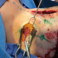
Fig. 14.1
The vessel is traced up from sites 5B to Site 5A with a handheld Doppler. This is done prior to infiltration of epinephrine to identify the vessel and localize the area of nerve compression
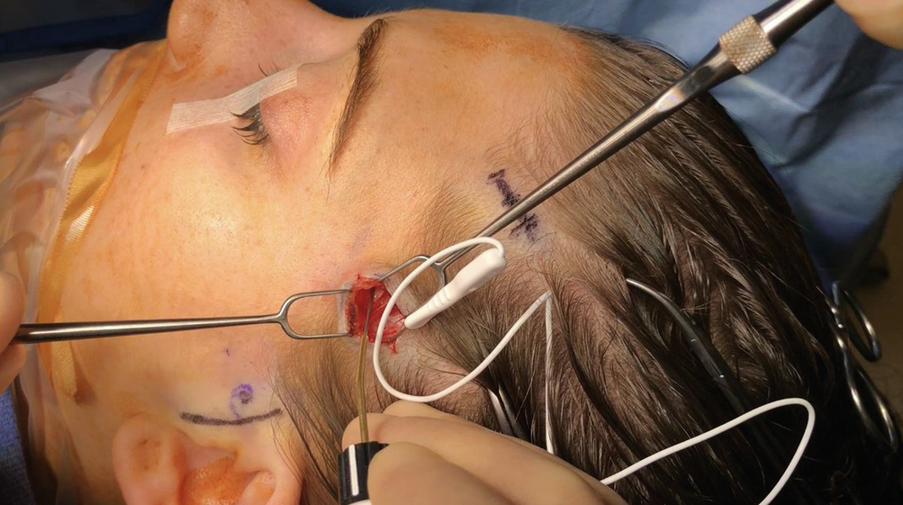
Fig. 14.2
A nerve stimulator is used to ensure the nerve is sensory in origin and not a branch of the facial nerve
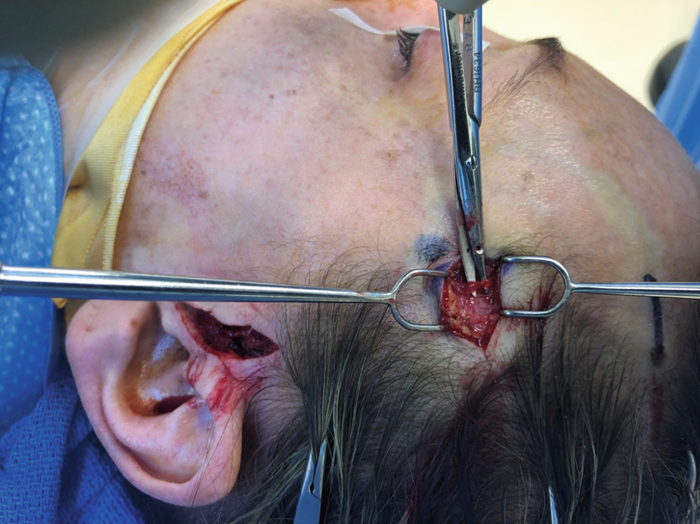
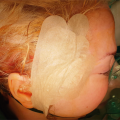
Fig. 14.3
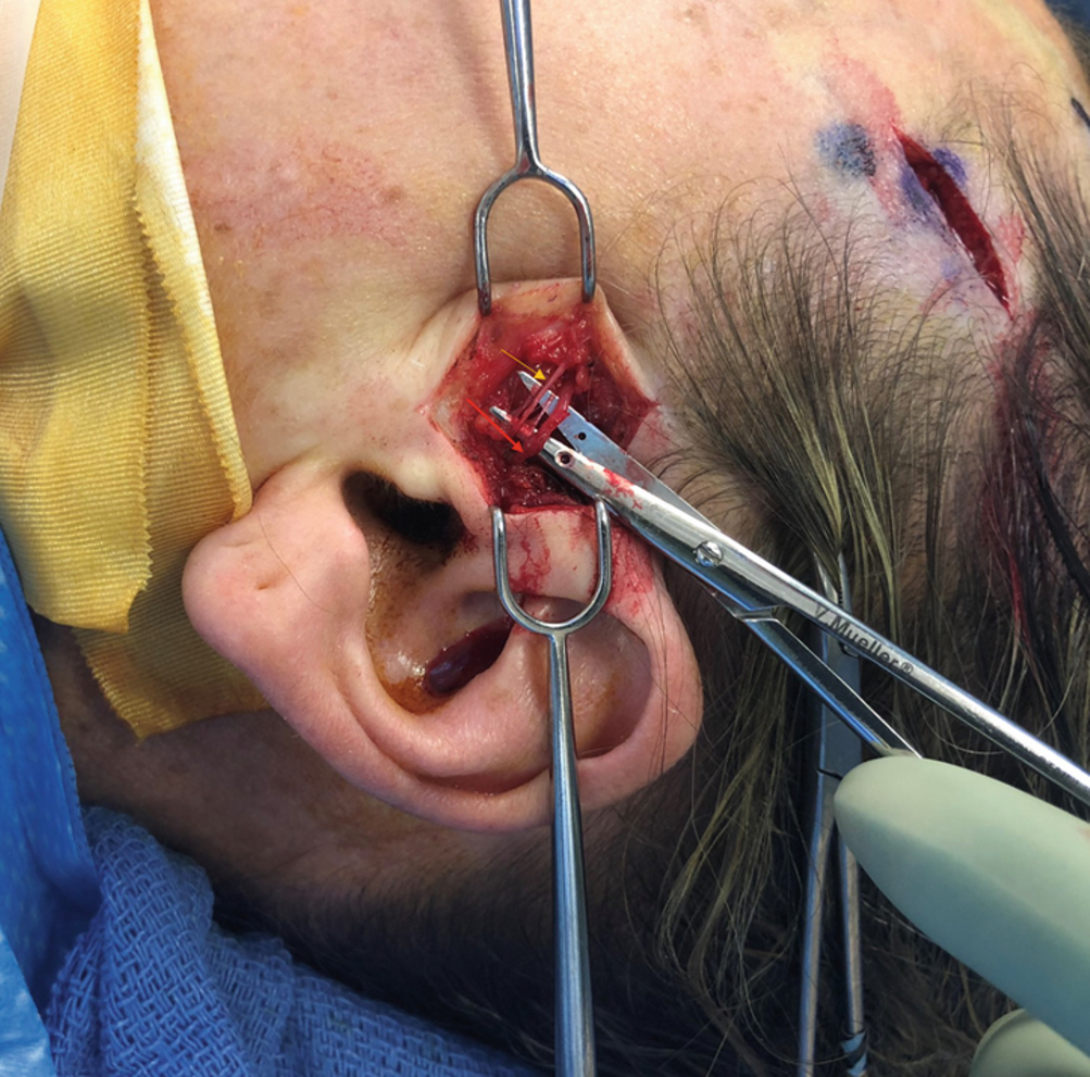
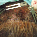
With the skin tented up with double hooks, the nerves and arteries are dissected out with blunt-tip scissors. A small spread technique parallel to the neurovascular structures is utilized to avoid any unnecessary trauma or bleeding to the surrounding tissue. In this image, the artery is dissected out first. Site 5A avulsion/decompression is performed prior to site 5B
Related posts:
 Anatomy of Craniofacial Nerves Regarding Migraine Surgery
 Neurotoxin: Basic Facts, Physiology and Pharmacology
 Anatomy of the Vascularization and Innervation of the Human Scalp
 Modification of Site 4 Decompression: Endoscopic Assisted Decompression of the Greater Occipital Nerve (GON) with Radical Excision of the Occipital Artery
 of Temporal Trigger Sites
 of Occipital Trigger Sites
Stay updated, free articles. Join our Telegram channel

Full access? Get Clinical Tree


