Preoperative Doppler of the temporal region
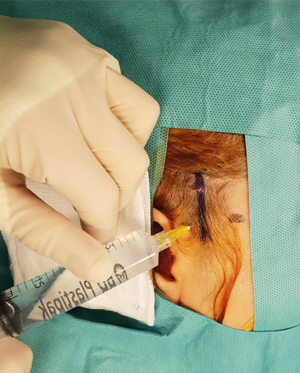
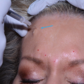
Local anesthesia
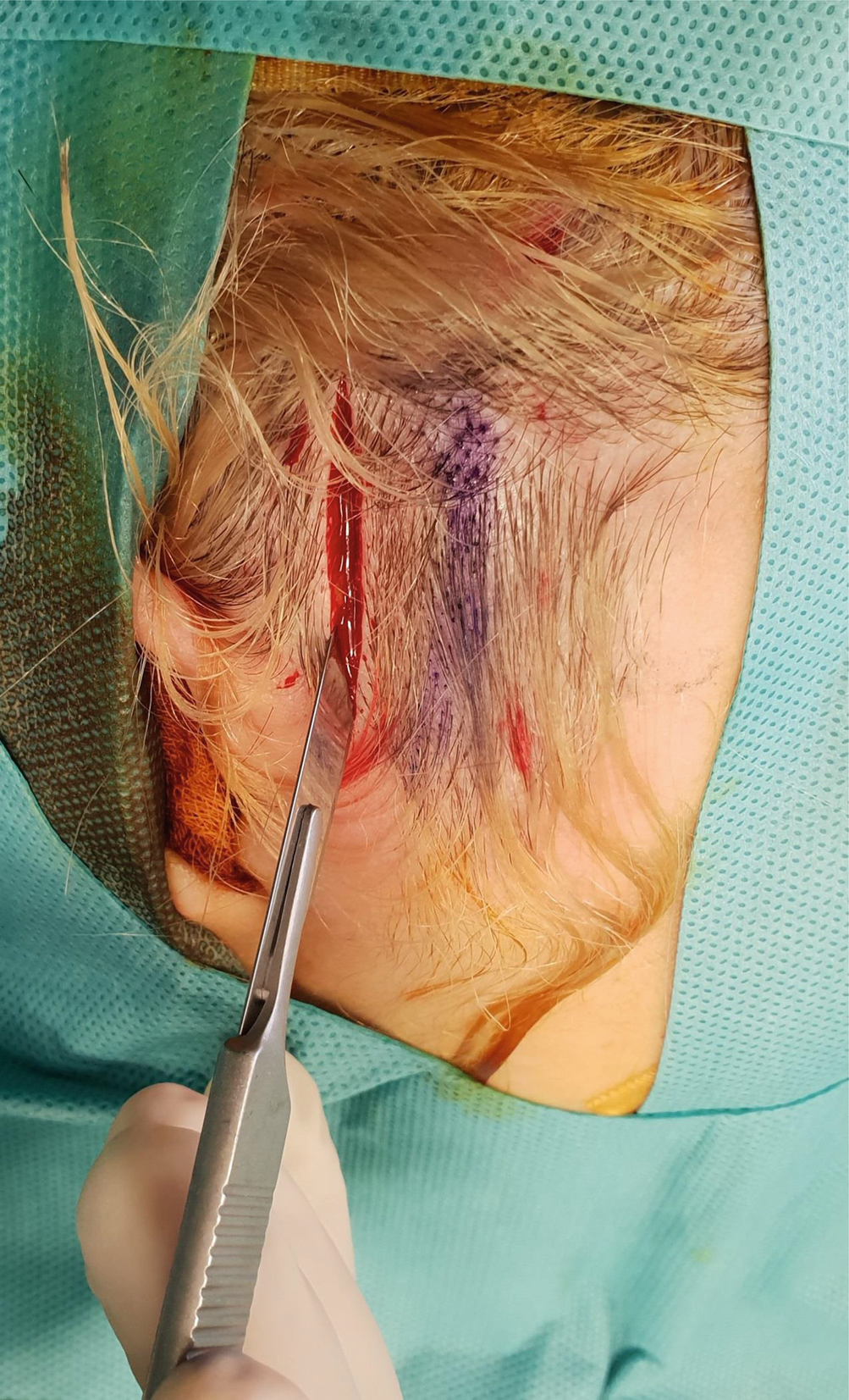
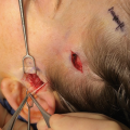
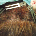
Skin incision
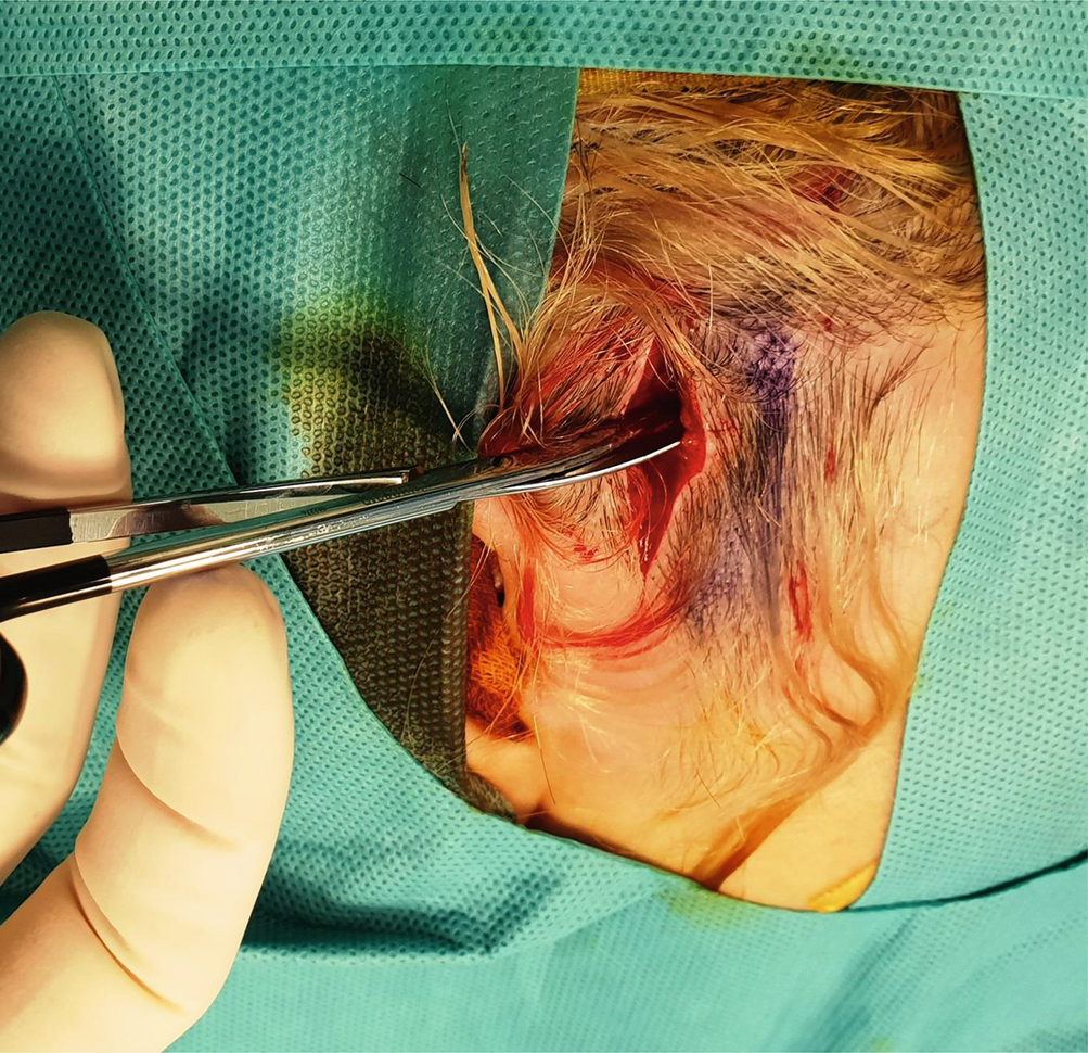
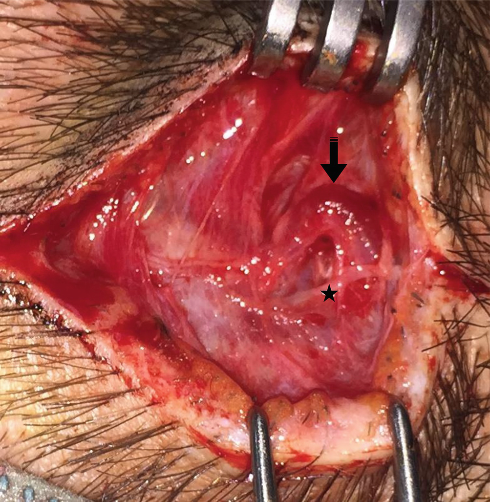
Subcutaneous undermining above the superficial temporal fascia
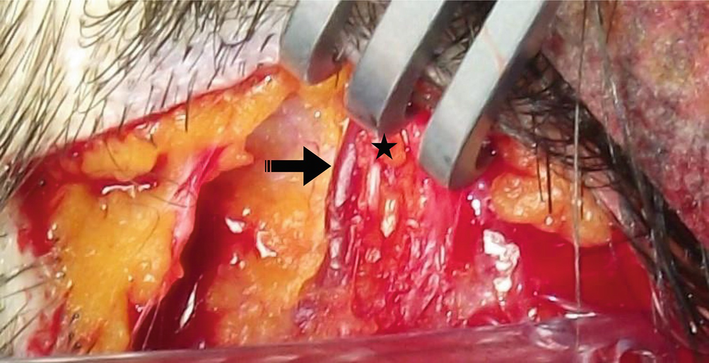
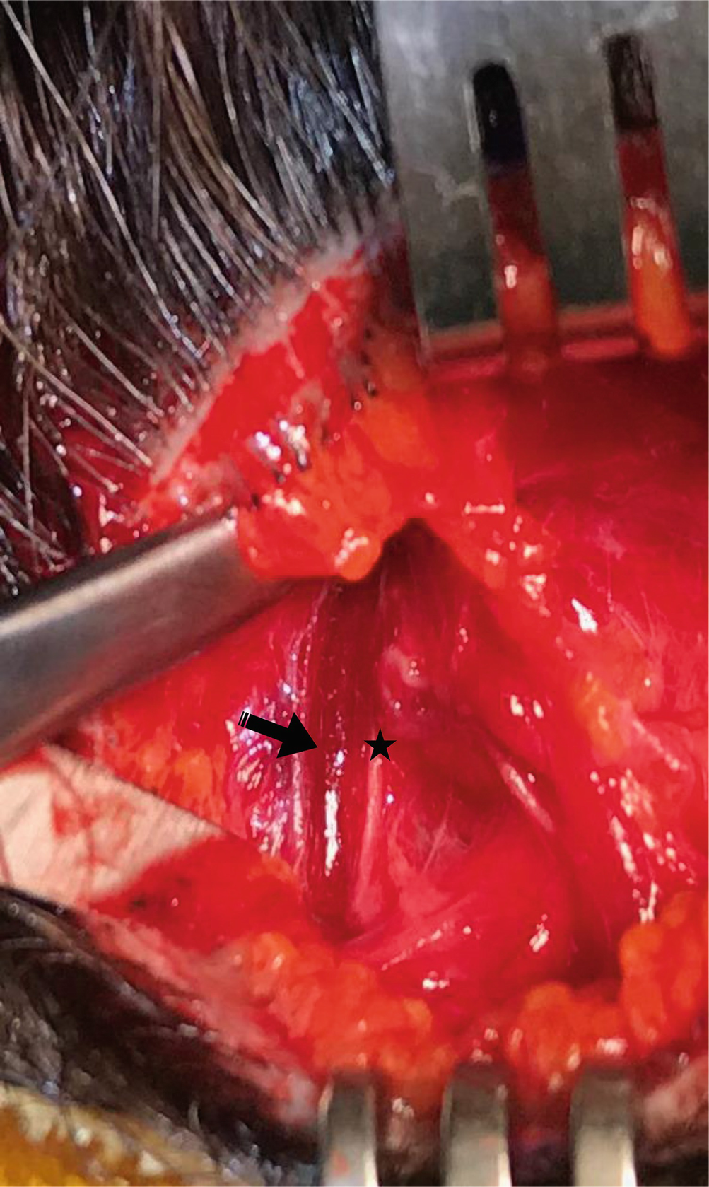
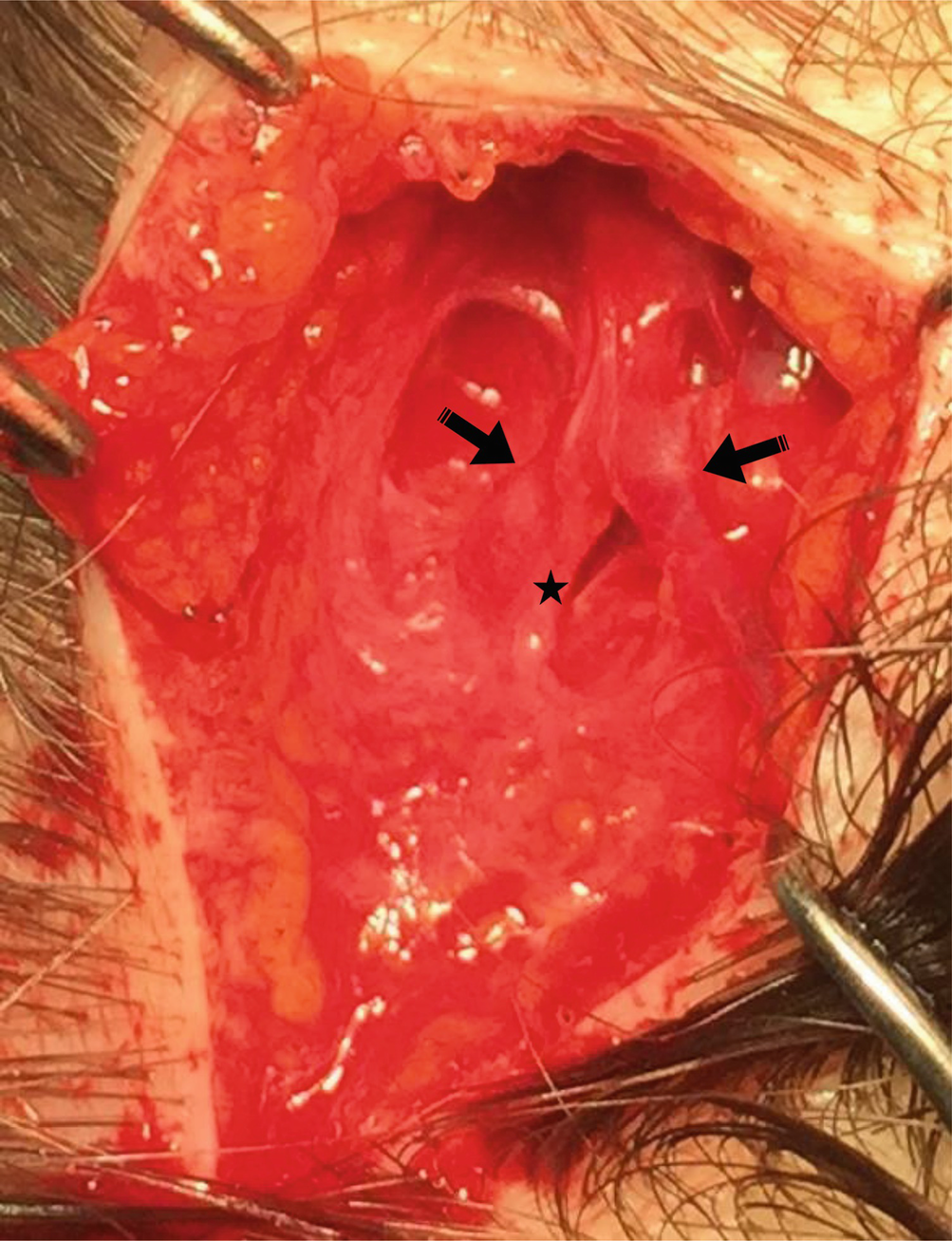
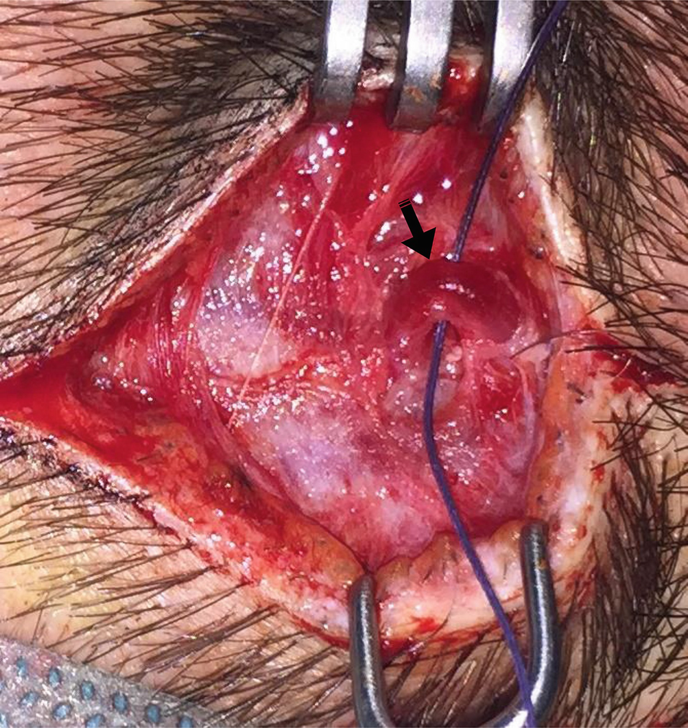
Superficial temporal artery (arrow) running anteriorly and parallel to the auriculotemporal nerve (star)
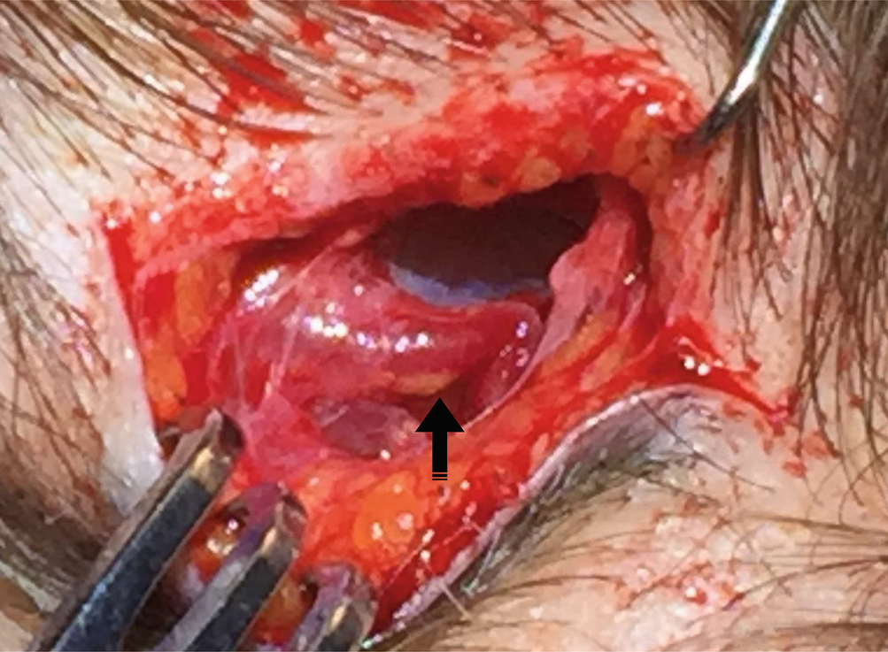
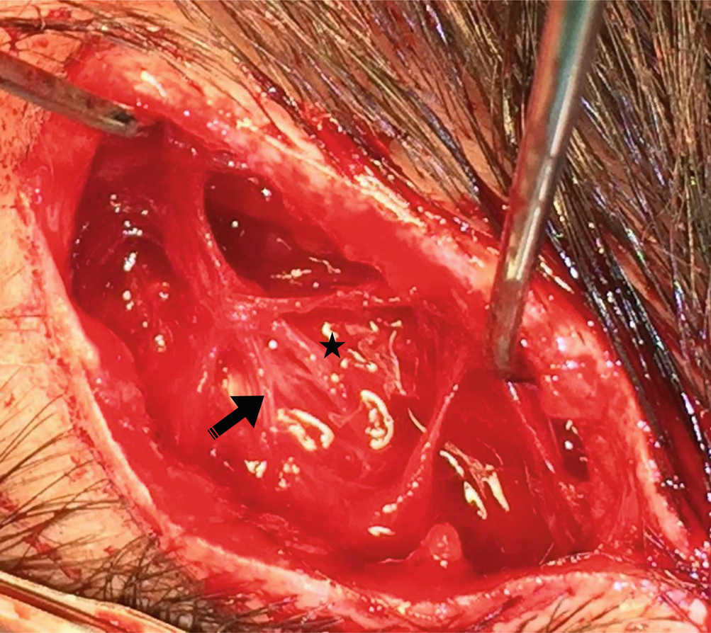
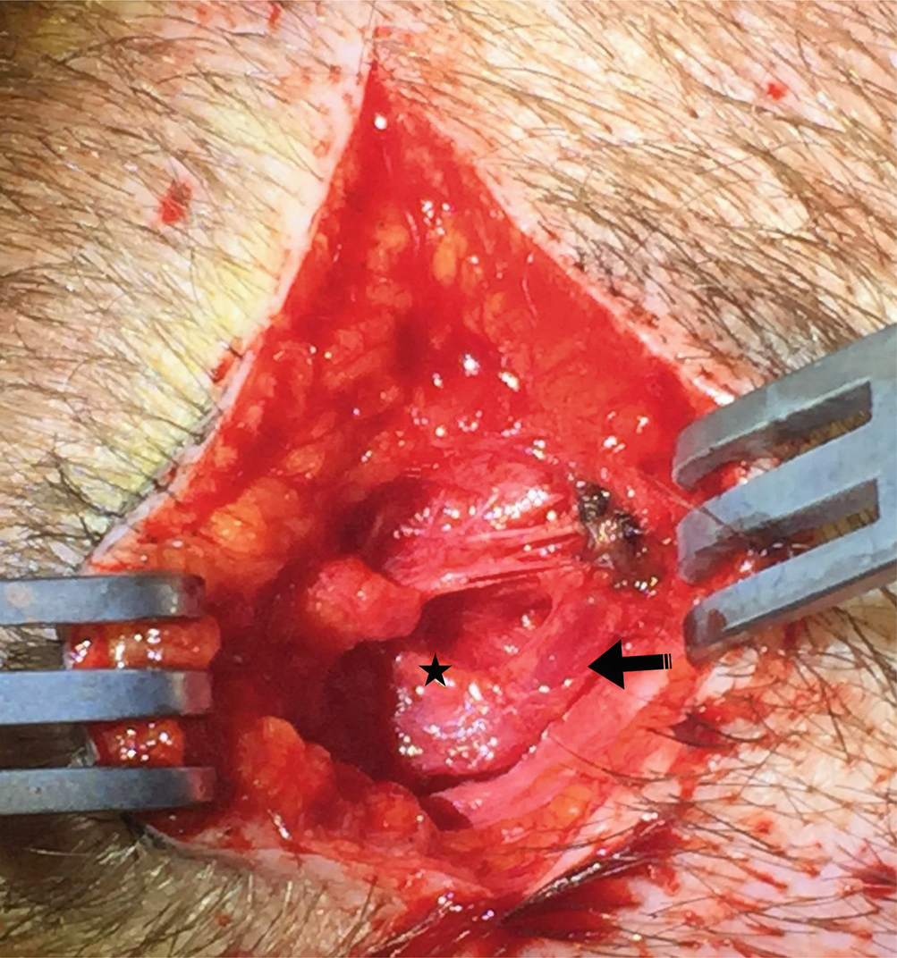
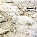
It is not uncommon to find an abnormally dilated superficial temporal artery (arrow)
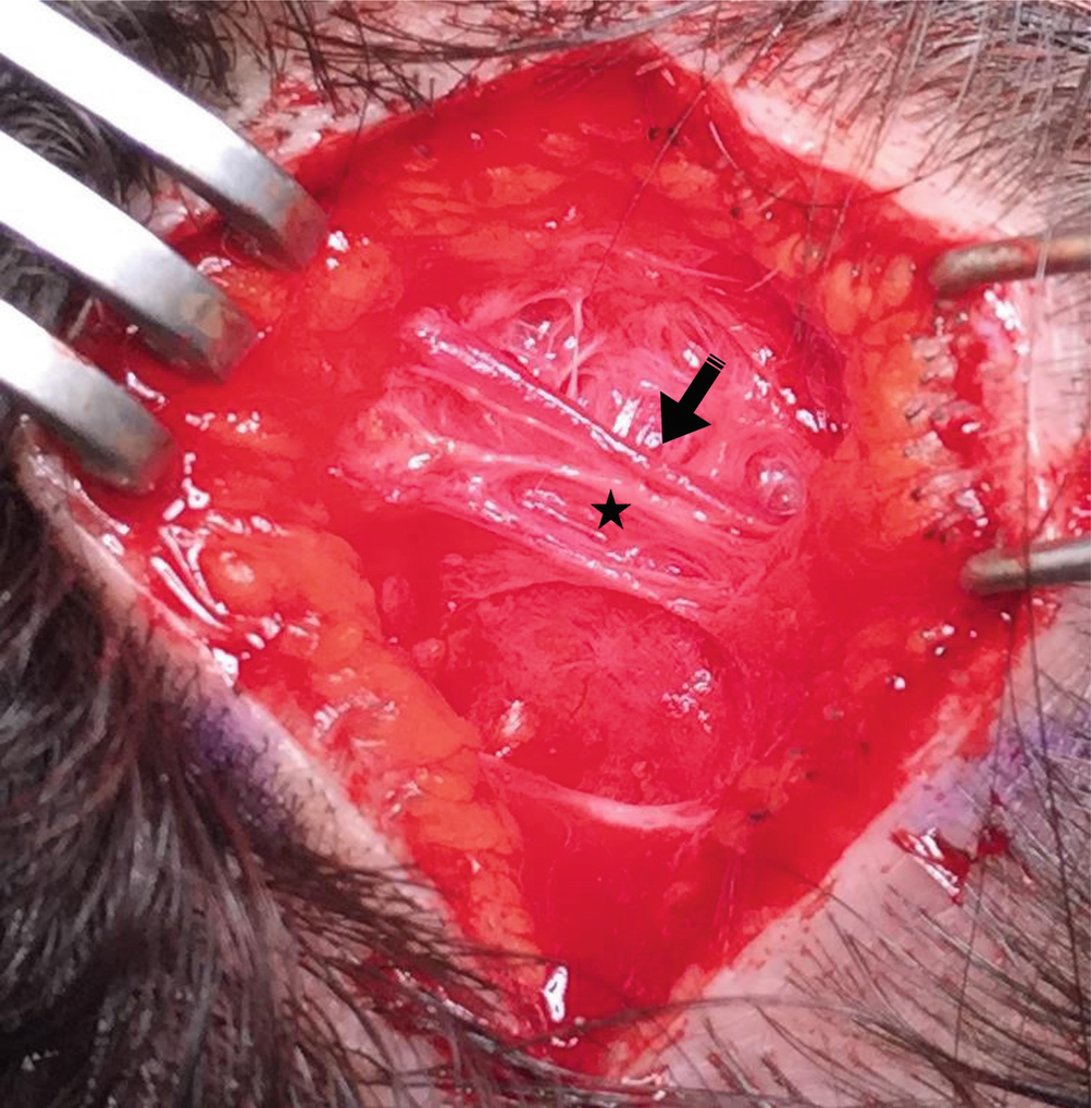
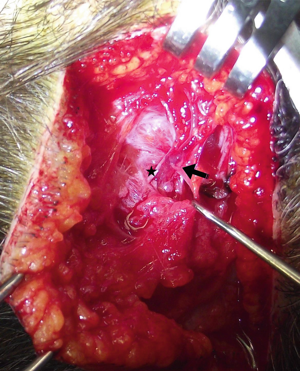
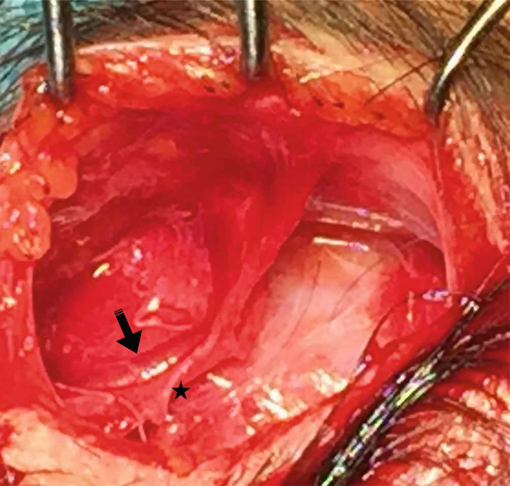
Dilated superficial temporal artery (arrow) crossing below the auriculotemporal nerve (star)
Dilated superficial temporal artery (arrow) crossing below the auriculotemporal nerve (star)
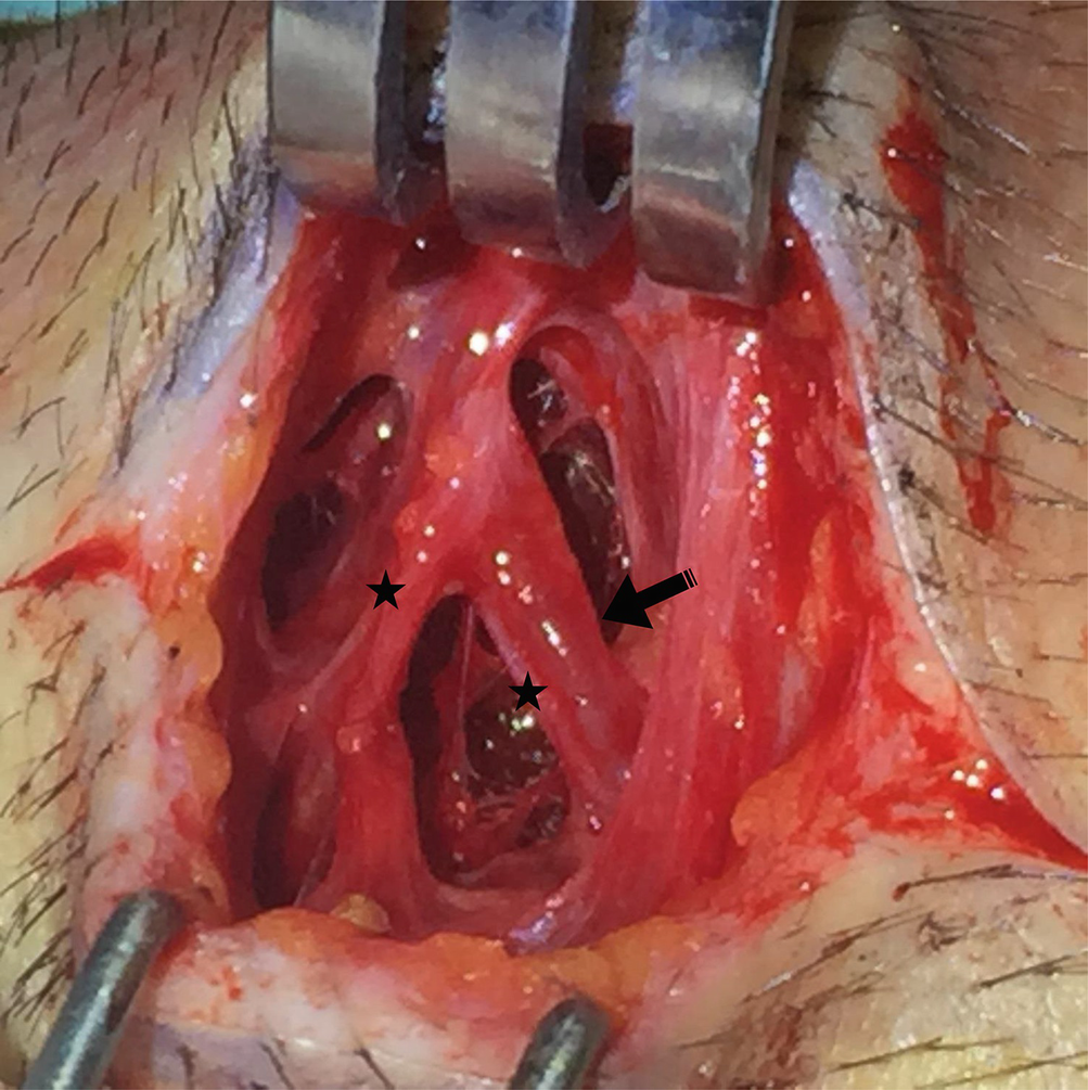
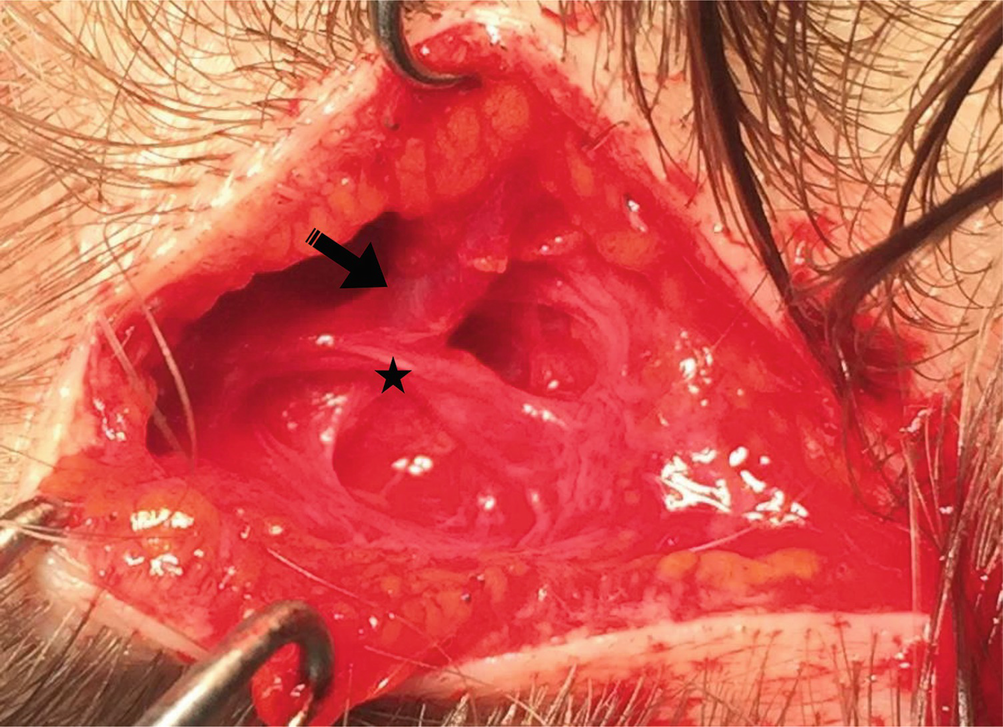
Dilated superficial temporal artery (arrow) running cephalic to the auriculotemporal nerve (star) is isolated from the surrounding superficial temporalis fascia
Superficial temporal artery (arrow) running parallel and in close relation to the auriculotemporal nerve (star)
Superficial temporal artery (star) running parallel and above to the auriculotemporal nerve (arrow)
Superficial temporal artery (arrow) running parallel close to the auriculotemporal nerve, which bifurcates distally (star)
Dilated superficial temporal artery (arrow) in close proximity to the auriculotemporal nerve (star)
Bifurcated and dilated superficial temporal artery (arrows) intertwined with the auriculotemporal nerve (star)
Dilated superficial temporal artery (arrow) lateral to the auriculotemporal nerve (star)
Dilated superficial temporal artery (arrow) running parallel to the auriculotemporal nerve (star), which appeared thinned as a consequence of chronic compression
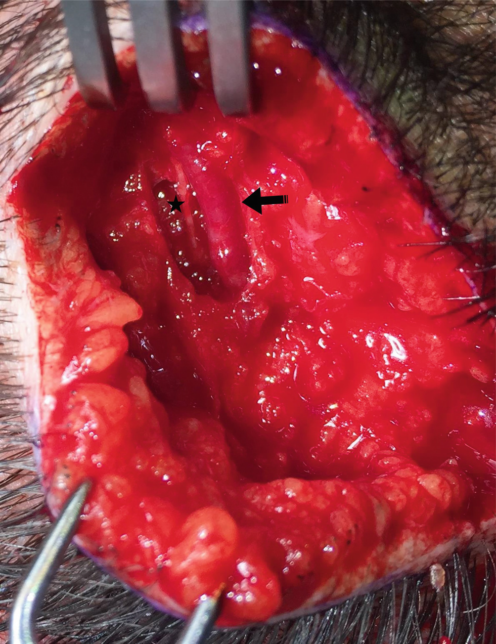
Dilated superficial temporal artery (arrow) running below the auriculotemporal nerve (star)
Dilated superficial temporal artery (arrow) running below the auriculotemporal nerve (star)
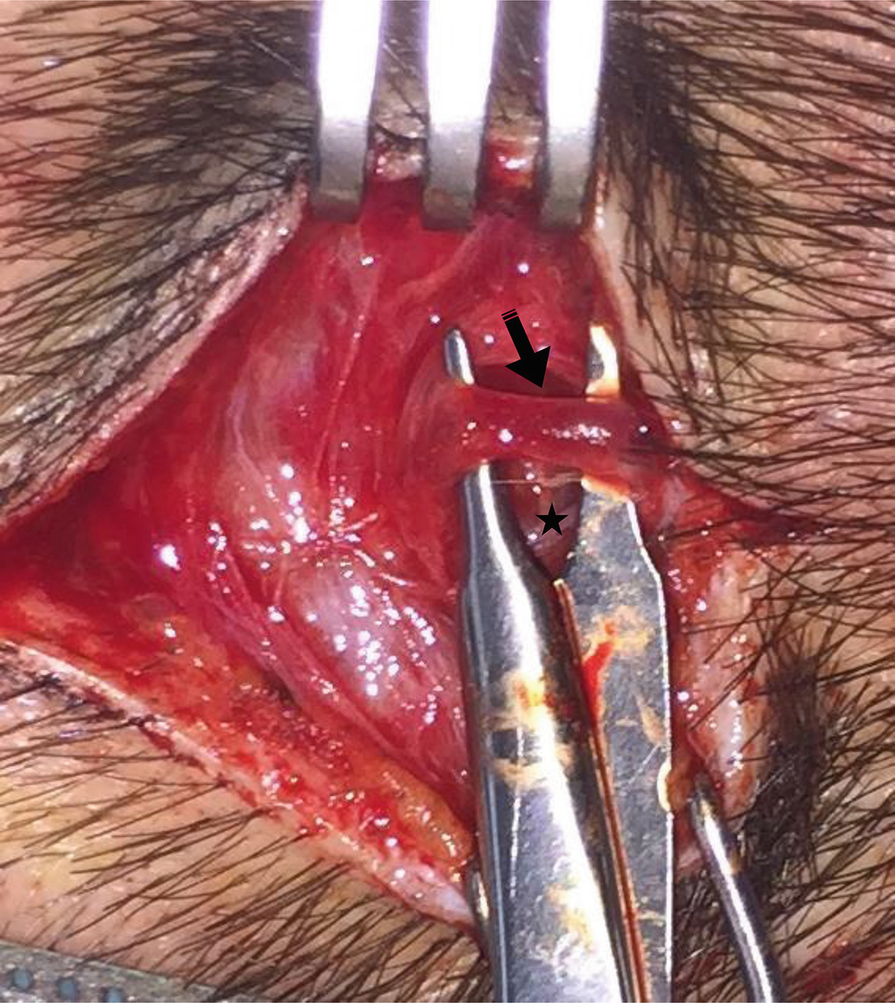
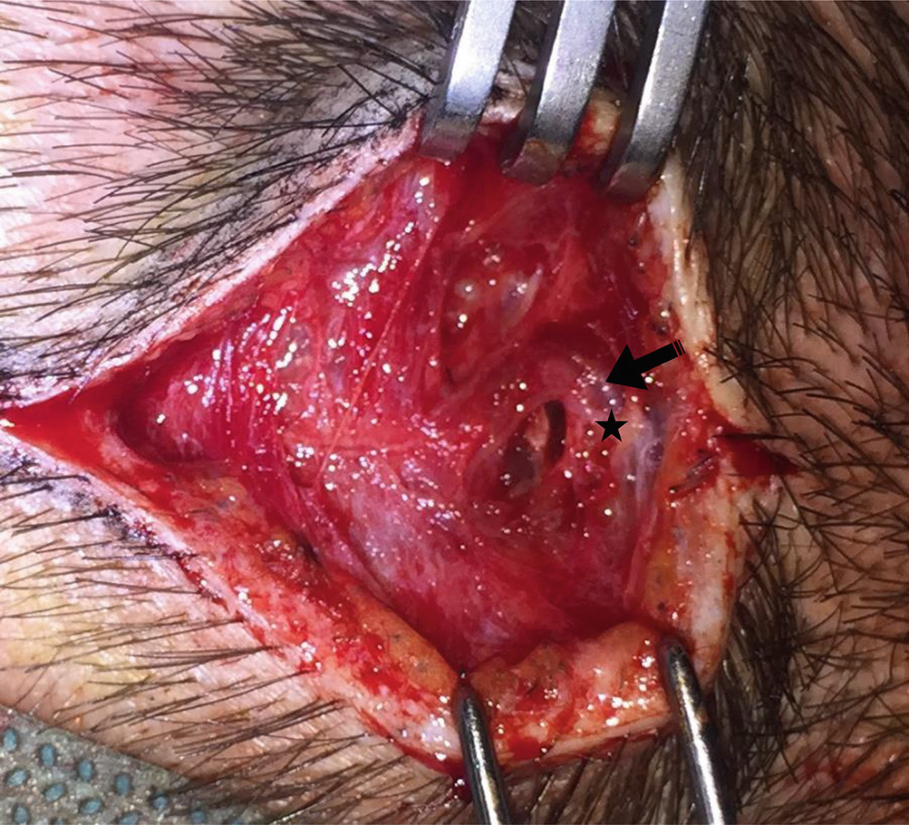
Superficial temporal artery (arrow) needs to be isolated from the surrounding superficial temporal fascia and from the auricolotemporal nerve that is usually in close proximity (star)
Related posts:
 Approach for Auriculotemporal Nerve Decompression, Amirlak Modification
 Anatomy of Craniofacial Nerves Regarding Migraine Surgery
 Regional, Targeted (ART) Botulinum Toxin Injection for Migraine and Chronic Headaches
 Anatomy of the Vascularization and Innervation of the Human Scalp
 of Frontal Trigger Sites
 of Occipital Trigger Sites
Stay updated, free articles. Join our Telegram channel

Full access? Get Clinical Tree


