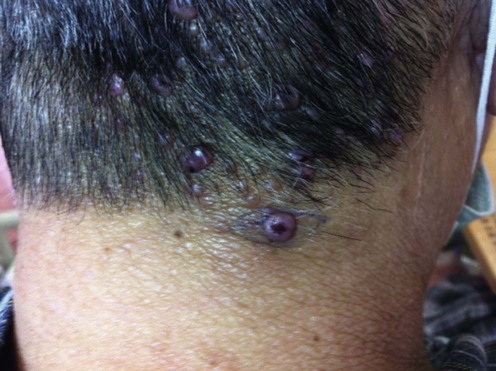
13 Angiolymphoid hyperplasia with eosinophilia William Y-M Tang and Loi-yuen Chan Evidence Levels: A Double-blind study B Clinical trial ≥ 20 subjects C Clinical trial < 20 subjects D Series ≥ 5 subjects E Anecdotal case reports Angiolymphoid hyperplasia with eosinophilia (ALHE) was first described by Wells and Whimster in 1969. It is a benign vascular proliferation of unknown etiology with a characteristic component of epithelioid endothelial cells. It is an uncommon disease, so data on its natural course and treatment response are based on a small number of patients. ALHE usually affects women in their third decade and presents as cutaneous papules or subcutaneous nodules, sometimes with inflammatory features, on the head, neck, and periauricular region. Involvement elsewhere is rare. Approximately 20% of patients have blood eosinophilia. Malignant transformation has not been observed. Although benign in nature, there may be disfigurement, bleeding, and pain. The etiology of ALHE is unknown, but neoplastic proliferation of vascular tissue, or reactive hyperplasia of vascular tissue secondary to trauma, infection, renin, or hyperestrogenic states have been proposed as causal factors. The previous alleged overlap with Kimura’s disease is incorrect: ALHE and Kimura’s disease are separate clinicopathological entities. Kimura’s disease is a chronic inflammatory condition of unknown etiology often affecting young male Orientals, and typically presents as cervical lymphadenopathy and subcutaneous nodules in the head and neck region. It is often associated with blood and tissue eosinophilia, and raised serum IgE. Management strategy Treatment is usually required for ALHE, as spontaneous remission is rare. Complete surgical excision is preferred for persistent lesions. Recurrence may occur if excision is incomplete. Laser therapy, radiofrequency ablation, and cryotherapy are alternative surgical options. Other treatments that have positive therapeutic effects include topical and intralesional corticosteroids, topical imiquimod, topical tacrolimus, isotretinoin, intralesional interferon-α2b, intravenous anti-IL-5 antibody, suplatast tosilate and photodynamic therapy. Systemic corticosteroid and radiotherapy would seem appropriate only for severely disabling disease unresponsive to less toxic therapies. Specific investigations Histopathology Imaging Microscopically, ALHE is characterized by a proliferation of capillaries and small vessels with plump, round, oval, or cuboidal endothelial cells that protrude into the lumen, creating a cobblestone appearance. There is also a perivascular lymphocytic and eosinophilic infiltrate. The location and extent of underlying vascular anomalies may be assessed by angiography, angiomagnetic resonance imaging, and angio-computed tomography. Owing to its predominant occurrence in females, hyper-estrogenic states may have a causative role in ALHE. However, successful treatment of ALHE using hormone therapy has not yet been reported. Angiolymphoid hyperplasia with eosinophilia associated with pregnancy: a case report and review of the literature. Zarrin-Khameh N, Spoden JE, Tran RM. Arch Pathol Lab Med 2005; 129: 1168–71. The authors report a 33-year-old woman who developed ALHE in her right ear during the second trimester of pregnancy. The lesion was completely excised. The authors also reviewed a total of five other ALHE cases associated with pregnancy. Lesions in some of these patients increased in size during pregnancy. One patient improved with cessation of oral contraceptive pills while another patient had her lesions reduced in size by half during the postpartum period. Two patients had skin biopsy which showed significant amounts of estrogen and progesterone receptors but not detected in the uninvolved skin. First-line therapies Surgery D Laser therapy D Corticosteroid, topical or intralesional E Cryotherapy E Angiolymphoid hyperplasia with eosinophilia and vascular tumors of the head and neck. Don DM, Ishiyama A, Johnstone AK, Fu YS, Abemayor E. Am J Otolaryngol 1996; 17: 240–5. A review of eight patients with confirmed ALHE showed that low-dose irradiation, intralesional corticosteroids, and cryotherapy were not successful. The authors suggested that the preferred treatment is complete surgical extirpation. Recurrence is common when the lesions are inadequately excised. Only gold members can continue reading. Log In or Register to continue Share this: Click to share on X (Opens in new window) X Click to share on Facebook (Opens in new window) Facebook Related Related posts: Discoid lupus erythematosus Mucoceles Tinea capitis Herpes genitalis Necrolytic migratory erythema Nevoid basal cell carcinoma syndrome Stay updated, free articles. Join our Telegram channel Join Tags: Treatment of Skin Disease Comprehensive Therapeutic Strategies Aug 7, 2016 | Posted by admin in Dermatology | Comments Off on Angiolymphoid hyperplasia with eosinophilia Full access? Get Clinical Tree Get Clinical Tree app for offline access Get Clinical Tree app for offline access
13 Angiolymphoid hyperplasia with eosinophilia William Y-M Tang and Loi-yuen Chan Evidence Levels: A Double-blind study B Clinical trial ≥ 20 subjects C Clinical trial < 20 subjects D Series ≥ 5 subjects E Anecdotal case reports Angiolymphoid hyperplasia with eosinophilia (ALHE) was first described by Wells and Whimster in 1969. It is a benign vascular proliferation of unknown etiology with a characteristic component of epithelioid endothelial cells. It is an uncommon disease, so data on its natural course and treatment response are based on a small number of patients. ALHE usually affects women in their third decade and presents as cutaneous papules or subcutaneous nodules, sometimes with inflammatory features, on the head, neck, and periauricular region. Involvement elsewhere is rare. Approximately 20% of patients have blood eosinophilia. Malignant transformation has not been observed. Although benign in nature, there may be disfigurement, bleeding, and pain. The etiology of ALHE is unknown, but neoplastic proliferation of vascular tissue, or reactive hyperplasia of vascular tissue secondary to trauma, infection, renin, or hyperestrogenic states have been proposed as causal factors. The previous alleged overlap with Kimura’s disease is incorrect: ALHE and Kimura’s disease are separate clinicopathological entities. Kimura’s disease is a chronic inflammatory condition of unknown etiology often affecting young male Orientals, and typically presents as cervical lymphadenopathy and subcutaneous nodules in the head and neck region. It is often associated with blood and tissue eosinophilia, and raised serum IgE. Management strategy Treatment is usually required for ALHE, as spontaneous remission is rare. Complete surgical excision is preferred for persistent lesions. Recurrence may occur if excision is incomplete. Laser therapy, radiofrequency ablation, and cryotherapy are alternative surgical options. Other treatments that have positive therapeutic effects include topical and intralesional corticosteroids, topical imiquimod, topical tacrolimus, isotretinoin, intralesional interferon-α2b, intravenous anti-IL-5 antibody, suplatast tosilate and photodynamic therapy. Systemic corticosteroid and radiotherapy would seem appropriate only for severely disabling disease unresponsive to less toxic therapies. Specific investigations Histopathology Imaging Microscopically, ALHE is characterized by a proliferation of capillaries and small vessels with plump, round, oval, or cuboidal endothelial cells that protrude into the lumen, creating a cobblestone appearance. There is also a perivascular lymphocytic and eosinophilic infiltrate. The location and extent of underlying vascular anomalies may be assessed by angiography, angiomagnetic resonance imaging, and angio-computed tomography. Owing to its predominant occurrence in females, hyper-estrogenic states may have a causative role in ALHE. However, successful treatment of ALHE using hormone therapy has not yet been reported. Angiolymphoid hyperplasia with eosinophilia associated with pregnancy: a case report and review of the literature. Zarrin-Khameh N, Spoden JE, Tran RM. Arch Pathol Lab Med 2005; 129: 1168–71. The authors report a 33-year-old woman who developed ALHE in her right ear during the second trimester of pregnancy. The lesion was completely excised. The authors also reviewed a total of five other ALHE cases associated with pregnancy. Lesions in some of these patients increased in size during pregnancy. One patient improved with cessation of oral contraceptive pills while another patient had her lesions reduced in size by half during the postpartum period. Two patients had skin biopsy which showed significant amounts of estrogen and progesterone receptors but not detected in the uninvolved skin. First-line therapies Surgery D Laser therapy D Corticosteroid, topical or intralesional E Cryotherapy E Angiolymphoid hyperplasia with eosinophilia and vascular tumors of the head and neck. Don DM, Ishiyama A, Johnstone AK, Fu YS, Abemayor E. Am J Otolaryngol 1996; 17: 240–5. A review of eight patients with confirmed ALHE showed that low-dose irradiation, intralesional corticosteroids, and cryotherapy were not successful. The authors suggested that the preferred treatment is complete surgical extirpation. Recurrence is common when the lesions are inadequately excised. Only gold members can continue reading. Log In or Register to continue Share this: Click to share on X (Opens in new window) X Click to share on Facebook (Opens in new window) Facebook Related Related posts: Discoid lupus erythematosus Mucoceles Tinea capitis Herpes genitalis Necrolytic migratory erythema Nevoid basal cell carcinoma syndrome Stay updated, free articles. Join our Telegram channel Join Tags: Treatment of Skin Disease Comprehensive Therapeutic Strategies Aug 7, 2016 | Posted by admin in Dermatology | Comments Off on Angiolymphoid hyperplasia with eosinophilia Full access? Get Clinical Tree


 Surgery
 Laser therapy
 Corticosteroid, topical or intralesional
 Cryotherapy


