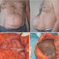
Fig. 4.1
Incision of the lateral abdomen that shows the reticular dermis and subcutaneuous tissue
4.2 The Subcutaneous Cellular Tissue
The adipose tissue that forms the subcutaneous tissue is divided into two layers by the superficial fascia (Figs. 4.2, 4.3, and 4.4).
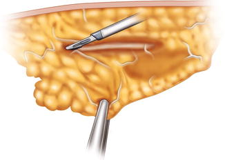
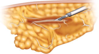
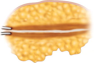
Fig. 4.2
Dissection of the areolar and lamellar layers and the superficial fascia
Fig. 4.3
Dissection of the superficial fascia in the lateral abdomen region
Fig. 4.4
Drawing of the two different layers: the areolar and the lamellar layers with also the trabecular superficial fascia
1.
The areolar layer is the superficial and is a loose connective tissue that has the function both to bind different tissue types together and provide flexibility and cushioning. It is formed by interlacing loosely organized fibers (including mostly collagenous ones, elastic, and reticular fibers) and blood vessels.
2.
The lamellar layer is composed of fibrous conjunctive tissue with a large amount of blood vessels coming from the musculoaponeurotic layer and fusiform fat cells.
Related posts:
 Abdominoplasty Combined with Cesarean Section: Discussion of the Evidence
The Impact of the Menstrual Cycle on Intraoperative and Postoperative Bleeding in Abdominoplasty Patients
Abdominoplasty Combined with Cesarean Section: Discussion of the Evidence
The Impact of the Menstrual Cycle on Intraoperative and Postoperative Bleeding in Abdominoplasty Patients
 Abdominoplasty Complications and Seroma: From Prevention to Effective Treatment
 Abdominoplasty as the Impetus for Selection of the Deep Inferior Epigastric Artery Perforator Free Flap for Non-breast Reconstruction
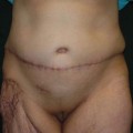
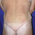 Circumferential Abdominoplasty
 Abdominal Wall Repair Post Hernia in Kidney and Liver Transplantation
Stay updated, free articles. Join our Telegram channel

Full access? Get Clinical Tree


