The Clinical Problem: Prominent Bulging Suborbicularis Oculi Fat Pad and Retro-Orbicularis Oculi Fat Pad ( Fig. 9.1 )
The aging lower eyelid can be a complex and difficult problem to improve aesthetically. There is considerable risk of over-resection of both fat and skin, with consequent pull-down ectropion and permanent hollowness. An anatomic understanding of the superficial and deep face fat compartments is crucial when attempting to relocate, reduce, or augment the suborbicularis oculi fat pad (SOOF) or retro-orbicularis oculi fat pad (ROOF) during periorbital rejuvenation.

Anatomy
Upper and Lower Eyelids
These are composed of an anterior lamella and posterior lamella separated by the orbital septum. The anterior lamella is formed by the skin and underlying orbicularis oculi muscle. The orbital septum, originating from a band of thickened periosteum at the orbital rim (the arcus marginalis), separates both lamellae and is often referred to as the middle lamella.
The posterior lamella is composed of the tarsal plate, eyelid retractors, and conjunctiva. Fibers from the capsulopalpebral fascia attach to the anterior lamella a few millimeters below the tarsus to form the lower eyelid crease. The forces imposed by the lower eyelid retractors and the canthal ligaments determine the position of the lower eyelid margin.
Orbit
The eyes are suspended in the orbital socket by the ocular muscles, the optic nerve and blood vessels, and periorbital fat, which often visibly bulge in the aging face. In the upper eyelid, there are two medial fat pads communicating directly with retro-ocular fat and one long lateral fat pad situated immediately anterior to the levator palpebrae superioris in the supraorbital sulcus. In the lower eyelid, there are three compartments, with the medial and intermediate pads separated by the inferior oblique muscle and the lateral pad held within its own fascial compartment.
Infraorbital Region
This is divided into medial and lateral parts by the course of the facial vein, typically coursing in a line 4 to 6 mm medial to the midpupillary line. The medial part is formed of two layers, the skin and the orbicularis oculi muscle, which is firmly attached to the underlying bone.
In the lateral part of the infraorbital region, seven different layers can be identified—the skin, subcutaneous fat layer, orbicularis oculi muscle, SOOF, deep fascia (continuation of the superficial lamina of the deep temporal fascia), prezygomatic fat layer, and periosteum.
The orbicularis oculi muscle is attached to the orbital rim by the orbicularis-retaining ligament. The zygomatic ligament is a true osseocutaneous ligament that originates from the periosteum of the zygoma and/or the zygomatic arch and attaches to the overlying skin.
The malar fat pad is located in the superficial fat layer of the midface, below the zygoma. This layer also comprises the nasolabial fat and jowl fat pads. Reflection of this subcutaneous fat layer and the orbicularis oculi muscle will expose the SOOF superolaterally and deep cheek fat inferomedially.
The SOOF is a layer of periorbital fat that lies deep to the superficial malar fat pad and the orbicularis muscle of the lower eyelid, extending superiorly to the lateral canthus. The tear trough forms the inferior boundary of the SOOF, and the lateral orbital thickening forms the superior boundary. SOOF has a horizontal medial component and a larger vertical lateral component. Symmetric SOOF elevation is important when considering a midface lift using a subciliary approach (Hamra).
The ROOF is analogous to the SOOF except that it is located in the upper eyelid deep to the orbicularis oculi and above the orbital fat pads in a separate layer. ROOF ptosis contributes to eyebrow ptosis in the aging face. Brow elevation lifts the ROOF; it is not a good idea to remove the ROOF ( Figs. 9.2, 9.3 and 9.4 ).
Age-Related Changes
Age-related changes in the lower eyelid and midface affect all tissues, including the skin, muscle, fat, and bone. Midface soft tissue descent, demarcation of the nasojugal tear trough and nasolabial folds, deflation of the facial soft tissues, and loss of bone mass are all changes associated with aging. The midface loses the uniformly rounded, full appearance due to laxity of the suspensory attachment of the zygomatic ligaments and shows an obliquely oriented Y shape grooving pattern. This pattern is formed by the palpebromalar groove superolaterally, the nasojugal groove medially, and the midcheek groove inferolaterally. Atrophy of the SOOF and its adjacent deep fat compartments in the midface are thought to be associated with the development of malar bags with aging. The degeneration and laxity of the superficial musculoaponeurotic system (SMAS) elastin fibers also causes malar bags, in addition to eyelid ectropion and orbital fat prolapse. Finally, the elongation of the area between the orbicularis-retaining ligament and the eyelid margin leads to prolapse of orbital fat over the inferior orbital rim and the appearance of eyelid bags. Weakness and stretch of the orbital septum also lead to the projection of orbital fat into the upper eyelid. Loss of elasticity leads to redundancy of the skin of both eyelids .
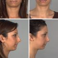
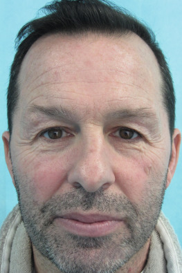
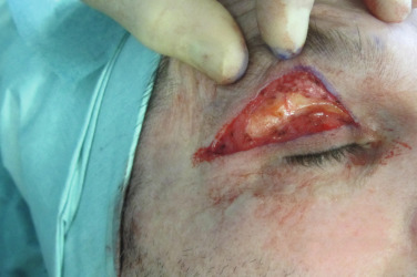
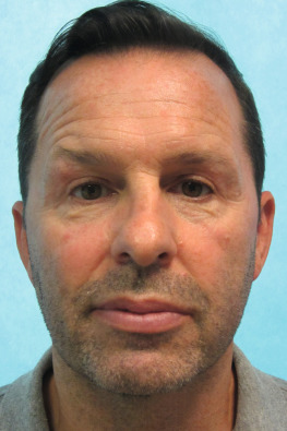
Case Study 1: Transconjunctival, Skin-Preserving, Suborbicularis Oculi Fat Pad Elevation ( Figs. 9.5 to 9.9 )
This 53-year-old woman presented with puffy lower eyelids, asymmetric mild hooding of the upper eyelids, malar fat pad ptosis, and prominent lower orbital sulci. There was no excess of lower eyelid skin. A transconjunctival blepharoplasty approach under general anesthesia was used to trim the lower fat pad bulge and thus reduce the shadow of the orbital sulcus. The postoperative pictures were taken 3 months after the procedure. The SOOF layer has been marginally raised, which helps elevate the superficial malar fat pad. The importance of keeping facial proportions by limiting surgery is clearly demonstrated in this case. If further elevation of the SOOF is required, skin excision of the lower eyelid and compensatory surgery elsewhere may be required.