7
Advanced Surgery for the Subaxial Cervical Spine in Children
Management of disorders in the subaxial cervical spine in children is a complex and challenging endeavor. The main challenge faced by pediatric spine surgeons is the broad range of patient ages they must manage and the various physiologies they represent. For example, on one end of the age spectrum, the pediatric spine surgeon sees teenagers with adult-type cervical spine physiology and pathology. Therefore, the surgeon must be knowledgeable about adult-type treatment principles regarding the cervical spine. On the other hand, complex, fascinating, and sometimes bewildering conditions occur at the other end of the age spectrum in newborn children or toddlers. These conditions may defy conventional management strategies. For patients between these extremes, surgeons must use their best judgment regarding the most appropriate treatment for a particular patient, borrowing heavily from their training in adult spine surgery.
This chapter synthesizes several important principles regarding advanced subaxial cervical spine surgery in children. It will begin by reviewing the clinical presentation of patients with these disorders and then will provide a detailed examination of their surgical and nonsurgical management. Finally, it will present several of the author’s preferred techniques for managing both straightforward and challenging cases of the pediatric cervical spine.
 Clinical Findings
The clinical presentation of cervical spine disorders in children is highly variable. Pain, weakness, sensory change, autonomic disturbance, or spine deformity may exist alone or in combination as the presenting complaints. Cervical spine pain is the most common presenting symptom and is almost universally found in the setting of a traumatic injury. Posttraumatic pain may be isolated to the neck area or may be radicular. Cervical spine pain, especially pain on motion, that lingers after a traumatic injury is abnormal and should be investigated. Like pain, weakness and sensory change may follow a radicular pattern, but they can also be part of a myelopathic syndrome involving a certain vertebral level. Autonomic disturbances, including bowel and bladder changes, may also occur but are uncommon. Urinary incontinence may be an early finding in a history suggestive of autonomic instability. Cervical spine and craniovertebral deformity, including torticollis or scoliosis, may be recognized at birth or noticed later in life. In addition, some congenital cervical spine deformities may be diagnosed as an incidental finding on a routine chest radiograph.
The physical examination of a known or suspected cervical spine disorder should proceed in a stepwise manner. General findings may be specific to a certain congenital vertebral syndrome or may suggest other syndromes associated with congenital vertebral anomalies. For instance, a low-lying hairline or web-neck deformity may suggest Klippel-Feil syndrome. Features consistent with Down syndrome are readily apparent. Abnormalities of the ears or palate may be associated with Goldenhar’s syndrome, and café au lait spots are indicative of neurofibromatosis type 1.
Examination of the extremities should be performed next. Generalized ligamentous laxity may suggest Ehlers-Danlos syndrome or Larsen’s syndrome. Foot deformities, such as high arches or cavus abnormalities, are associated with Friedreich’s ataxia. The association of foot and spine deformities strongly suggests spinal dysraphism or a generalized neuromuscular disorder.
Physical examination of the cervical spine is centered on the assessment of pain and mobility. Pain in the form of paravertebral muscle spasm is commonly found in posttraumatic injuries. Localized “trigger points” in the myofascial planes usually indicate a soft tissue injury and do not necessarily indicate cervical instability is present. Patients with significant posttraumatic instability have a deep, unrelenting pain in their cervical spine and are hesitant to move their neck. Given that, it is important to note how much motion is present in the cardinal planes of motion (flexion, extension, lateral bending, rotation) in each patient. The presence of Lhermitte’s-type symptoms on flexion or extension should also be noted. Scoliosis of the cervical spine should be described by the curve’s apex and location.
A complete neurological examination should be performed as well. Particular emphasis should be placed on the cranial nerve examination, sensorimotor testing, and deep tendon and plantar reflexes. These tests form the foundation for neurological localization in spinal cord disease. Any decrease in muscle bulk or change in muscle tone should also be sought and may aid in localizing value. The patient’s gait and station may provide subtle clues for the presence of lower extremity or truncal weakness.
 Management of Subaxial Cervical Spine Lesions
Conservative Management
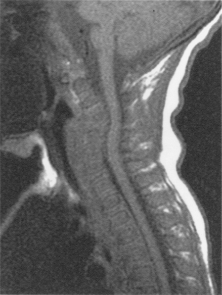
Figure 7–1 Midsagittal T1-weighted magnetic resonance imaging scan, depicting a congenitally narrow spinal canal in a 10-month-old boy. Note severe congenital narrowing from C3 down to C5.
Conservative management of subaxial cervical spine abnormalities in children is used when the patient fails to meet criteria required for operative intervention. Such circumstances include traumatic injuries where the bony and ligamentous damage is not severe enough to result in progressive deformity or neurological insult, or very young patients where a congenital abnormality is present but not progressively deteriorating (Fig. 7–1). The decision not to proceed with surgery is sometimes harder to make than the decision to proceed to the operating room. If the decision is not to operate, most patients are kept in a standard hard cervical collar and followed carefully with appropriate serial radiographs. The radiographic gold standard for determining whether a cervical spine is stable is plain spine x-rays with flexion and extension views. These views must be obtained after the patient’s posttraumatic paravertebral spasm has subsided, usually 2 to 3 weeks after the injury. Very young patients with congenital subaxial cervical spine abnormalities may require a custom cervical collar or external orthosis to provide support and stability while the child is growing. Again, appropriate serial radiographs are required to determine whether the child’s spine and spinal cord remain out of danger.
Surgical Management and Decision Making
The decision to operate on a child with a cervical spine lesion below C2 is based on several factors. Evidence of cervical spine instability, neurological deficit caused by compressive lesion, and progressive deformity are the main criteria on which the decision is based. Radiographic evidence of cervical spine instability is by no means absolute, but the following findings are helpful guidelines: vertebral body angulation greater than 15 degrees, vertebral body subluxation greater than 5 mm, or unilateral or bilateral locked facets. Pang (unpublished data) has shown evidence that a kyphotic angulation of greater than 7 degrees represents significant ligamentous injury and predisposes the juvenile cervical spine to further kyphosis and instability.
Neurological deficits caused by unstable cervical spine injuries should lead to surgical stabilization; however, it can sometimes be difficult to determine whether a cervical spine lesion is unstable. For example, many spinal cord deficits caused by the so-called spinal cord injury without radiographic abnormality (SCIWORA)–type injury are clearly not caused by an unstable lesion. In these cases, radiographic studies are normal, and the patient is usually treated in a conservative fashion, perhaps even in a hard cervical spine collar for 1 to 3 months. On the other hand, some cervical spinal cord injuries caused by SCIWORA-type mechanisms have evidence of ligamentous injury on magnetic resonance imaging (MRI) and normal flexion and extension cervical spine films. Such patients should be followed carefully with serial cervical spine flexion and extension films to rule out late instability. We have seen several patients over the years develop late instability in these circumstances. As already noted, identifying patients with greater than 7 degrees of kyphotic angulation on a plain lateral cervical spine x-ray can provide a helpful guideline for managing children with incipient instability. Those patients are at high risk of developing further angulation and ultimately might require surgical stabilization.
Other circumstances that call for careful decision making are injuries in which major supportive structures of the spine, such as the transverse ligament of C2 or subaxial disk space, are completely destroyed by a penetrating injury. Gunshot wounds and knife injuries can produce these lesions. Although static films and MRI scans may show good anatomical continuity, the known effects of missing these important structures should compel the surgeon to either stabilize the patient surgically or follow the patient very carefully with serial dynamic x-rays.
Related posts:
 Advanced Occipitocervical Surgery in Children
 Common Pathological Conditions of the Pediatric Craniovertebral Junction and Cervical Spine
 Advanced Atlantoaxial Surgery in Children
Stay updated, free articles. Join our Telegram channel

Full access? Get Clinical Tree


