This discussion focuses primarily on lipoatrophy and periorbital deflation in relation to adjunctive fat grafting of the brow and upper eyelid. Like with all clinical information for cosmetic and reconstructive surgeons in this multidisciplinary review of rejuvenation of the brow and upper lid, the authors present anatomy, evaluation, patient expectations, technique, and complications – here, specifically in terms of fat grafting and its associated aspects of fat transfer and relocation and autologous fat, along with hyaluronic acid fillers. Fat harvest and preparation are also described in detail.
Key points
- •
Lipoatrophy and volume loss are significant components of upper lid and brow aging.
- •
The aging process of the upper eyelid may lead to deep hollowing of the upper lid with increased upper eyelid show and a skeletonized supraorbital rim appearance without significant dermatochalasis.
- •
Traditional blepharoplasty, even with fat preservation, does little to rejuvenate patients with severe lipoatrophy and skeletonization. This population requires volume restoration instead of soft tissue removal.
- •
Fat transposition techniques, autologous fat grafting, and hyaluronic acid fillers are reliable choices for volume augmentation of the upper eyelid.
- •
Autologous fat is biocompatible, naturally integrates into the host tissues without producing an inflammatory reaction, and is potentially permanent.
- •
Fat grafting may be used alone or in conjunction with other procedures such as blepharoplasty and browlift techniques as part of the treatment of periorbital aging.
Video of Fat Transfer Showing Technique of [CR] Accompanies this Article at http://www.plasticsurgery.theclinics.com/
Introduction
Lipoatrophy and volume loss are now recognized as significant components of upper lid and brow aging Traditional upper lid blepharoplasty techniques that focus on soft tissue excision of skin and fat do not treat volume loss. Therefore, recent technical advances aim to provide volume restoration for upper periorbital rejuvenation in select patients. These advances include autologous fat transfer techniques and other novel maneuvers of local fat transposition.
Patterns of upper lid and brow aging
The youthful brow–upper lid complex is full and convex. The upper lid crease is sharp, and pretarsal show, the amount of exposed upper lid skin visible when the eye is open, is minimal. The lateral upper lid and brow are particularly full and round and transition smoothly without orbital rim skeletonization into the adjacent temple esthetic unit ( Fig. 1 ).
Solar damage, gravitational forces, and lipoatrophy together cause upper lid and brow aging. Solar damage leads to skin degeneration with fine rhytids and increased elastosis. Gravitational forces initiate slow downward descent of soft tissue with resultant laxity and ptotic appearance. Lipoatrophy leads to volume loss and an overall deflated form. These 3 components–skin degeneration, laxity, and deflation–each manifest in varying degrees across patients. This variability leads to distinct presentations of upper periorbital aging.
The present discussion focuses primarily on lipoatrophy and periorbital deflation. Several recent studies have described patterns of upper lid fat pad changes during the aging process. It seems that the nasal fat pad volume resists atrophy, whereas the central pad atrophies extensively. Clinical experience suggests the rate and degree of this atrophy vary considerably, with some patients presenting in their fifth and sixth decades with severe changes of atrophy and others maintaining upper lid volume indefinitely.
With this in mind, a simplified methodology may place patients in 2 distinct categories—those with dermatochalasis and those with hollowing.
Those in the “dermatochalasis category” present with pronounced upper lid skin degeneration and laxity but no clinically significant lipoatrophy. This category contains most patients who seek upper lid rejuvenation. Findings include a blunted and obscured upper lid crease, with loss of pretarsal show as a result of upper lid skin redundancy and accumulation of orbital fat ( Fig. 2 C–D).
Conversely, those in the “hollowing category” have pronounced, perhaps premature, upper lid lipoatrophy and deflation that are severe compared with the impact of elastosis and laxity. In such cases, there is deep hollowing of the upper lid, particularly at the central fat pad, with increased upper eyelid show and a skeletonized supraorbital rim appearance without significant dermatochalasis (see Fig. 2 E–F). On initial inspection, one often suspects such patients have previously undergone an overly aggressive upper lid blepharoplasty with fat excision.
Although upper periorbital aging represents more of a spectrum than a dichotomy, this delineation may be useful in considering treatment regimens for a given patient’s presentation.
Volume restoration for upper lid and brow rejuvenation
Traditional blepharoplasty aims to rejuvenate the upper periorbita by excising excess skin and postseptal fat. This approach may be valuable for patients in the dermatochalasis category as described. For these patients, the removal of excess skin, and sometimes a degree of fat, may lead to a more youthful pretarsal show and defined supratarsal crease ( Fig. 3 ).
However, blepharoplasty, even with fat preservation, does little to rejuvenate patients in the hollowing category, with little dermatochalasis but severe lipoatrophy and skeletonization. This population requires volume restoration instead of soft tissue removal. Techniques include pedicled fat transposition procedures, akin to those developed by Loeb and Hamra, within the lower lid to smooth the lid–cheek junction, and autologous fat grafting.
Pedicled Fat Transposition
Shorr and colleagues describe transposing pedicled fat from the central compartment of the upper lid into the lateral lid and brow deep to the orbicularis muscle to create youthful lateral fullness with subjective success without complications in 31 patients at a 2-year follow-up. However, because the central fat pad undergoes most age-related atrophy, there may be risk in further depleting it surgically. Such endeavors could accelerate the deepening of the central sulcus over time.
Orbital Fat Relocation
Park and colleagues describes an “orbital fat relocation” procedure in which both fat pads are dissected to the Whitnall ligament and relocated anteroinferiorly between the conjoined tendon of the levator aponeurosis, the orbicularis muscle, and the skin flap. Although the follow-up was only 4 months, the group reported good esthetic results in 50 Korean and Chinese patients without complications.
Medial Fat Pad Transposition
Massry has described a technique in which the medial fat pad, which is resistant to atrophy, is transposed laterally in a pedicled fashion into the medial compartment and affixed to the arcus marginalis to slow or prevent postoperative hollowing after routine upper lid blepharoplasty. In 65 patients, he reports no new formation of the superior sulcus deformity during an 11-month follow-up. He reports 2 cases of temporary mechanical ptosis early in the series that is attributed to incomplete freeing of the nasal fat pad causing undue tension on the levator aponeurosis at the time of fat transfer.
Hyaluronic Acid Filler
Instead of using fat grafting, Morley and colleagues and others describe the use of hyaluronic acid filler to treat upper lid hollowing or excessive postblepharoplasty eyelid show with an 85% success rate. Hyaluronic acid treatment may have a niche for less severe cases with modest medial “A”-shaped hollowing and in those patients open to serially repeated treatments.
Autologous Fat Transfer
After falling out of favor because of unreliable results and the introduction of safe commercial facial fillers, the general use of autologous fat transplantation has again gained interest among plastic surgeons. The efforts of Coleman and others have refined the procurement process, in part stimulating this revival because clinical and animal data now support fat inosculation and long-term survival. Many believe fat to be the ideal filler: it is autologous and biocompatible, it naturally integrates into the host tissues without producing an inflammatory reaction, and it is potentially permanent.
Several groups have reported on fat transfer for upper lid and brow rejuvenation with promising results. The remainder of this article reviews the relevant anatomy, perioperative considerations, and complications of upper lid and brow autologous fat transfer.
Anatomy of eyelid
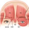
The extremely thin skin of the eyelid is firmly connected to the underlying orbicularis oculi (OO) muscle through fine connective tissue attachments. The OO is divided into pretarsal, preseptal, and orbital components. The medial canthal tendon is a continuation of superficial and deep heads of the pretarsal OO inserting onto the anterior and posterior lacrimal crest, respectively. Similarly, the lateral canthal tendon is a coalescence of OO laterally inserting onto the tubercle of Whitnall on the medial aspect of the lateral orbital wall.
Deep to the thin OO is the orbital septum (OS). The OS is a thin fibrous sheet extending from the arcus marginalis of the orbital rim to its fusion with the levator aponeurosis (LA) just cephalad to the superior tarsal border. The LA is an extension of the levator palpebra superioris that arises at the undersurface of the lesser wing of the sphenoid at the orbital apex. After 40 mm of anterior intraconal extension, the levator palpebra superioris transitions into the LA approximately10 mm behind the OS. After the LA fuses with the OS, some LA fibers descend to insert into the lower third of the anterior surface of the tarsal plate, the pretarsal OO, and the overlying skin. The latter attachment, which is approximately 10 mm from the lid margin, forms the upper lid crease in the Western eyelid.
Between the OS and the LA, there are 2 preaponeurotic fat collections, the nasal and central fat pads, that are separated by the superior oblique muscle tendon. The nasal fat is white, akin to intraconal fat, and postulated to be of neural crest lineage, perhaps underpinning its resistance to atrophy. This fat pad is an extraconal extension of intraconal fat and is not separated from orbital fat by the LA. Central fat is yellow, similar to adipose tissue throughout the body, and of mesodermal origin. It is subject to significant volume loss with aging and is separated from intraconal orbital fat by the LA. The lacrimal gland occupies the lateral aspect of the orbit and is divided into orbital and palpebral lobes by a lateral extension of the LA.
Patient evaluation
For all patients, it is critical to elucidate one’s motivation for esthetic surgery, a history of any previous periorbital pathologic conditions or procedures, and the precise physical aspects for which modification is desired. Photography is crucial and is performed with each consultation to guide conversation. Often, youthful photographs of the patient play a role in effectively identifying and communicating the patient’s specific periorbital aging pattern.
Global Treatment Strategy
Typically, patients present for a consultation to discuss facial aging in general, and the upper lid–brow complex is only a small focus of their overall attention. In these cases, it is important to clarify and prioritize a patient’s concerns to effectively create a global treatment strategy with intelligent timing of the various procedures and operations she or he may desire.
Eye and Brow Aging Assessment
The key to the upper eyelid and brow examination is identifying the relative impact of skin degeneration, soft tissue laxity, and lipoatrophy on the patient’s presentation of periorbital aging. This assessment will guide the surgeon to selecting the optimal maneuver for rejuvenation.
This examination therefore includes an assessment of the patient’s pretarsal show, presence of dermatochalasis, position of the superior sulcus, solar damage and rhytid formation, the degree of lipoatrophy and skeletonization, and postseptal fat compartments that are most affected. Further, the degree of volume loss overlying the lateral and lid brow region, and the affect this has on a smooth transition into the temple region, is appraised. The overall location of the brow is noted in relation to the supraorbital rim so as to determine to the potential complementary role of brow-lifting procedures.
For those patients with a significant lipoatrophy and resultant periorbital hollowing, particularly with volume depletion of lateral lid–brow complex, fat augmentation may be considered. In patients with little dermatochalasis and acceptable brow position, this may be performed independently. Fat transfer may also be considered in conjunction with other procedures, such as fat transfer to the temple, blepharoplasty, or brow lift, based on the clinical findings and desires of the patient. Fat transfer to the temple is of particular importance in patients with concomitant temple lipoatrophy, which is typically the case, because adding volume to the lateral lid and brow without also treating the temple will only exaggerate the appearance of temporal hollowing.
Related posts:

Stay updated, free articles. Join our Telegram channel

Full access? Get Clinical Tree


