Chapter 54 Table 54.1 Medial geniculate artery flap
Additional Free Vascularized Bone Grafts
Flap |
|
Tissue | Reliable vascular pedicle that is based on the medial geniculate artery; descending branch originates from the femoral artery in Hunter’s canal; variation includes the superior geniculate artery, which courses posterior to the medial femoral condyle; skin flap can be based on the saphenous artery perforators or the perforating vessels that originate directly from the medial geniculate artery |
Course of the vessels | — |
Dimensions | — |
Extensions and combinations | — |
Anatomy |
|
Neurovascular pedicle | — |
Artery | — |
Veins | — |
Length and arc of rotation | — |
Diameter | — |
Nerve | — |
Surgical technique | — |
Preoperative examination and markings | — |
Patient position | — |
Dissection | — |
Advantages |
|
Vascular pedicle | — |
Flap size and shape | Versatile source of small vascularized bone grafts; may be harvested as periosteal flap only, a corticocancellous bone flap, an osteocutaneous flap, or an osteomyocutaneous flap; long pedicle possible; muscle can be included; hyaline cartilage can be harvested from the trochlear nonarticular portion of the knee as a substitute for articular surface defects (i.e., as a replacement for the proximal pole of the scaphoid) |
Combinations | — |
Tissue | — |
Dissection | — |
Donor site | — |
Further options | |
Disadvantages |
|
Flap size | — |
Donor site morbidity | — |
Dissection | — |
Flap | — |
Pedicle | — |
Pearls and pitfalls |
|
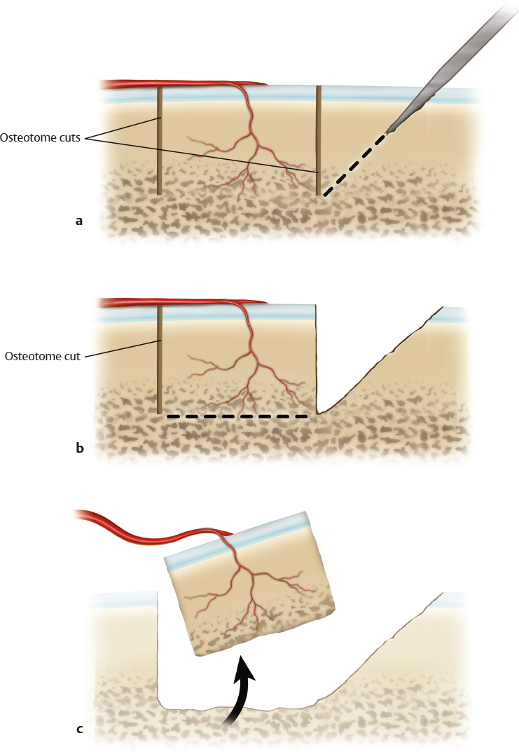
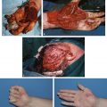
Dissection | Dissection should proceed distal to proximal, with a tourniquet in place; periosteal vessels and optimal condylar vascularity will be found in the distal inferior quadrant of the knee; begin with a longitudinal incision over the mid portion of the medial femoral condyle; divide the skin and dissect to the vastus medialis oblique fascia; incise the fascia and reflect the vastus medialis anteriorly; identify the periosteal perforators on the condylar surface (more superficial layers of fascia that contain vessels can be confused for the medial geniculate artery, but these are not the periosteal perforators); follow the vessels from distal to proximal along the adductor longus tendon; the medial geniculate artery and vein originate from the femoral artery and vein; saphenous branch for skin flap posterior in thigh is a possibility to use as an osteocutaneous flap. |
Extensions and combinations | — |
Contouring and correction | — |
Clinical applications | Use cautery to incise the rich periosteum around bone, being sure to leave the periosteum attached to the cortical surface of the condyle; use an oscillating saw or an osteotome to cut the bone flap; leverage bone from the condyle, and keep it attached to the periosteal sleeve from the surface of the femoral diaphysis; trace the vessels proximally for the pedicle length desired; close the donor site over a drain; bone putty or allograft paste may be placed in the femoral condyle to augment the local site after the harvest of living bone |