4
Acne
Bárbara R. Ferreira, José C. Cardoso, José P. Reis, and Américo Figueiredo
Department of Dermatology, Coimbra Hospital and University Centre, Coimbra, Portugal
Introduction and Epidemiology
Celsus was one of the most important Roman medical writers, known for his first‐century medical work, “De Medicine.” Celsus also first described a dermatosis characterized by lesions on the face of young people that, according to the description, may correspond to acne. He used terms like “varus” and “varius” in order to highlight the polymorphism of the lesions. Later, Aetius Amidenus, a physician in Constantinople, first used the word “acne,” in the sixth century, underlining that the lesions occurred on the face at the “acme” of life, pointing to “puberty” [1]. In the field of the history of dermatology, we have also suggested in a recent paper on a controversial disease in psychodermatology, “Morgellons disease,” that some past reports incorrectly identifying Morgellons could correspond to “comedones,” and then to acne, highlighting the impact of acne throughout time [2].
Acne is a very common inflammatory disorder of the pilosebaceous unit that belongs to the group of psychophysiological dermatoses due to its relationship with psychological stress and has multifactorial etiopathogenesis, deserving a holistic approach. In fact, a link has been suggested between acne, psychosocial issues, and several environmental factors, such as Western diet and pollution, which has led to the emergence of a discussion on complementary treatments [3]. The global prevalence is 9.4%; acne is the eighth‐most prevalent disease worldwide [3] and one of the most common dermatoses [4]. Important epidemiological studies conducted in 2011 and 2012 reported that the prevalence may be different according to ethnic group (for instance, African American with 37% and Caucasian with 24% prevalence), although this may not be so significant, since environmental factors may have a deep contribution too. Acne is most commonly observed between 16 and 20 years [5, 6] and boys have higher risk for the most severe forms. Acne seems to be more common in girls at the lower age groups and, at puberty, acne is more prevalent in boys [3]. Thereafter, in adults, women are more affected [3], with a prevalence of 12% [7]. Adolescents are thus by far the biggest group affected by this dermatosis, with a prevalence ranging from 35% to 90% [8], which points to the need to pay particular attention to the psychosocial issues related to this age group, namely due to the physical changes that occur at that time and the deeper concerns of peer group integration and of self‐image, as well as the higher sensitivity to stigmatization and feelings of shame [9].
Thus, this chapter intends to present an integrative approach to acne, taking into account the relationship between psychosocial and environmental factors, the subtleties of the adolescent psychodermatology, and management according to the subtleties of each affected age group.
Clinical Features and Classification
Acne vulgaris is easily diagnosed by its clinical features. The dermatosis typically has a chronic evolution and distribution on the body areas where there is a higher quantity of pilosebaceous glands, particularly the face, chest, and back, as it is an inflammatory disease of the pilosebaceous unit. The first lesion of acne is a microcomedo, which is a microscopic lesion. Through the clinical evolution of the dermatosis, we may find non‐inflammatory or inflammatory lesions [4]. The former corresponds to comedonal acne, where opened and closed comedones, which are skin‐colored papules, with and without a clear follicular opening respectively, can be seen. The latter includes erythematous papules and pustules as well as nodules and cysts that can be filled with pus or serosanguinous content. In addition, patients may also present secondary features, namely: erosions; excoriations, in the setting of acne excoriée; ulcers and crusts, in the context of acne fulminans; and scars, especially as a consequence of the severe forms. As a complication, some patients may rarely exhibit a clinical variant of acne called solid facial edema, or Morbihan’s disease, that clinically presents with induration and erythema of the face, and whose etiopathogenesis may be linked to chronic inflammation together with genetic predisposition. Commonly, post‐inflammatory hyperpigmentation can also be secondarily observed [10]. Acne should then be classified according to the following points: type of cutaneous lesion observed, severity, age of onset, factors of worsening or of induction of the dermatosis and associated disorders or syndromes [4, 10].
Thereby, considering the type of cutaneous lesions (primary and/or secondary lesions), acne can be classified as comedonal, papulopustular, nodular, nodulocystic, and conglobate. The latter may be considered the ending part of the spectrum of nodulocystic acne, with a predominance of highly inflammatory lesions and sinus tracts. This form can be part of the follicular occlusion tetrad together with hidradenitis suppurativa, dissecting cellulitis of the scalp, and pilonidal cysts [4]. The male gender is much more affected and, despite the severity of the dermatosis, systemic findings are not present [11].
According to the severity, acne can be classified as mild, moderate, or severe and this classification also takes into account the type and number of the cutaneous lesions described above. More precisely, both mild and moderate forms include comedonal and papulopustular lesions, but, in the former, comedones are the main lesions and nodules are not observed. The latter has comedones, papules, and pustules, and may have some nodules, but cysts and sinus tracts are not observed. In mild acne, there are fewer than 20 comedones and fewer than 10 papules or pustules on the face. In moderate acne (Figure 4.1), the number of comedones on the face may range from 10 to 40 as well as the number of papules and pustules. Nodules can be occasionally seen in moderate acne, but the number of these lesions on the face should not be higher than 10 [4]. Concerning the severe forms, here we observe comedones, papules, pustules together with nodulocystic acne (Figure 4.2) or even acne with clinical criteria for acne conglobata. Acne fulminans, previously called “acne maligna” or “acute febrile ulcerative acne conglobate,” is the most severe form of acne, commonly affecting boys in adolescence, in contrast with acne conglobate which typically occurs in early adulthood. Clinically, it is characterized by a sudden onset of severe cutaneous lesions in a patient who had mild to moderate acne; there are painful and hemorrhagic nodules which can evolve into ulceration, commonly on the trunk and healing with significant scars [12, 13]. Acne fulminans can occur with or without systemic findings and abnormalities in laboratory parameters, such as fever, arthralgia, leukocytosis, and increased erythrocyte sedimentation rate and C‐reactive protein. Osteolytic lesions can be observed in its most severe presentation. The etiopathogenesis is not fully understood. Testosterone therapy, intake of anabolic androgen steroids, and isotretinoin, especially when started with high doses, have been suggested as triggers interfering with the activity of Propionibacterium acnes (P. acnes) [13]. Acne fulminans should be classified as “acne fulminans without systemic symptoms,” “acne fulminans with systemic symptoms,” “isotretinoin‐induced acne fulminans with systemic symptoms,” and “isotretinoin‐induced acne fulminans without systemic symptoms,” the latter being the most common. Overall, acne fulminans is more common in Caucasians and has genetic predisposition; a history of two years of evolution and macrocomedones are also typical [12].
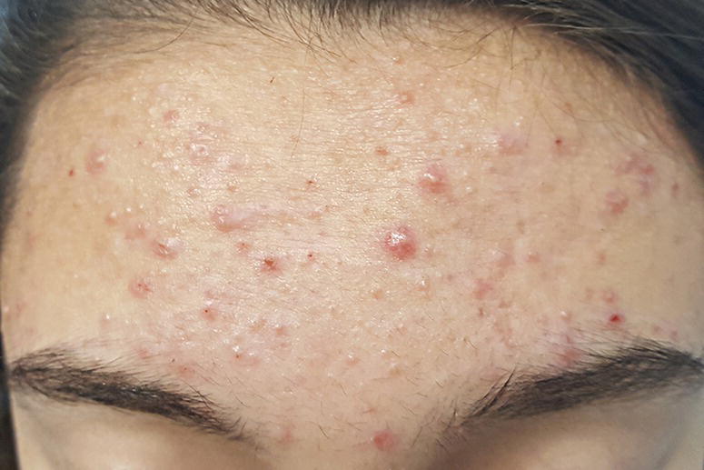
Figure 4.1 Moderate acne, with comedonal and papulopustular lesions.
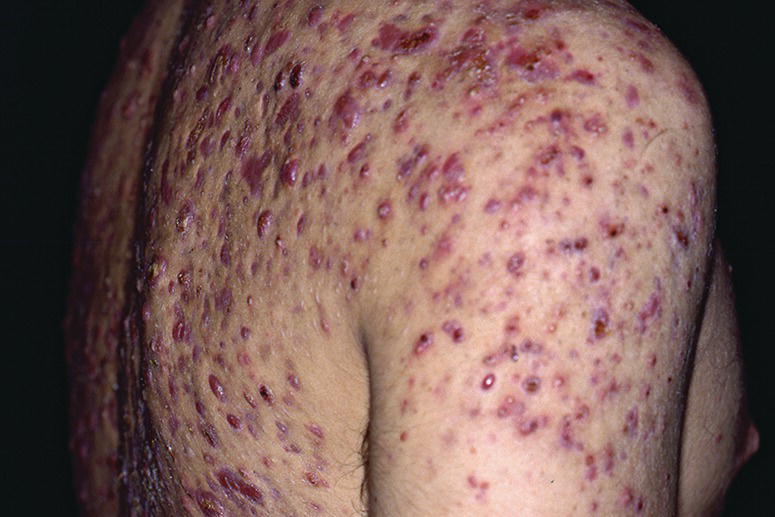
Figure 4.2 Nodulocystic acne.
Considering the age of onset, acne can be pediatric (neonatal, infantile, mid‐childhood, preadolescent), adolescent, or adult acne [14]. Adult acne is that observed in patients above the age of 25, both those who also had acne during puberty and those with late‐onset acne [4]. Adolescent acne includes patients between the age of 12 and adulthood. Pediatric acne is used for patients from birth up to the age of 11: neonatal acne from birth to the fourth week of life; infantile acne from the first month of life up to the age of first year of life; mid‐childhood acne from the first to the sixth year of life; and preadolescent acne from the seventh year of life to the age of 11 years old. Neonatal acne is characterized by an inflammatory component, with erythematous papules and pustules, and, rarely, comedones. Neonatal cephalic pustulosis is a more pustular form of the same disease, without comedones, according to some authors, although some discussion still exists whether they are different conditions due to these subtleties on the clinical presentation [14]. The etiopathogenesis of neonatal acne is still being discussed and some authors have linked it to the stimulation of the sebaceous glands by the maternal androgens, while neonatal cephalic pustulosis would be more closely related to the transient inflammatory response to Malassezia furfur or Malassezia sympodialis. Both neonatal acne and infantile acne are more common in boys and on the face, typically on the cheeks. In contrast with neonatal acne, in infantile acne comedones are prominent as well as nodules and cysts and its etiopathogenesis is explained by the physiological and transient increase in androgen levels (production of testosterone and dehydroepiandrosterone [DHEA]) [10, 14]. Preadolescent acne is the first cutaneous finding of pubertal maturation, typically with comedones on the frontal region, and the etiopathogenesis is explained by the increasing levels of adrenal androgens, namely DHEA [14].
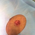
Furthermore, acne can also be classified in variants that take into account some factors which induce acneiform lesions or worsen the dermatosis. In this context, we can consider the following variants: contact acne, drug‐induced acne, and acne excoriée. Contact acne is the result of persistent occlusion of the follicular opening, leading to development of comedones, and includes acne mechanica, acne cosmetica, and occupational acne. The first is the result of repetitive local trauma, such as by helmets and collars. Acne cosmetica is the term used for acneiform lesions caused by certain cosmetics [10]. Occupational acne is caused by exposure to a wide range of substances in the workplace. For example, occupational exposure to some halogenated aromatic hydrocarbons is called “chloracne” and is considered one of the most sensitive markers of systemic poisoning by them. It is relevant to mention, furthermore, that chloracne can also occur as a result of non‐occupational exposure, namely through contaminated food, and represents an important matter in public health since the chloracnegens are lipophilic and persist for years in the body fat. Clinically, patients may present both comedones and inflammatory lesions. The clinical picture is characteristic because, firstly, the lesions appear on the face and neck and, afterwards, they develop on the trunk, genitalia, and extremities, and tend to be chronic [15]. Interestingly, the first example provided in the literature of drug‐induced acne was that it is caused by iodides and chlorinated hydrocarbons. From a clinical point of view, drug‐induced acne can be differentiated from acne vulgaris since the first can be observed not only on the face, neck, and chest, but also beyond the seborrheic areas [16]. Besides, more frequently, follicular erythematous papules and pustules are predominant and comedones are not characteristic [10]. Drug‐induced acne seems to be more resistant to conventional acne treatment. The drugs that have a recognized relationship with acneiform eruptions are the previously described halogens, corticosteroids, testosterone, anabolic steroids, isoniazid, lithium, and anticancer drugs such as epidermal growth factor receptor (EGFR) inhibitors, BRAF inhibitors and mitogen‐activated protein kinase (MAPK) inhibitors. Some studies have also highlighted an association between acneiform eruptions and other drugs, namely, cyclosporine A, azathioprine, tricyclic antidepressants, and tacrolimus [16]. Acne excoriée “des jeunes filles” (Figure 4.3) is an example of a skin‐picking disorder that typically affects young women, who scratch the papules, pustules, and comedones, and is within the obsessive‐compulsive spectrum [17].
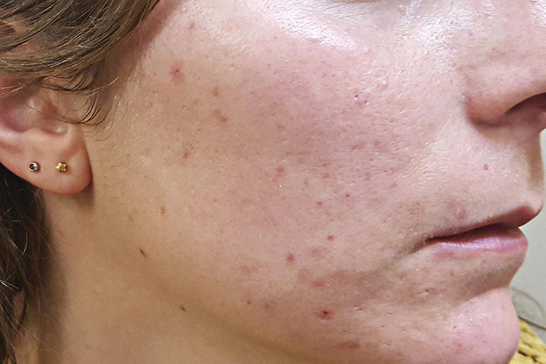
Figure 4.3 Acne excoriée.
Finally, acne should also be clinically analyzed as a cutaneous sign of systemic disease [18]. Some patients with acne may also have criteria for some syndromes, the following being the most traditionally described: synovitis, acne, pustulosis, hyperostosis, osteitis or SAPHO Syndrome, involving synovitis, acne from mild to severe forms, pustulosis (the most common observed dermatosis), hyperostosis, and osteitis; pyogenic arthritis, pyoderma gangrenosum, and acne or PAPA syndrome, with pyogenic sterile arthritis, pyoderma gangrenosum and acne conglobata; pyoderma gangrenosum, severe acne, and hidradenitis suppurativa or PASH; and pyogenic arthritis, pyoderma gangrenosum, severe acne, and hidradenitis suppurativa or PAPASH [11, 19]. Although the etiopathogenesis is not fully understood, it is possible that SAPHO could be triggered by P. acnes, causing important cellular and humoral inflammatory responses. PAPA has been linked to mutations in PSTPIP1 (proline‐serine–threonine‐phosphatase interacting protein 1) gene, also called the gene of CD2‐binding protein 1 (CD2BP1), modifying the activity of pyrin and causing neutrophilic infiltration [19]. Mutations in PSTPIP1 were also described in some patients with PASH, but the genetic background seems to be wide and not fully clarified [20].
Furthermore, acne fulminans was occasionally seen in the setting of Marfan syndrome and late‐onset congenital adrenal hyperplasia [12]. Congenital adrenal hyperplasia corresponds to the presence of a wide range of genetic disorders of the cortisol and/or aldosterone enzymatic pathways, with the deficiency of 21‐hydroxilase being the most common. As a consequence, there is glucocorticoid deficiency, mineralocorticoid deficiency, and higher levels of androgens, the latter explaining the early development of acne [18]. Acne can also be part of the seborrhea‐acne‐hirsutism‐androgenetic alopecia syndrome (SAHA), the polycystic ovarian syndrome (PCOS), the hyperandrogenism‐insulin resistance‐acanthosis nigricans (HAIR‐AN) syndrome, and the Apert syndrome. SAHA results from high circulating levels of androgens or higher sensitivity of the pilosebaceous unit to androgens in women. In PCOS there are clinical and biochemical signs of hyperandrogenism, disorder of ovulation, and polycystic ovaries, probably related to genes also linked with obesity. This condition should be suspected in women with severe acne or acne with late onset or acne which is resistant to conventional treatment. HAIR‐AN is considered a type of PCOS, where the insulin resistance leads to high levels of insulin. Insulin and insulin‐like growth factor (IGF‐1) stimulate epidermal thickness through its binding to receptors on keratinocytes and fibroblasts. The activity of sebaceous glands is also increased by IGF‐1, stimulating the development of acne lesions. The high levels of insulin also interfere with steroidogenesis and increase the production of androgens that also contribute to epithelial thickness and a higher density of melanin, subsequently explaining acanthosis nigricans [21]. Apert syndrome is a congenital and autosomal dominant disorder due to the mutation of the growth factor receptor 2 (FGFR2) and is defined by craniosynostosis, syndactyly, and abnormalities of the skin and other organs. Inflammatory acne in adolescence that is also observed beyond the seborrheic areas is the typical skin finding. There is also a mosaic cutaneous form of Apert syndrome observed along the lines of Blaschko, which is called Munro’s acne nevus [22]. Table 4.1 summarizes the clinical spectrum of acne and the factors to consider in its classification.
Table 4.1 Acne: spectrum of cutaneous lesions and classification.
| Acne: clinical features and classification | |
| Primary cutaneous lesions (papules, pustules, nodules) | Comedonal acne; Inflammatory acne: papulopustular ± nodulocystic; acne conglobata |
| Severity (number and type of primary lesions) | Mild; moderate; severe. |
| Age of onset | Pediatric (neonatal, infantile, mid‐childhood, preadolescent) acne; adolescent acne; adult acne |
| Factors of worsening/induction | Contact acne; drug‐induced acne; acne excoriée |
| Associated disorders/syndromes | Examples: SAPHO, PAPA, PASH, PAPASH, Congenital adrenal hyperplasia, Apert syndrome |
| Secondary cutaneous lesions/complications | Morbihan’s disease; Acne fulminans (inflammatory acne+ulcers and crusts); Acne scars; Acne excoriée |
SAPHO syndrome: synovitis, acne, pustulosis, hyperostosis, osteitis; PAPA syndrome: pyogenic sterile arthritis, pyoderma gangrenosum and acne conglobata; PASH: pyoderma gangrenosum, acne and hidradenitis suppurativa; PAPASH: pyogenic arthritis, pyoderma gangrenosum, severe acne and hidradenitis suppurativa
Perspective of Multifactorial Etiopathogenesis and Relevance of an Integrative Approach
The etiopathogenesis of acne is centered in the pilosebaceous unit and involves four main mechanisms: high sebum synthesis by the sebaceous gland; the activity of P. acnes; disorder of the keratinization in the follicle, with hyperproliferation and a reduction in the shedding of intra‐follicular keratinocytes, leading to obstruction and, then, to the formation of microcomedones (the first cutaneous lesion); and the role of inflammatory mediators, namely proteases, lipases, and chemotactic factors released by P. acnes, activating mediators of both innate and adaptive immunity [23]. Furthermore, androgens, stress‐related hormones, the endocannabinoid system, diet, lifestyle, and environmental factors together with genetics can also help to understand the development and evolution of this dermatosis.
The Sebaceous Gland: Sebum Synthesis and Acne – An Overview
The sebaceous glands are holocrine glands. They secrete through the disruption of the glandular cells to the upper part of hair follicles and contribute to the production of the cutaneous lipid film together with lipids of keratinocyte origin. The density of sebaceous glands is higher on the face, back, and chest and they are not observed on the palms, soles, and dorsal surface of feet. The face, particularly the T‐zone, is where they show the highest density. Besides these anatomic considerations, the activity of sebaceous glands may differ according to age and gender [24]. This may explain why acne presents different prevalence in the male and female gender throughout life, as described above in the clinical features. The lipid composition of sebum differs from that of keratinocyte origin and includes 57% triglycerides and free fatty acids, 26% wax esters, 12% squalene, and 2% cholesterol, and in the pilosebaceous unit it is made up of triglycerides, squalene, and wax esters [24, 25]. Knowledge of the physiology and pathophysiology of the sebaceous gland and lipid production is relevant to attain an integrated approach to the dermatosis. For instance, patients with acne have higher cutaneous levels of squalene, which can also contribute to the development of comedones. Isotretinoin, a 13‐cis‐retinoid acid, and spironolactone, an antiandrogenic drug, have a sebostatic role, modulating these mechanisms [24]. Moreover, sebaceous glands are also modulated by sexual hormones and adrenal corticosteroids, a fact that is important to integrating this dermatosis in the context of associated disorders and syndromes. For instance, it was observed that sebaceous glands in acne show a high manifestation of activity of 11‐beta‐hydroxysteroid dehydrogenase (HSD) type I. This is important to understand, at a biochemical level, the connection between acne and psychological stress, because this enzyme converts cortisone to cortisol and modulates lipid synthesis, and this was correlated with acne severity in some studies [2426–28]. Actually, a large number of studies showed that stress response‐related hormones, particularly corticotrophin releasing hormone (CRH), adrenocorticotrophic hormone (ACTH) and melanocyte stimulating hormone (MSH) are expressed in sebaceous glands and modulate lipid synthesis in sebocytes [29, 30]. Dihydrotestosterone, testosterone, and a wide range of mediators and hormones, like CRH and IGF‐1, increase the activity of sebocytes, which, together with P. acnes, stimulate the release of inflammatory mediators in the perifollicular dermis, contributing to the development of comedones and inflammatory lesions [23].
Propionibacterium acnes
Propionibacterium acnes (P. acnes) is a rod‐shaped gram‐positive that is commensal in the pilosebaceous unit [31]. However, some strains of P. acnes are pathogenic. It has been shown that patients with acne do not significantly differ from those without the dermatosis in terms of the quantity of P. acnes; rather, the strains are different [32]. In a recent study, the phylotypes IA‐2, IB‐1, and IC were identified as being associated with acne, which could be of interest for further research on type‐specific therapies [33, 34]. Pathological strains may be more prone to secrete lipases that hydrolyze the triglycerides of sebum, bind Toll‐like receptor (TLR) 2 and TLR4 on sebaceous glands, leading to an increase in antimicrobial peptides, such as granulysin and cathelicidin, and trigger an inflammatory mechanism with high levels of interleukin (IL)‐1, IL‐6, IL‐8, and tumor necrosis factor‐α (TNF‐α), and recruitment of neutrophils and macrophages, with rupture of the follicle wall. This inflammatory mechanism is, then, enhanced by macrophages through higher production of IL‐8 and IL‐12 and also T‐helper 1 (Th1) response. Besides, P. acnes stimulates hyperkeratinization through an increase in the levels of integrin and filaggrin [31]. P. acnes also secretes proteases and hyaluronidases that, together with lipases, enhance destruction through the stimulation of pro‐inflammatory mechanisms in the pilosebaceous unit, the activation of the classical and alternative complement pathways and recruitment of neutrophis [35]. Furthermore, other microorganisms, namely Streptococcus epidermidis, may interact with P. acnes in patients with acne and may also have a role in the etiopathogenesis of the dermatosis [32].
Disorder of Follicular Keratinization
The disorder of follicular keratinization corresponds to hyperkeratinization, resulting from a proliferation of epithelial follicular cells with retention of keratinocytes that leads to a keratin plug placed in the follicular infundibulum and, then, to the formation of the precursor cutaneous lesion of acne, the microcomedo. Several mechanisms contribute to the etiopathogenesis of the microcomedo: hormones, particularly, the androgens, and the lipid content, strains, and activity of P. acnes and inflammatory mediators, such as IL‐1 and leukotriene B4, the latter leading to the recruitment of inflammatory cells through a peroxisome proliferator‐activated protein (PPAR), namely, PPAR‐α [31, 36]. Macrophages and CD4+ lymphocytes stimulate the pilosebaceous vasculature, also contributing to the process of follicular hyperkeratinization, together with the higher local levels of squalene and matrix metallo‐proteinases present in the sebum [36].
Innate and Adaptive Immunity
The etiopathogenesis of acne involves both innate and adaptive immunity [37, 38]. The innate system involves the cutaneous barrier, disturbed through an imbalance of the lipid content, and the TLRs, particularly, the TLR2 and TLR4, with subsequent involvement of keratinocytes, neutrophils, macrophages, natural killer, and dendritic cells, like Langerhans cells, as well as the release of antimicrobial peptides and activation of matrix metallo‐proteinases, the latter being correlated with the development of scars [31]. Pathological strains of P. acnes are important in the activation of these mechanisms [32, 38]. Langerhans cells will then present the antigens to CD4+ T cells and, later, an infiltrate of CD8+ cells also occurs, and these mechanisms involve adaptive immune response, particularly on the Th17 axis, also shared with psoriasis, involving the cytokines IL‐6, IL‐1ß, and tumor growth factor‐ß (TGF‐ß). Retinoids, zinc, and dihydroxyvitamin D3 have an effect on the Th17 pathway. Besides, isotretinoin downregulates TLR2, controlling the migration of monocytes and neutrophis [37]. SAPHO syndrome shows good response to anti‐IL‐1 [39]. Further research on acne and associated disorders and syndromes could be targeted to cytokines involved in this pathway.
Androgens
Androgens can be produced by gonadal, adrenal, and sebaceous glands [10]. DHEA, testosterone, and dihydrotestosterone (DHT) regulate genes involved in the synthesis of the lipid content by the sebaceous glands [40]. The first is converted into testosterone by 3ß‐HSD and 17ß‐HSD and then into DHT by 5α‐reductase, the major androgen that interferes with the sebaceous gland [10]. In addition, androgens also stimulate comedogenesis [31]. Androgens interfere with sebaceous glands beginning in the neonatal period. The levels are higher during the first year of life and then decrease up to the adrenarche, when the levels of DHEA‐sulfate, of adrenal production, increase, correlating with the development of comedones. One‐third of women with post‐adolescent acne have hyperandrogenia. Hyperandrogenia should be suspected in women with hirsutism and androgenetic alopecia and children who develop acne between the age of two up to seven years and have typically severe acne which is resistant to treatment [10]. As explained above, in the management, syndromes that also present hyperandrogenism should be excluded, such as SAHA, PCOS, and congenital adrenal hyperplasia [21].
Endocannabinoids
Endocannabinoids are endogenous lipid mediators that are also expressed in the skin and have a wide range of functions, especially in the pilosebaceous unit, controlling sebum production and having an anti‐inflammatory effect. This has raised interest in researching its usefulness for acne treatment [41].
CRH‐ACTH: Cortisol Axis, Psychological Stress, and Acne
Psychological stress was reported to worsen acne in 50% of patients [42]. Psychological stress modulates immunity, sharing mechanisms with several chronic dermatoses, considering that the skin has a peripheral hypothalamo‐pituitary‐adrenal (HPA) axis. Psychological stress induces the production of CRH in the paraventricular nucleus of hypothalamus, leading to the secretion of ACTH and cortisol. However, corticosteroid receptors become insensitive to the persistent effects of cortisol and the effect of catecholamine on macrophages becomes preponderant, stimulating the secretion of TNF‐α, IL‐1, and IL‐6. In the brain, these cytokines induce or exacerbate symptoms of depression, leading to the persistence of these mechanisms, both at a central and peripheral level [43, 44]. Sebaceous glands express receptors for neuroendocrine mediators related to stress, since they are involved in the peripheral HPA axis. They have receptors for CRH and other components of stress response, such as vasoactive intestinal polypeptide, neuropeptide Y, and calcitonin gene‐related peptide, which interfere with the synthesis of lipid content as well as the androgen metabolism by the sebaceous gland [45]. Furthermore, in the setting of HPA axis stimulation there is a local increase of substance P, which stimulates peptidases and lipid production, and then comedogenesis, by the sebaceous gland [44, 45]. Thereafter, there is proliferation of P. acnes and high inflammatory responses involving mast cells [46]. The sebaceous gland is also influenced by CRH in response to stress, increasing sebum production [36]. Besides, CRH also promotes the conversion of precursors of androgens into testosterone and psychological stress also induces a slower wound healing process and repair of the active cutaneous lesions of acne [47].
Diet, Lifestyle Habits, and Environmental Factors
A field of recent heated discussion in the etiopathogenesis of acne is its relationship with lifestyle and diet [3]. The absence of acne in native people in Papua New Guinea and in Paraguay raised interest to examine the link between acne vulgaris and the Western diet, particularly food with high glycemic load, leading to hyperinsulinaemia and high levels of IGF‐1 [36]. Randomized controlled dietary intervention studies showed that there is a strong link between acne and diet. It was documented that a lower glycemic diet was related to improvement in acne, both for non‐inflammatory and inflammatory cutaneous lesions, with correlated reduction in inflammatory markers. Besides, omega‐3 fatty acids and γ‐linolenic acid were shown to improve inflammatory cutaneous lesions of acne after 10 weeks [35]. Melnik et al. reported that there is overstimulation of mammalian target of rapamycin complex (mTORC) 1 in the pilosebaceous unit by Western diet through dairy‐origin essential amino acids. In addition, an insulinotropic Western diet disturbs the activity of forkhead box transcription factor O1 (FoxO1). As a consequence, mTORC1 is stimulated and enhances hyperkeratinization and hyperplasia and function of the sebaceous gland, so that acne vulgaris can be considered as part of the family of mTORC1‐driven diseases of civilization, as well as cancer [48]. Furthermore, patients with acne vulgaris exhibit lower levels of adiponectin which is also connected with a diet with high glycemic load, and this may facilitate the hyperactivity of mTORC1 [49].
The link with vitamin deficiency was also recently studied. Vitamin D deficiency was significantly more prevalent in patients with acne, as it was previously documented for obesity, a finding that needs further study. Vitamin D could modulate the expression of inflammatory cytokines, namely IL‐6 and IL‐8, and matrix metalloproteinases in the pilosebaceous unit. Moreover, it was observed that vitamin D inhibits P. acnes and the Th17 pathway [50].
Related posts:

Stay updated, free articles. Join our Telegram channel

Full access? Get Clinical Tree


