Capsular contracture is the most frequent complication following breast augmentation, prompting many revision surgeries. Its prevention has evolved through precise surgical technique, antimicrobial measures, and the use of innovative materials. Advances in breast reconstruction—from submuscular and dual-plane to prepectoral placement—have shaped modern contracture management. Acellular dermal matrices and biosynthetic scaffolds like poly-4-hydroxybutyrate (P4HB) have proven effective in reducing contracture rates, with P4HB offering benefits such as lower costs and improved pocket stability. Today, the combination of prepectoral positioning, advanced matrices/scaffolds, and strict antimicrobial protocols allows for consistent and successful prevention and treatment of capsular contracture.
Key points
-
•
Capsular contracture remains the most common complication after breast augmentation and a leading reason for revision surgery.
-
•
Prevention strategies include precise, atraumatic technique, antimicrobial protocols, and use of sleeves or barriers.
-
•
Evolution in breast reconstruction techniques—from subcutaneous to submuscular, dual-plane, and prepectoral placement—has influenced contracture management.
-
•
Acellular dermal matrices and biosynthetic scaffolds like poly-4-hydroxybutyrate (P4HB) significantly reduce contracture rates and improve pocket stability, with P4HB offering advantages in cost and complication profile.
-
•
Combining prepectoral positioning, advanced matrices/scaffolds, and antimicrobial steps has led to consistently low contracture rates in aesthetic breast surgery.
Abbreviation
| ADM | acellular dermal matrices |
| P4HB | poly-4-hydroxybutyrate |
Introduction
Capsular contracture has been the number one complication following primary breast augmentation for the past 2 decades. It has historically been the most common complication of aesthetic and reconstructive breast surgery, and it remains the primary reason for most revisionary surgeries. A litany of data has shown that capsular contracture can be minimized in primary augmentation by technically precise, atraumatic bloodless dissection, appropriate anti-microbial technique, use of various pocket irrigants, and minimizing points of contamination using sleeves or barriers. Additionally, treatment of the capsule and addition of sight changes, as well as use of biosynthetic supportive matrices and scaffolds, have become commonplace and typically quite successful in the treatment and prevention of capsular contracture. ,
An inordinate amount of the techniques we employ in aesthetic plastic surgery have been learned from their successes in the reconstructive realm. Many of the techniques we utilize in the treatment of capsular contracture were learned from our reconstructive evolution, and from a historical perspective, it is important to see how use of new planes and supportive matrices in prosthetic breast reconstruction have led to our current concepts in the treatment of capsular contracture.
History of prosthetic breast reconstruction
The first breast reconstructions, performed in the early 1960s with the introduction of the silicone implant, involved the placement of the implant in a subcutaneous pocket, beneath the mastectomy skin but over the pectoralis major muscle. This subcutaneous approach was simple, quick, and preserved the integrity of the pectoralis muscle, but was associated with a number of complications. Implant malposition (bottoming out), visibility, and palpability, rippling/wrinkling, implant exposure subsequent to skin breakdown, and capsular contracture were some of the most commonly reported complications. With the realization that these complications arose from insufficient soft-tissue coverage, breast reconstruction technique evolved to moving the implant from the subcutaneous to submuscular position.
In submuscular placement, the implant is placed under the pectoralis major muscle without releasing the inferior origin of the muscle. Full muscle coverage of the implant is achieved by recruitment of muscle flaps (serratus anterior and rectus abdominis sheath) for lateral and inferior coverage of the implant. Full-muscle coverage eliminated the soft-tissue coverage limitations of the subcutaneous approach, but it resulted in unnatural-looking breasts. The inferior restrictions imposed by the submuscular pocket prevent lower pole expansion that results in poor breast projection, ptosis, and definition of the breast shape. In addition, the recruitment of muscle flaps introduced donor site morbidity.
In order to address the inferior restriction of full muscle coverage, the partial muscle coverage or dual-plane technique was introduced. In partial muscle coverage, the implant is covered partially superiorly by the pectoralis major and partially inferiorly by only the mastectomy flap. The inferior subcutaneous coverage not only allows for lower pole expansion but also eliminates the need for flap recruitment for lower pole coverage. Partial muscle coverage, however, introduced a new problem. Without its inferior attachment, the pectoralis major is free to migrate superiorly, which causes “window-shading.” The lack of lower pole support gave way to inferior malposition. Moreover, subcutaneous coverage at the lower pole reintroduced the same inherent problems associated with subcutaneous implant placement.
To address the problem of lower pole coverage, acellular dermal matrices (ADMs) were introduced in breast reconstruction in 2008. In this widely utilized modification of the partial muscle coverage technique, the ADM sutured at the lower pole provides the additional support needed at the inferior pole. With lower pole ADM placement, complications associated with subcutaneous coverage are minimized without restricting lower pole expansion.
Dual-plane reconstruction with ADM was (and still continues to be) successful and provided beautiful aesthetic outcomes, but with any contraction of the pectoralis major, animation deformity on the chest can immediately be noted.
As a means to alleviate muscle-related issues, we resorted to moving the implant from the subpectoral back to the prepectoral (subcutaneous) position in certain revision cases, followed by primary reconstructions. We then utilized our bioengineered breast concept to overcome the limitations of subcutaneous placement and improve the aesthetic outcome. Using form-stable implants, we reinforced the entire breast pocket with ADM to mimic muscle coverage and performed fat grafting to enhance the thickness and the gliding ability of the subcutaneous pocket. This produced consistent outcomes with the resolution of animation deformity and implant-related complications in appropriately selected patients.
Dr Maxwell’s bioengineered breast concept of 2008 made placement of devices on top of the pectoralis major muscle a reality. Prepectoral breast reconstruction was thus born. What started with many questions and trepidations became a consistent and reproducible means of performing breast reconstruction, offering patients less invasive reconstructions with easier recoveries and improved cleavage, with no animation deformities. Complications were no different than with dual-plane, and, most importantly, capsular contracture rates remained exceedingly low.
Matrices and scaffolds
The use of ADM was thought to play a significant role in the seemingly soft breasts that resulted in both the reconstructive and eventually the aesthetic breast space, and although it was utilized in an off-label fashion, ADM became a key component in capsular contracture prevention and treatment. ,,,,,, Through the inhibition of cytokines IL-1B, IL-6, IL-8, and VEGF, ADMs can reduce capsular contracture by acting both as a protective barrier between the implant and capsule and also by minimizing the inflammatory process responsible for capsular contracture. Histological studies have shown that ADMs significantly reduce the concentration of myofibroblasts within periprosthetic capsules. Additionally, ADM is associated with significantly decreased granulation tissue formation, capsular fibrosis, fibroblast concentration, and foreign body inflammatory giant cell reactions.
Over time, however, our prepectoral ADM-supported breast reconstructions began to change. Given the dermal nature of the matrix, we began to see the pockets stretch due to implant weight and gravity. This resulted in increased rippling and wrinkling requiring fat grafting, flipping of implants (AP malposition), lower pole stretch deformities requiring skin resection, and, combined with the increased costs of ADM, United States, we looked for a suitable alternative.
After using Galaflex (poly-4-hydroxybutyrate, P4HB) for aesthetic revision cases for soft tissue support in malposition cases, it was trialed and then became my scaffold of choice for primary and revisionary breast reconstructions. P4HB is a biologically derived scaffold derived from Poly-4-hydroxybutyrate. It provides a lattice for tissue ingrowth and is hydrolized to H2O and CO2 and is fully bioresorbed at 18 to 24 months. A recent study showed that during the bioresorption of P4HB, there is an in vivo immune response with up-regulation of both anti-inflammatory and anti-microbial peptides. This is early data, and more clinical studies are warranted; however, given the likely mechanism of capsular contracture consisting of biofilm and inflammation leading to scar formation, P4HB’s use in the breast is safe and also promising. With the use of a less flexible, stiffer product, we saw far fewer stretch deformities, AP malposition, and capsular contracture rates remained near zero, as with ADM, which was previously observed. Decreased costs compared to ADM made P4HB a good substitute that offered the same positive characteristics of ADM (low capsular contracture rates, pocket stability, allows prepectoral positioning) without some of the negative ones (high costs, high seromas, lower pole stretch, AP malposition, higher rippling and wrinkling).
The evolution of prosthetic reconstruction from dual plane to prepectoral and from ADM to P4HB has led to the simultaneous use of prepectoral positioning and use of P4HB in the prevention and treatment of capsular contracture in aesthetic patients. With good data for each of these two entities, combined with previous data on 14-point anti-microbial considerations, we are in a time where capsular contracture can be minimized by applying several technical considerations.
Surgical Treatment
Prevention of capsular contracture in primary augmentation
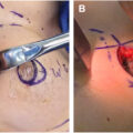
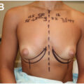
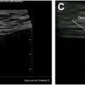
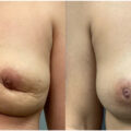
For patients undergoing primary breast augmentation, scaffolding in the form of P4HB may be used not only for lower pole support, but also for (off-label) prevention of encapsulation. The majority of my primary augmentations in 2025 are placed in the prepectoral space. These are all supported with a sling of P4HB in the lower pole. Precise, non-bloody, sharp dissection is used to create the pocket below the pectoralis major fascia. Appropriate pocket irrigation with 0.5% povidone-iodine solution is performed. An insertion sleeve is used to deliver the silicone implant into the pocket. Once the implant is in the pocket, a 15 cm × 20 cm piece of rectangular lightweight P4HB (Galaflex Lite) is cut in half along the long axis. The scaffold is soaked in the irrigation. The implant is retracted anteriorly, and the long edge of the scaffold is placed below the implant. The implant is then released, and the remainder of the scaffold is pushed above the implant using a Levine breast augmentation retractor (assi, Westbury, New York) centrally, medially, and laterally, creating a sling or gutter along the inframammary fold. I do not inset the scaffolding as it is not necessary and creates unwanted contours or pleats in the scaffolding. Inset can also potentially overtighten the lower pole, changing the curve or shape of the lower pole. I have not seen any cases of malposition in the absence of an inset, and it also saves significant surgical time. The soft tissues are closed above the scaffolding, and the incision is closed in 2 layers ( Figs. 1–3 ).