As the demand for breast implant removal continues to rise, surgeons need to consider whether to remove the surrounding capsule at the time of explantation and what type of capsulectomy should be performed. Following explantation, post-explantation deformities such as ptosis, nipple retraction, skin laxity, and soft tissue deficits may require aesthetic contouring with tissue rearrangement and/or fat grafting. The risks and benefits of these procedures should be discussed with each patient and the most appropriate procedures should be selected based on the patient’s desires and surgeon’s clinical judgment.
Key points
-
•
The demand for breast implant removal is rising due to safety concerns.
-
•
Surgeons must address management of the capsule at the time of breast explantation surgery, using consistent definitions and evidence-based data.
-
•
Breast appearance after implant removal with or without capsulectomy can be variable and may require contouring procedures and volume restoration.
-
•
Autoaugmentation mastopexy utilizes breast parenchymal flaps in place of the implants to improve breast contour but will not provide additional volume.
-
•
Fat grafting may be performed as an adjunct to mastopexy, or as a stand alone procedure for contouring following explantation.
Abbreviations
| BIA-ALCL | breast implant associated-anaplastic large cell lymphoma |
| BIA-SCC | breast implant associated-squamous cell carcinoma |
| BII | breast implant illness |
| BSCC | Breast Surgery Collaborative Community |
| SSBI | systemic symptoms associated with breast implants |
Introduction
Breast augmentation is one of the most common plastic surgery procedures performed worldwide. According to The Aesthetic Society’s National Databank Statistics, it was the second most frequently performed surgery after liposuction in 2023. However, the Aesthetic Society statistics also show that the demand for breast implant removal, or explantation, is rising with an increase of 40.2% between 2019 and 2023.
Indications for explantation
The indications for breast implant removal traditionally have included device-related complications such as implant rupture, capsular contracture, and infection. Patient preference for implant removal includes age-related and weight-related changes, postpregnancy or breast-feeding changes, as well as a desire for “a natural look.” More recently, concerns about breast implant-related cancersbreast implant associated-anaplastic large cell lymphoma (BIA-ALCL) and breast implant associated-squamous cell carcinoma (BIA-SCC)—and discussion of systemic symptoms associated with breast implants (SSBI) or breast implant illness (BII) on social media platforms have prompted concern surrounding the safety of breast implants. SSBI and BII may be used interchangeably in this article. In October 2021, the Food and Drug Administration placed a boxed warning on breast implant packaging addressing these concerns to assist patients in making informed decisions about breast implant placement.
Origins of Capsule
While breast implant removal itself is relatively straightforward, the management of the surrounding capsule, formed by means of a foreign body reaction to the implant, has been subject to controversy. Immediately after implant placement, capsule formation begins with the accumulation of cellular debris and progresses through protein absorption, acute inflammation, chronic inflammation to fibrous encapsulation. Histologically, the capsules contain 3 layers: an inner layer of fibroblasts and macrophages; an intermediate layer of loose connective tissue fibrils rich in vasculature; and a dense vascular outer collagen layer. Although most capsules remain benign, some undergo varying degrees of fibrosis and/or can develop a seroma or mass. Capsular contracture occurs when the collagen fiber capsule tightens and compresses the breast implant causing a firm and sometimes distorted breast. It can occur in 2% to 15% of primary breast augmentations and 5% to 22% of secondary breast augmentations. It is thought to be due to a chronic inflammatory state surrounding the breast prosthesis within the pocket due to microbial contamination, persistent blood in the pocket, traumatized tissue, and silicone leak ( Fig. 1 ).
Baker 1 capsule.
Surgical management of capsule
Surgical management of capsular contracture varies but may involve either capsulotomies or capsulectomies depending on severity. Surgical treatment of the capsule may also be warranted in cases of implant rupture, where there is a thickened capsule or silicone embedded in the capsule, and capsular malignancies. In recent years, patient-driven requests for capsulectomies have been on the rise for SSBI (BII) and as prophylaxis for BIA-ALCL although evidence for this remains elusive ( Fig. 2 ).
Calcified capsule.
Capsulotomy
A capsulotomy is a surgical procedure that involves making an incision to release the fibrous capsule around the breast implant to alleviate symptoms of breast firmness, pain, and distortion. Capsulotomy is the simplest and safest of the procedures used to manage the capsule in explant patients. The advantages of a capsulotomy include preservation of support for the overlying skin envelope when soft tissue coverage is thin, improved circulation for the overlying breast skin soft tissue envelope especially when a mastopexy is planned, and improved pocket control and support for the implant if an implant replacement is planned. Capsulotomies are not adequate in cases of capsular malignancies or calcified capsules where complete removal of capsular tissue is indicated.
Partial Capsulectomy
Partial capsulectomy involves the removal of a portion of the breast implant capsule. It is generally considered when the capsule is adherent to the dermis or rib perichondrium to avoid serious injury to the skin or chest wall. Partial capsulectomy may also be performed in revision breast implant surgeries, to improve shape and remove irregular contours. Retained capsule may be a nidus for cyst or seroma formation, particularly in patients with histologic evidence of synovial metaplasia where synovial fluid may be produced. In these cases, a secondary operation to remove the remaining capsule may be considered.
Total Capsulectomy
Total capsulectomy (total precise capsulectomy) is performed with complete removal of the anterior and posterior capsules, not necessarily done as a single unit or in one piece. With total intact capsulectomy complete removal of the breast implant capsule is performed as a single unit. These are more extensive procedures that carry a higher risk of bleeding and other perioperative complications compared to partial capsulectomy or capsulotomy and patients should be informed of these risks. Total capsulectomy with explantation increases the risk of breast deformities as more soft tissue is removed with the implant. Total capsulectomy of subpectoral implants requires more dissection and presents a greater risk for pneumothorax, muscle edge bleeding and injury to deeper vascular structures. It can be challenging in cases where the capsule is very thin and fragile. There is no scientific evidence that supports total capsulectomy in the absence of grade 4 capsular contracture, breast cancer, or BIA-ALCL. If the capsule is calcified, has a mass present, or is embedded with silicone product, removal of the capsule is warranted. Other indications for total capsulectomy, such as implant rupture, capsular contracture, and SSBI (BII) are controversial and the subject of debate ( Fig. 3 ).
Total intact capsulectomy.
In patients concerned with SSBI (BII), total capsulectomy may be considered based on the clinical scenario and in consultation with the patient, considering the risks and benefits. Previous studies have reported symptomatic improvement with breast implant removal and capsulectomy, in which the degree of capsulectomy remains under substantial discussion. Recent prospective blinded studies by Glicksman and colleagues , reported statistically significant improvement in symptoms of patients with self-reported SSBI (BII) following explantation, regardless of the type of capsulectomy performed as well as in the absence of capsulectomy.
En Bloc Capsulectomy
En bloc capsulectomy is defined as an oncologic procedure in which a margin of normal-appearing tissue around the capsule is removed as part of the specimen that also contains the implant, an intact implant capsule, and associated masses. Currently available evidence supports the use of en bloc capsulectomy only in patients with an established diagnosis of BIA-ALCL or other capsular malignancies before surgical intervention. BIA-ALCL is associated with textured implants with the most common symptoms being seroma, pain or swelling of the breast, capsular contracture, overlying skin rash, and a mass around the implant. The capsule, periprosthetic fluid and any associated masses should be completely removed with the implants based on evidence provided by Clemens, who demonstrated better overall survival and event-free survival outcomes compared to radiation therapy, chemotherapy, partial capsulectomy, or no capsulectomy. Currently, there is no evidence whether a partial or total capsulectomy has any effect on risk mitigation in patients that have textured implants, but no disease. For nononcologic indications, there is currently no evidence of additional benefit in en bloc capsulectomy as compared to total capsulectomy. En bloc capsulectomy for nonmalignant conditions requested by the public appears to be at the suggestion of social media rather than based on scientific evidence.
Current consensus statement on capsulectomy definitions
In June of 2024, a consensus statement on the terminology and management of breast implant capsules was issued by the Breast Surgery Collaborative Community (BSCC), a consortium of patient advocates, board-certified plastic surgeons and others with a vested interest in enhancing breast surgery safety, to offer clarity for women seeking guidance on removal of breast implants related to health concerns. The BSCC consensus statement defines 4 types of capsulectomy: partial capsulectomy, total capsulectomy, total intact capsulectomy, and en bloc capsulectomy.
The BSCC notes that the informed-consent process for patients considering capsulectomy should be a shared decision-making process with their surgeon, including a discussion of potential benefits and risks. This should include patient concerns regarding SSBI (BII), the risk of breast implant-associated cancers involving the capsule (such as BIA-ALCL, BIA-SCC, and so forth), and any potential future risks, especially resulting from textured implants and tissue expanders. Importantly, the risk of the capsulectomy procedure itself, including the patient’s medical history, must also be weighed against those risks noted. Thorough discussion with the patient must occur to ensure the most appropriate procedure is performed on each patient based on the surgeons’ evaluation and clinical judgment, the patient’s clinical scenario and concerns, and currently available evidence-based data to guide the ultimate recommendation and treatment course.
Outcomes after explantation
Appearance of the breasts after implant removal with or without capsulectomy will depend on several factors including the amount of breast tissue, duration of augmentation, size of implants placed, number of procedures performed, number of pregnancies, breast feeding, age of the patient, and skin quality. Many patients assume that their breasts will revert to their preaugmentation appearance after implant removal, but they fail to consider how their bodies have changed. Anatomic changes after breast implant placement include stretched skin, thinner and atrophic breast parenchyma and, with subpectoral placement, loss of muscle volume and strength. In addition, many patients have gained weight since their augmentation and now have more breast volume than before augmentation. All these variables will influence postexplanation deformities, which may range from debilitating to quite minimal.
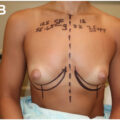
In addition to volume loss and wrinkled skin, the breasts may appear deflated with central hollowing and upper pole deflation ( Fig. 4 ). The nipple-areola complex may appear retracted ( Fig. 5 ) and breast tissue may be displaced laterally, medially, or inferiorly based on the position of the breast implants. Implant removal will unmask breast asymmetries, tubular breast deformities, and chest wall abnormalities that had been camouflaged by the implant. Inframammary fold abnormalities may also be evident, especially in dual plane augmentation or where the fold was lowered at the time of the initial surgery. Assessment of the patients flexing their pectoralis muscle may help determine how high the fold will move after explant and better help plan explant incisions ( Figs. 6 A, B and 7 ).
Breast appearance demonstrating volume loss with central hollowing and upper pole deflation after implant deflation.
Nipple-areola complex showing retraction following breast volume loss and deflation.
( A , B ) Augmented patient at rest and at flexion with dual plane augmentation
Patient from Fig. 6 after implant removal with tubular ptotic breasts revealed with high inframammary folds and wide cleavage line.
Surgical planning
If the capsule itself is not thickened or calcified, it can be left in situ. This may contribute to a better cosmetic outcome as the capsule not only can contribute some thickness to the tissues, but it can prevent the unpredictable scarring down of the breast to the chest wall in the immediate postoperative phase. If the capsule must be removed, early tissue massage and intermittent suction, if drains are used, can be helpful in minimizing this deformity. If, however, the patient needed a mastopexy prior to the augmentation, they will probably still benefit from mastopexy after implant removal. If they did not have ptosis prior to augmentation, many of these patients will see mild to moderate ptosis resolve once the breast is no longer weighed down by the implant. If the patient’s breasts have been subjected to other life factors such as pregnancies or breast feeding, or weight changes, they may now have ptosis where they previously did not ( Fig. 8 A–D ).
( A–D ) ( A ) Favorable result where capsule is not removed. ( B ) Favorable result with tissue contraction where capsule is removed. ( C ) Favorable outcome after explant only still upper pole deflation. ( D ) Unfavorable outcome with progressive contraction after explant with capsulectomy.
Incisions
Related posts:


Stay updated, free articles. Join our Telegram channel

Full access? Get Clinical Tree


