This article chronicles a 30 year evolution in breast augmentation, highlighting advances in implants, techniques, and anatomic understanding while reaffirming foundational surgical principles. It traces the transition from subglandular to submuscular placement following the 1992 Food and Drug Administration moratorium on silicone implants, and the subsequent return to the inframammary fold (IMF) approach after 2006. Detailed anatomy of the IMF is emphasized as critical for preventing complications such as malposition. Major advancements include stronger, more cohesive implants, improved imaging and surgical precision, infection-reducing techniques, and high-resolution ultrasound diagnostics, setting a new standard for safe, precise, and esthetically refined breast augmentation.
Key points
-
•
Anatomy of the inframammary fold is a structure we look at everyday but really do not understand.
-
•
A better understanding of the inframammary fold will allow for improved long-term outcomes with fewer complications, including fold malposition.
-
•
The inframammary fold allows for the most direct approach for a bloodless, precise pocket to perfectly match and fit the dimensions.
-
•
The fold approach allows for precise access to the subglandular, subfascial, or dual-plane breast implant pocket placement.
-
•
A partial submuscular approach allows for creation of a fascial shelf to better support an implant.
-
•
Deeper position allows for the least visibility and palpability of the device.
Abbreviations
| FDA | Food and Drug Administration |
| IMF | inframammary fold |
| N-IMF | nipple to inframammary fold |
Introduction and background
Presented in this article is a 30 year journey through breast augmentation. Although there have been some very significant advancements in devices, technologies, and techniques, little has changed from the basic principles laid down by our mentors as we are all standing on the shoulders of giants. In the United States, during the 1980s and early 1990s, the inframammary fold (IMF) approach was the dominant incision, much as it is today, with the subglandular pocket often preferred at that time. Most surgeons practicing then may recall breast augmentation with this approach under local anesthesia with benzodiazepines and intravenous narcotics, often followed by the patient’s groggy approval of her immediate outcome. With the improvement of devices with less visibility and capsular contracture, we may in fact be heading back to this time.
With the Food and Drug Administration (FDA) moratorium on silicone breast implants for cosmetic procedures in 1992, there was a significant shift to saline-filled implants with more inherent visible rippling and palpability, requiring a shift to the submuscular plane for increased implant coverage. Saline implants could now be placed initially unfilled, leading to other surgical incisional approaches including the transaxillary and periareolar approaches. This allowed implants to be placed through smaller various incisions and then filled in situ. With the lifting of the moratorium in 2006 and the introduction of shaped and textured implants, the incision preference again has shifted back to the inframammary incisional approach, which provides greater accuracy among other benefits. With the focus on research and reduction of capsular contracture techniques and Biofilm concepts, , along with the emphasis of bloodless pocket dissection under direct vision, the most common surgical approach has returned and remained the inframammary crease incision.
Anatomy of the inframammary fold
The IMF is a structure that most plastic surgeons look at every day, but do not really understand. Appreciating the detailed anatomy of the IMF is absolutely critical for excellent, consistent outcomes in breast surgery with and without breast implants. Now that capsular contracture is being significantly lowered by reducing the bacterial load and skin contact with a device, and improved bloodless techniques, implant malposition, particularly fold malposition is becoming the most common complication in implant surgery. Having direct control over the fold has many advantages particularly if the surgeon desires to reinforce the fold with suture or scaffolds. In fact, both capsular contracture and malposition continue to be the most common complications in breast revision research. These histologic and anatomic details have been presented and published previously and remain critical to understand and implement into surgical practice. ,,,,
The best detailed anatomy has come out of Robert Acland’s laboratory from Louisville that many plastic surgeons fondly remember as sponsoring an excellent microvascular laboratory training center. Mutan and colleagues provide histologic confirmation of what happens clinically in this anatomic location ( Fig. 1 ).
( A , B ) The inframammary fold is a structure plastic surgeons look at nearly every single day, but do not really understand. Histologically, Mutan and colleagues have shown that the resting fold that we mark at the skin inflection point is 1.5 to 2.0 cm higher than the true fold because of the oblique fascial fibers that extend to the deeper fascia lying below this resting fold.
There is a fusion of the superficial and deep fascia of the breast that inserts into the region of the IMF at an oblique angle that continues inserting below the resting inframammary crease. This creates what has been termed the “resting” fold and the “true” fold. Surgeons will typically mark the resting fold prior to surgery believing this will be the final fold location and foundation of the breast. Secondary to these downward oblique fibers (see Fig. 1 A, B), the true fold is, on average, 1 to 1.5 cm lower than this resting fold. This relationship may be demonstrated by putting downward pressure on the lower pole of the breast elucidating the actual true fold and base of the breast. This also explains why the resting fold over time will ride up on the lower pole of the breast. In an overly tight breast reduction, augmentation mastopexy incision, or in fold malposition, the lower incision rides up higher on the breast. When the implant is completely engaged in the pocket, without any reinforcement or support, and without any scar contracture, it settles into the true fold, while and the resting fold rises. This is just not an arbitrary lowering of the fold, but a histologic, anatomic, and clinical truth.
Whatever tissue-based planning system for implant selection is chosen utilizing implant dimensions or implant volume, the ideal nipple to inframammary fold (N-IMF) distance needs to be determined preoperatively. This calculated distance is then applied to the breast, measuring the N-IMF on maximal stretch. , Typically, 5 mm is added or subtracted based on the degree of skin stretch, with excessive skin tightness or laxity. If the N-IMF distance is too long for the predicted implant size/volume, a small wedge may be excised to approximate the distance at the time of the primary procedure. If the natural N-IMF is too short, the distance may be lowered. If lowered more than the true fold level, it should be reinforced with suture or scaffolding to set and help secure this new fold position. If the ideal N-IMF distance is calculated approximately 1.5 cm lower than the resting fold, or actually within the true fold, this is the absolute best-case scenario ( Fig. 2 A, B ). Additionally, the implant size and volume may also be adjusted to choose the best implant “fit” to the patient.
( A , B ) The resting fold is the dashed line above and is marked in the sitting or standing position where the breast attaches to the body. It is also termed the inflection point when gentle pressure is applied to the breast to create the junction with the skin. The true fold is reproduced when the skin is lifted and the breast tissue is forced into the lower breast pocket. This is where the implant will ultimately rest, which is why the incision if made in the resting fold rides up on the lower pole of the breast if overfilled with breast tissue (breast reduction) or implant. With the inframammary incision is made at the true fold and breast fascia of the upper skin flap is grasped with a forceps and distracted downward, it identifies where the resting fold is located.
Major advancements
There have been some major advancements both in implant technology and breast augmentation techniques, over the past few years that have brought about a new standard, state-of-the-art in breast augmentation. Some of these major advancements include
-
•
A new generation of breast implants including
-
○
Stronger barrier proof shells, strong enough to tolerate incredible pressures
-
○
Higher percentage implant fills that decreases inherent implant wrinkling and visible rippling that decreases internal shell folds, that can increase implant fold flaws and permanent implant creases ultimately decreasing shell failure rates
-
○
Improved rheology and higher cohesivity of the internal silicone gel fillers
-
○
New surface modifications that may decrease future capsular contracture rates
-
○
Future increase in shell elasticity and elimination of implant polarity (the implants having a flat bottom and rounder top) that would allow for the implant conforming to whatever surface it is on and responding to external pressures and forces
-
○
-
•
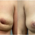
Continued improvements in photographic breast imaging and simulation allowing for surgeons and patients to better navigate implant selection and allowing for patients to visualize their breasts postoperatively with different implant sizes and projections ( Fig. 3 A, B )