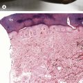
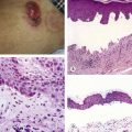
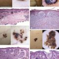
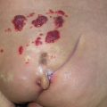
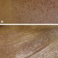
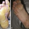
Fig. 20.1 Similar clinical appearance of two different dermal tumors. A Infantile hemangioma. B Rhabdomyosarcoma. From Eichenfield LF, Frieden IJ, Zaenglein AL, Mathes E. Neonatal and Infant Dermatology, 3e. London: Saunders, 2014.
Table 20.1
Dermally-based lesions
Entity Classic morphologic clues* Histopathology Vascular tumors Infantile hemangioma
• History: Not present at birth, grows rapidly over first couple of months
• (see Fig. 20.1A )
•
•
Congenital hemangioma
• History: Present at birth
• Site: Predilection for pressure points
•
• ( Fig. 20.2 )
•
Tufted angioma
• ( Fig. 20.3A,B )
•
•
Glomuvenous malformation (glomangioma)
•
• ( Fig. 20.3C,D )
•
Fibrous tumors Myofibroma
• Site: Often on the head/neck or trunk
•
•
• (see Fig. 20.4 )
•
Infantile digital fibroma
• Site: Typically on the 2nd toe
• ( Fig. 20.5 )
•
Hematologic processes Mastocytoma
• History: Intermittent blistering
•
• ( Fig. 20.6 )
•
Table 20.2
Characteristic papulonodules in children/adults
Entity Classic morphologic clues* Histopathology Adnexal tumors Pilomatricoma ( Fig. 20.7 )
•
•
•
•
Apocrine hidrocystoma (see Fig. 21.4 )
• Site: Eyelid
•
•
Syringoma (see Fig. 2.1D )
• Site: Eyelids
•
•
Sebaceous hyperplasia (see Fig. 2.1E )
• Site: Face
•
•
Vascular tumors Cherry angioma ( Fig. 20.8 )
•
Pyogenic granuloma ( Fig. 20.9 )
• Site: Predilection for head/neck, fingers
•
•
Only gold members can continue reading.
Log In or
Register to continue
Related
Stay updated, free articles. Join our Telegram channel
Join
Full access? Get Clinical Tree
Get Clinical Tree app for offline access



 Get Clinical Tree app for offline access