28 Spiral Thigh Lift

Abstract
One of the most challenging procedures in postbariatric plastic surgery is the thigh lift or thighplasty surgery. It is challenging for the following reasons: the thigh deformity is variable; the thigh contour is heavily influenced by the adjacent anatomic regions, such as the abdomen, waist, and buttocks; the tissues are heavy and subject to considerable movement; and the location of the thigh incisions adjacent to the perineum make them more prone to contamination. The goal of a successful thighplasty procedure should be reduction of the thigh excess and thigh descent. Two major approaches are the medial inner thigh lift and the medial vertical thighplasty, with the latter being the preferred technique for most weight-loss patients. This chapter presents our spiral thigh lift technique, which is universally used in all our postbariatric patients to achieve a circumferential thigh lift when performed in conjunction with a lower body lift. When combined with a vertical thighplasty for significant thigh laxity and excess, we refer to the procedure as the T-thighplasty.
Introduction
Massive weight loss (MWL) results in a broad spectrum of anatomic deformities. The nature of the weight gain and the subsequent weight loss deformities are determined by the patient’s gender, age, prebariatric weight, actual weight loss, and genetic predisposition. With massive weight gain, both the upper and the lower body accumulate adipose depositions that are not only dependent on the amount of calorie intake, but also determined by the patient’s gender and genetic predisposition. In addition, the patient’s tissue quality and zones of adherence play an important role in determining the anatomy of the local fatty depositions. In the lower body, hypertrophy of the adipose tissue results in a three-dimensional expansion of subcutaneous tissues of the lower trunk, thighs, and legs. This results in stretching of the superficial fascial system (SFS) and variable dermal breakage of skin. Consequently, zones of adherence and demarcations become loose, and the skin develops striae. With MWL, the subcutaneous tissue becomes deflated and loose due to reduced SFS and dermal elasticity. Thus, patients present with significant changes in the form, shape, and contour of their abdomen, waist, lower back, buttock, thighs, and legs.
Thigh Aesthetics
The ideal thigh contour complements an aesthetically beautiful and healthy figure. The thighs are demarcated from the abdomen via the inguinal crease that may be extended posteriorly as the superogluteal V-shaped demarcation of buttock and lower back tissue. Medially, the thighs are separated from the perineum by the groin crease, which extends into the inguinal crease anteriorly. The inferior gluteal fold defines the thigh origin posteriorly and demarcates the lower border of the buttocks. The thighs should be proportionate to the buttocks, abdomen, and legs. When in proportion, the trunk and lateral thighs follow an hourglass silhouette with pleasing concavity of the waist, rising to a smooth convexity of hips extending over the outer thighs.
The thighs are asymmetric cones, with the upper lateral aspect fuller than the medial. There is a gentle convexity of the anterior thigh, which conforms to the large muscle mass, in contrast to the flattened posterior thigh. The inner thighs are soft and slightly convex. The lower lateral thigh skin is firmly adherent to underlying muscular fascia.
Anatomic Deformity
With MWL, the thighs become deflated. The thigh deformity varies by genetic predisposition, extent of weight loss, and residual obesity. As women have less muscular and more adipose development of their thighs than men, the changes are more profound in women. For those female patients who have lost most of their excess weight, there is a characteristic presentation. Except for the lower lateral thigh, the skin is diffusely loose and flaccid. Almost all MWL patients pre sent with vertical thigh laxity. The upper lateral thighs bulge into what is commonly referred to as saddlebags, abruptly ending at the mid-lateral thigh. Looseness of the upper posterior thigh is usually less dramatic. The excess tissue of the medial thighs forms cascading rolls, like a hanging curtain, suspended from the inguinal ligament anteriorly and the inferior gluteal fold posteriorly. These folds progressively diminish toward the knees. The anterior thighs have stacked waves of skin. In severe cases, the deflated thigh appears to widen circumferentially. The dysmorphic changes of the thigh and buttocks often result in loss of the lateral buttock to thigh demarcation. The postbariatric patient also has unique dysmorphic features of the gluteal and perigluteal regions. It is important to look at these changes in combination and treat this area as a unit.
Surgical Techniques
Thighplasty is the aesthetic reshaping of the thigh following removal of excess skin and fat. We prefer to use the term thigh lift to denote elevation of the thigh in a vertical manner and reserve the term thighplasty for circumferential (transverse) reduction of the thigh. The new contour should be attractive, the scars inconspicuous, the groin crease reconstructed meticulously, and the complications minor. The current thighplasty techniques for MWL patients include the following:
Medial inner thigh lift for correction of the loose upper medial thigh1
Lower body lift to correct the lateral thigh
Vertical thighplasty to correct for the excess circumferential laxity2
Spiral Thigh Lift
Although the traditional crescent-shaped medial inner thigh lift is an effective technique for the upper inner thigh in nonbariatric patients, it does not address the need for a circumferential thigh lift in the MWL patient. Many current techniques, as exemplified by Lockwood,1 are at best a partial remedy for the thigh deformity because they do not address the anterior or posterior thigh laxities. Since 2005, we have been performing a modified inner thigh lift approach that we have termed the “spiral thigh lift.” The spiral thigh lift technique provides circumferential thigh elevation when performed with a lower body lift concomitantly. This procedure not only elevates the thigh as needed, but also decreases the width of the thigh to a limited degree by advancing the lax thigh tissue superiorly.
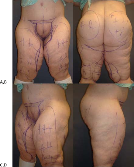
Patients are marked preoperatively at first supine and then standing. The markings start with the upper transverse incision between the medial thigh and labia majora or scrotum as the supine patient abducts the thighs. It is extended anteriorly and vertically across the mons pubis to the lower abdominoplasty incision. Posteriorly, it is extended transversely within the inferior gluteal fold. This identifies the upper incision of the spiral thigh lift.
Next, the laxity in the upper anterior and medial thigh is evaluated by advancing the lax upper medial thigh tissue superiorly toward the groin crease, marking the lower incision on the inner thigh. This is extended posteriorly to meet the upper incision of the posterior thigh lift at the lateral aspect of the inferior gluteal fold. This marking identifies the lower incision of the spiral thigh lift. One of the authors (D.J.H.) uses the abdominoplasty excision to elevate the anterior thigh. In the technique of the other author (S.A.M.), the anterior thigh laxity is then assessed by pinching the anterior thigh tissue toward the level of the inguinal ligament. The anterior thigh marking is then made as an extension of the medial markings extended over the lateral thigh and posteriorly as the inferior incision of the lower body lift. The inferior marking of the lower body lift is made at a height determined by the extent of buttock and lateral thigh soft tissue redundancy. The superior marking for the buttock-thigh lift are posterior extensions of the superior abdominoplasty markings. The superior line delineates a gullwing-shaped incision. Between these two incisions lie the tissues that would normally be used for autologous buttock augmentation in our practice ( Fig. 28.1 ).3,4
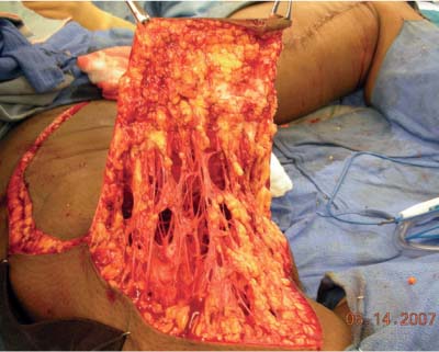
The procedure is started while the patient is in the prone position. The upper and lower markings of the posterior thigh lift are incised through the subcutaneous tissue to the superficial muscular fascia. The superior incision is then made to the gluteal aponeurosis, which is continuous with the superficial muscular fascia. The redundant posterior thigh tissue is then excised. In this plane, the lower buttock tissue is dissected from the ischial tuberosity. The posterior thigh is then suspended to the ischial tuberosity by multiple interrupted sutures through the thigh fascia lata, SFS, and ischial periosteum. Elevation of the posterior thigh to this level and closure of the buttock SFS recreate the inferior gluteal fold.
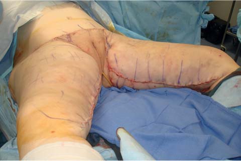
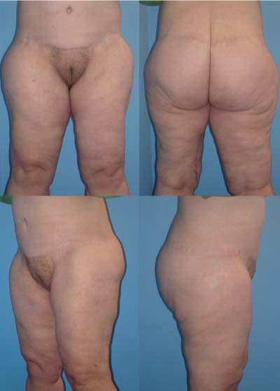
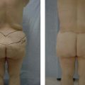
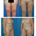
The spiral thigh lift is continued after turning the patient to the supine position. The marked excess upper medial thigh is then liposuctioned to preserve the neurovascular and lymphatics between the mons pubis and the femoral triangle. This radical removal of fat between the dermis and muscular fascia is called excisionsite liposuction (ESL). We prefer ultrasonic-assisted lipoplasty (UAL) in many cases for its retention of connective tissue. The redundant excess skin over the ESL is then excised in the subdermal plane ( Fig. 28.2 ). Accurate preoperative markings are essential, as the ESL commits the surgeon to a minimal width of excision. The medial thigh lift is completed after elevating the thigh to the level of the groin crease. Again, similar to the posterior thigh lift, the lower incision of the medial thigh is extended to the thigh muscular fascia and Colles fascia. The anteromedial thigh is then lifted in a vertical direction to the level of the groin crease. This advances the anterior thigh incision to the planned groin incision level and closes the medial thigh defect. The muscular fascia of the medial thigh is then sutured with multiple nonabsorbable sutures to the periosteum of the pubic tuberosity and to Colles fascia. The groin crease is recreated during the closure of the deep SFS of the thigh and the groin region. This not only restores the natural infolding of the crease area, but also prevents migration of the incisions, resulting in symmetrically placed scars ( Figs. 28.3, 28.4, and 28.5; see also the video clip). We have termed our approach a “spiral thigh lift” because the incisions follow a spiral line from the inferior gluteal fold, through the groin crease, and to the circumferential lower body lift incision. The notion of the spiral thigh lift is complementary and inclusive of the lower body lift and abdominoplasty. The combined procedures of spiral thigh lift, lower body lift, and abdominoplasty produce synergy, capitalizing on the biomechanics of skin tension ( Fig. 28.6 ).5,6
The spiral thigh lift differs from the conventional medial inner thigh lift in several ways. Whereas the conventional thigh lift is an ideal procedure for the nonbariatric patient who has mild-to-moderate laxity of the inner thigh, the spiral thigh lift is a more extensive procedure that corrects laxity of the thigh tissue posteriorly, medially, and anteri-orly. Together with the lower body lift, it offers a complete circumferential thigh lift. Also, the vectors of correction differ between the two procedures. The medial inner thigh lift relies on a superomedial vector of lift1,4 with advancement of the inferior thigh incision toward the groin incision. On the other hand, the spiral thigh lift employs a purely vertical vector with elevation of the inferior thigh tissue superiorly. As a result, the procedure combines elements of lift with some degree of circumferential thigh reduction. Conceptually, the spiral thigh lift is a more secure lift because the thigh tissues are secured posteriorly, medially, and anteri-orly. Thus, it should obviate labial spreading that is a known complication of the conventional inner thigh lift.1,2
Related posts:
 29 Fascio-Fascial Suspension Technique for Medial Thigh Plasty
 30 Anterior Proximal Extended (APEX) Medial Thigh Lift
 32 Combination Circumferential Abdominoplasty with Medial Thigh Lift
 31 Medial Thigh Contouring: Cones and Cylinders
 4 Anesthesia and Safety Considerations in the Perioperative Period
 36 Facial Rejuvenation: Open Technique
Stay updated, free articles. Join our Telegram channel

Full access? Get Clinical Tree


