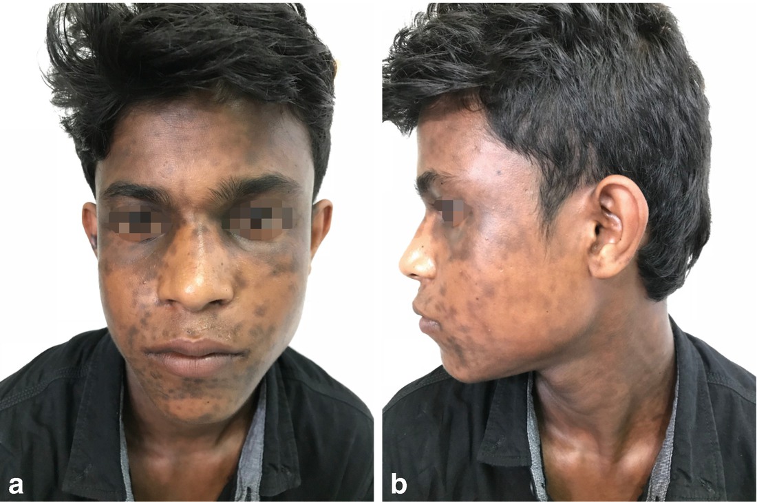
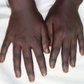
A 24 year male presented with multiple grey brown macules on face, neck and upper trunk since 6 months. It was insidious in onset and gradually progressive. It was not preceded by any erythema over the lesions and was asymptomatic. There was no history of any application of cosmetics. On examination patient had discrete as well as confluent slate grey coloured hyperpigmented macules with ill-defined border distributed over face, neck and upper limb (Fig. 7.1). The surface was smooth and non-scaly. Oral cavity, nails, palms and soles were not affected.
Figure 7.1
Blue-grey hyperpigmented macules on face (a) and neck (b) in a 24 year old male
Based on the case description and figure, what is your diagnosis?
1.
Lichen planus pigmentosus
2.
Erythema dyschromicum perstans
3.
Reihl’s melanosis
4.
Macular amyloidosis
5.
Postinflammatory pigmentation
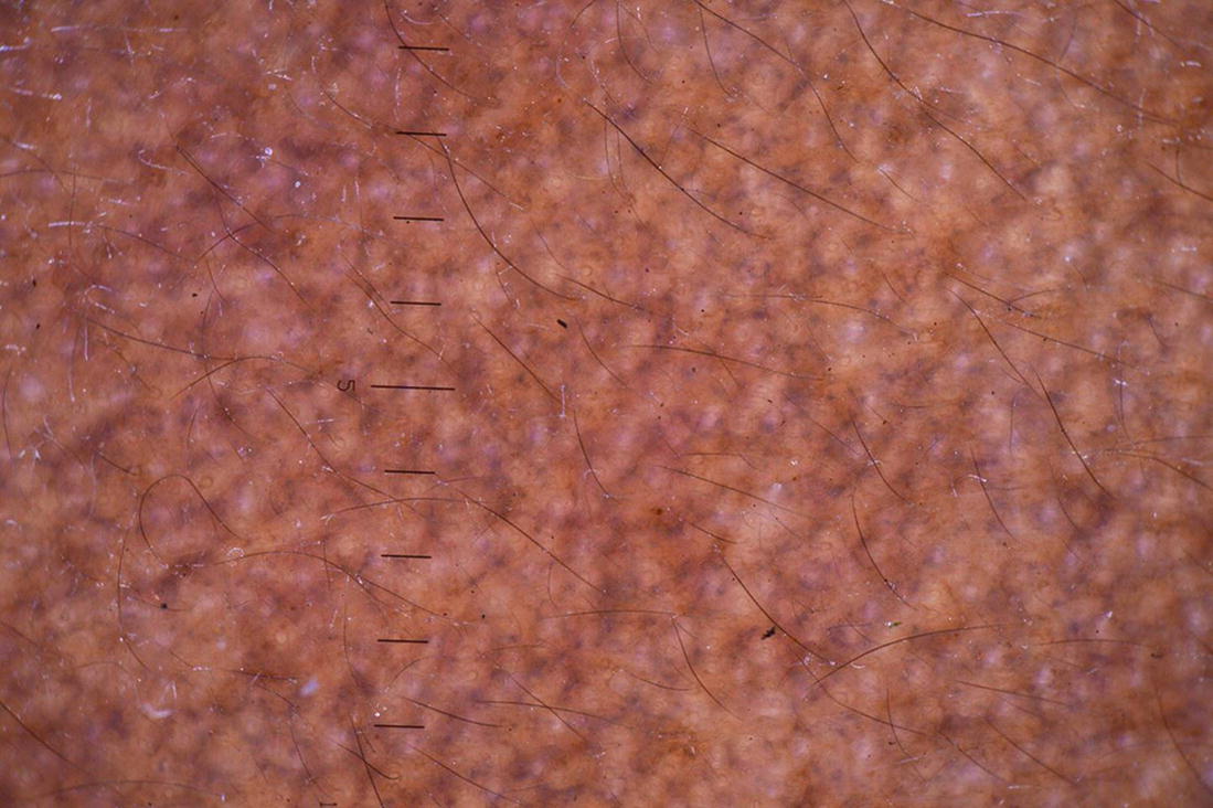
Dermoscopy reveals grey to blue pigment dots forming a net like pattern with reticular pigment network (Fig. 7.2). Histopathology showed marked pigmentary incontinence, focal basal cell degeneration with minimal perivascular lymphocytic infiltrate in superficial dermis.
Figure 7.2
Grey to blue pigment dots which are bigger in size and forming a net like pattern and reticular pigment network in dermoscopy (Courtesy: Dr. Shekhar Neema)
Diagnosis
Lichen planus pigmentosus
Only gold members can continue reading. Log In or Register to continue
 Years Old Male with Multiple Hyperpigmented Macules on Trunk
 Gray Pigmented Macule on Right Cheek
 Female with Multiple Pigmented Macules on Face
 Young Female with Generalized Mottled Pigmentation
 Young Boy with Generalized Hyperpigmentation
 Female with Freckles Like Pigmentation on Face and Extremities


