Source: Adapted from Nahhas AF et al. J Clin Aesthet Dermatol. 2017 Apr; 10(4):37–46. Huang C and Boyce SM. Surgical margins of excision for basal cell carcinoma and squamous cell carcinoma. Semin Cutan Med Surg. 2004; 23:167–173. Adapted from Ad Hoc Task Force, et al. AAD/ACMS/ASDSA/ASMS 2012 appropriate use criteria for Mohs micrographic surgery. J Am Acad Dermatol 2012; 67:531. Location Moderate risk/area “M”: cheeks, forehead, scalp, neck, jawline, and pretibial surface. High‐risk tumor features High‐risk patient features Tips: Adapted from AJCC Cancer Staging manual, Eighth Edition (2017). Balch CM et al. Final version of 2009 AJCC melanoma staging and classification. J Clin Oncol 2009; 27:6199–6206. Gershenwald JE et al. Melanoma staging: evidence‐based changes in the American Joint Committee on cancer eighth edition cancer staging manual. CA Cancer J Clin 2017;67:472–492. Clark level is no longer recommended as a staging criterion and is used for staging tumors ≤1 mm2 only if mitotic rate cannot be determined. Breslow tumor thickness is measured in mm from the top of the granular layer of the epidermis (or the base of an ulcer) to the deepest point of tumor invasion using an ocular micrometer. Gershenwald JE et al. Melanoma staging: evidence‐based changes in the American Joint Committee on Cancer Eighth Edition cancer staging manual. CA Cancer J Clin 2017;67:472–492. *There are no Stage III subgroups in clinical staging. Source: Adapted from NCCN Practice Guideline in Oncology‐ v.2.2016 Melanoma. *Sentinal lymph node should be performed at the time of Wide Local Excision. Consider in tumor <1 mm if initial bx with mitotic rate ≥1 mm2, ulceration, positive deep margin, angiolymphatic invasion, or young age. The yield and clinical significance of SLNBx in Stage IA is unknown and is generally not recommended. **Followup: At least annual skin exam for life, educate patient in monthly self‐skin and lymph node exam. (Bichakjian et al. Guidelines of care for the management of primary cutaneous melanoma. JAAD 2011; 65:1032–1047) ***Evaluation: Routine imaging/lab tests not recommended in Stage 0/IA disease. In Stage IB, may consider nodal basin ultrasound prior to SLNB for patients with equivocal regional lymph node physical exam (NCCN guidelines 2.2016). CT, PET, and MRI may be performed to evaluate specific sxs. ┼Regimens include dacarbazine, temozolomide, paclitaxel, vinblastine, IL‐2, IFNα‐2b, and combinations thereof (see NCCN guidelines). Source: Adapted from National Comprehensive Cancer Network stratification of low versus high‐risk cSCC. National Comprehensive Cancer Center. NCCN Clinical Practice Guidelines in Oncology; Squamous Cell Carcinoma (V2.2018). 2018; www.nccn.org, 1 February 2018. Area H = “mask areas” of face (central face, eyelids, eyebrows, periorbital, nose, lips [cutaneous and vermilion], chin, mandible, preauricular and postauricular skin/sulci, temple, and ear), genitalia, hands, and feet. Area M = cheeks, forehead, scalp, neck, and pretibia. Area L = trunk and extremities (excluding pretibia, hands, feet, nail units, and ankles). High‐risk histologic subtype: Acantholytic (adenoid), adenosquamous (showing mucin production), desmoplastic, or metaplastic (carcinosarcomatous) subtypes. BWH classification provides better prognostication in patients with localized disease (stratifies T2 into a and b) Use of antibiotic prophylaxis for endocarditis indicated for surgical procedure on infected tissue in patients with high‐risk cardiac lesion or as detailed below One hour prior to surgery: (all p.o. doses). History of HSV infection of the orofacial area is an indication for prophylaxis for facial resurfacing, chemical peels, dermabrasion, PDT, and orofacial surgery. Treat for 7–14 days with acyclovir, valacyclovir, or famciclovir to suppress viral reactivation during reepithelialization. Source: Adapted from CDC. MMWR Recomm Rep. 2002 Oct 25;51(RR‐16):1–48. Reversibly inhibit nerve conduction by blocking sodium ion influx into peripheral nerve cells = prevent depolarization of nerves. The patient The anesthetic agent Add 1 cc 8.4% NaHCO3 to 10 cc Lidocaine 1% with epi The injection technique 1% Lidocaine with epinephrine 1:100 000 Lidocaine 0.05–0.1% + Epinephrine 1:1 000 000 Max Tumescent is 35–50 mg/kg Peak Lidocaine level at 12–14 hours Source: Cavef et al. Arch Derm 2007;143:1074–1076. *Risk of methemoglobinemia. Also, may create artefactual vacuolization/swelling of the upper epidermis and basal layer damage/clefting. Source: Adapted from Snow SN and Mikhail GR. Mohs Micrographic Surgery Second Edition. Chapter 14. Table 14–3. †Epinephrine is pregnancy category C. Low doses (diluted 1:300,000 can be used during pregnancy) *Bupivacaine and mepivacaine: pregnancy category C due to potential for fetal bradycardia From Robinson JK (ed.). Atlas of Cutaneous Surgery, first edition. WB Saunders: 1996, p. 2, figure 1.2, with permission. From Robinson JK (ed.). Atlas of Cutaneous Surgery, first edition. WB Saunders: 1996, p. 2, figure 1.4, with permission. From Robinson JK (ed.). Atlas of Cutaneous Surgery, first edition. WB Saunders: 1996, p. 2, figure 1.3, with permission. Courtesy of Dr. Quan Vu Courtesy of Dr. Quan Vu From Robinson JK (ed.). Atlas of Cutaneous Surgery, first edition. WB Saunders: 1996, p. 186, figure 3.1, with permission. From Robinson JK (ed.). Atlas of Cutaneous Surgery, first edition. WB Saunders: 1996, p. 3, figure 1.5, with permission. From Robinson JK (ed.). Atlas of Cutaneous Surgery, first edition. WB Saunders: 1996, p. 3, figure 1.5, with permission. From G Bernstein. J Dermatol Surg Oncol, 1986; 12(7):725, figure 6, with permission. From RG Wheeland RG (ed.). Cutaneous Surgery, first edition. WB Saunders: 1994, p. 61, figure 5.13, with permission. From Leventha: Fractures, dislocations, and fracture‐dislocations of the spine. In Canale et al (eds.) Campbell’s Operative Orthopaedics, tenth edition. Mosby: 2003, Figure 35.1, with permission. Modified from Min RJ, et al. Duplex ultrasound evaluation of lower extremity venous insufficiency. J Vasc Interv Radiol 2003 14: 1233–1241, with permission. From RK Scher and CR Daniel. Nails: Therapy, Diagnosis, Surgery, second edition. WB Saunders: 1997, p. 13–14, figures 2.1, 2.2a, 2.2b, with permission. From Wheeland RG (ed.). Cutaneous Surgery, first edition. WB Saunders: 1994, p. 51, figure 5.6, with permission. Cosmetic result of wound healing by secondary intention according to anatomical site.From Zitelli JA. Wound healing by secondary intention. JAAD 1983;9(3) 407–415; with permission From S Burge, R Rayment. Simple Skin Surgery. Blackwell Scientific Publications: 1986; with permission. Modifications of rhombic flaps Webster 30º Narrower flap, easier to close secondary defect Less reorientation of tension vectors Dufourmentel Compromise between Limberg and Webster flap Extend dotted lines then bisect them Second incision parallel to defect midline Bi‐rhombic flap Zitelli modified bilobe flap Causes of graft failure *Gut made from mucosa/submucosa of sheep or beef intestine. *Electrocautery = not electrosurgery, no electric current, uses heat conduction. IR: infrared; R: red; O: orange; Y: yellow; G: green; B: blue; UV: ultraviolet; Mel: melanin; Hb: hemoglobin; KTP: Potassium‐titanyl‐phosphate. Selective photothermolysis: selective heating of a target chromophore occurs:
Part 2
Surgical and Cosmetic Dermatology
SURGICAL DERMATOLOGY
Skin cancer
Surgical margin guidelines
Tumor Type
Tumor characteristics
Excision Margin
Melanoma
(see melanoma guide pg.)
In‐situ
0.5–1 cm
or Slow Mohs
consider SLN Bx for > 0.8 mm or ulceration
≤1 mm
1.01–2 mm
>2 mm
1 cm
1–2 cm
2 cm
Basal Cell Carcinoma (BCC)
Low risk BCC
Well‐defined borders
Small size
Area L < 20 mm
Area M < 10 mm
Area H < 6 mm
Nodular or superficial subtype
Primary tumor
3–4 mm
High Risk BCC
Poorly defined margins
Larger Size
Area L > 20 mm
Area M > 10 mm
Area H > 6 mm
High risk tumor or patient features (see indication for mohs below)
Mohs or 5–10 mm
Squamous Cell Carcinoma (SCC)
Low risk SCC
Well‐defined borders
Small size
Area L < 20 mm
Area M < 10 mm
Area H < 6 mm
Well differentiated histology
Primary tumor
4–6 mm
High Risk SCC
Poorly defined margins
Larger Size
Area L > 20 mm
Area M > 10 mm
Area H > 6 mm
High risk tumor location (ear, lip)
High risk tumor or patient features (see indication for mohs below)
Mohs or 6–10 mm
Dermatofibrosarcoma protuberans (DFSP)
NCCN favors Mohs over WLE
2–4 cm to level of deep fascia
Merkel Cell Carcinoma
NCCN favors WLE. Can do Mohs if it does not interfere with SNLBx
1–2 cm to investing fascia layer
Advised SLNBx.
Indication for Mohs micrographic surgery
Melanoma ‐ AJCC TNM classification
Major changes in AJCC eighth edition
T classification
Tx
1° tumor cannot be assessed
T0
No evidence of 1° tumor
Tis
Melanoma in situ
T1
≤1.0 mm
a: <0.8 mm with no ulceration
b: 0.8–1.0 mm with no ulceration
or <1.0 mm with ulceration
T2
1.0–2.0 mm
a: no ulceration
b: + ulceration
T3
2.0–4.0 mm
a: no ulceration
b: + ulceration
T4
>4.0 mm
a: no ulceration
b: + ulceration
N classification
Survival %
5 yr
10 yr
Nx
Nodes cannot be assessed/not performed
N0
No regional lymphadenopathy/metastases detected
N1
1 node
a: no MSI, node clinically occult
b: no MSI, node clinically detected
84
76
75
71
0 node
c: MSI present
81
75
N2
2–3 nodes
a: no MSI, node clinically occult
b: no MSI, node clinically detected
79
71
71
71
1 node
c: MSI present, node detectable or occult
69
59
N3
4+ nodes
a: no MSI, node all clinically occult
b: no MSI, >1 node clinically detected or matted
60
64
46
57
2+ more nodes
c: MSI present, node clinically detectable or occult
52
43
Microsatellite instability (MSI) = any in‐transit, satellite, locally recurrent, or microsatellite metastases
M classification
M
Site
Serum LDH
Mx
Distant mets cannot be assessed
N/A
M0
No distant mets
N/A
M1a
Distant skin, soft tissue including muscle, and/or nonregional lymph node
(0) Normal
(1) Elevated
M1b
Lung mets
(0) Normal
(1) Elevated
M1c
Non‐CNS visceral mets
(0) Normal
(1) Elevated
M1d
CNS mets
(0) Normal
(1) Elevated
Clark level
Level I
Confined to the epidermis (MIS)
Level II
Invasion past basement membrane into the papillary dermis
Level III
Tumor filling papillary dermis to the junction of the superficial reticular dermis
Level IV
Invasion into the reticular dermis
Level V
Invasion into the subcutaneous tissue
Breslow depth
Melanoma ‐ AJCC TNM staging and survival
Clinical staging
Pathologic staging
Survival (%)
T
N
M
T
N
M
5 yr
10 yr
IA
T1a
0
0
T1a
0
0
99
98
IB
T1b
T2a
0
0
T1b
T2a
0
0
99
96
96
92
IIA
T2b
T3a
0
0
T2b
T3a
0
0
93
94
88
88
IIB
T3b
T4a
0
0
T3b
T4a
0
0
86
90
81
83
IIC
T4b
0
0
T4b
0
0
82
75
IIIA
Any T*
N1–3
0
T1–2a
T1–2a
N1a
N2a
0
93
88
IIIB
T0
T1–2a
T1–2a
T2b–3a
N1b–c
N1b–c
N2b
N1a–2b
0
83
77
IIIC
T0
T0
T1a–3a
T3b–4a
T4b
N2b–c
N3b–c
N2c–3c
Any N
N1a–2c
0
69
60
IIID
T4b
N3a–c
0
32
24
IV
Any T
Any N
M1
AnyT
Any N
M1a
M1b
M1c
M1d
9–27
16
3
6
Melanoma: treatment guidelines
Breslow depth (mm)
Margin (cm)
SNL*
Physical exam**
Workup***
Adjuvant treatment
In situ
0.5–1
No
q6 mo x 1 yr
then yearly
—
<1
1
No*
q6–12 mo × 5 yr
then yearly
—
1.01–2.00
1–2
Yes
—
2.01–4.00
2
Yes
q3–6 mo × 2 yr, q3–12 mo × 3 yr, then yearly
>4
2
Yes
Stage III SLN +, micromet
WLE (as above)
LND or clinical trial
q3–6 mo × 2 yr, q3–12 mo × 3 yr, then yearly
Stage III Clinical + nodes, macromet
WLE
FNA or bx of + LN, then LND
Stage III in‐transit
WLE + FNA or Bx of in‐transit lesions
Yes
q3–6 mo × 2 yr, q3–12 mo × 3 yr, then yearly
Stage IV
FNA or bx
Yes
q3–6 mo × 3 yr, q4–12 mo × 2 yr, then yearly
Squamous cell carcinoma (SCC)
NCCN stratification of low versus high risk cutaneous SCC
Parameters
Low risk
High risk
Clinical H&P
Location/size (including peripheral rim of erythema)
Area L < 20 mm
Area M < 10 mm
Area L ≥ 20 mm
Area M ≥ 10 mm
Area H
Borders
Well defined
Poorly defined
Primary vs. recurrent
Primary
Recurrent
Immunosuppression
−
+
Site of prior radiation or chronic inflammatory process
−
+
Rapidly growing tumor
−
+
Neurological symptoms
−
+
Pathology
Degree of differentiation
Well or moderately differentiated
Poorly differentiated
High‐risk histological subtype
−
+
Depth (thickness or Clark’s level)
<2 mm or I, II, III
≥2 mm or IV, V
Perineural, lymphatic, or vascular involvement
−
+
Recommended TX
SCC TNM classification: tumor classification AJCC vs BWH
AJCC eighth ed
Brigham and Women’s Hospital
Tx
1° tumor cannot be assessed
1° tumor cannot be assessed
Tis/T0
SCC in situ
SCC in situ
T1
<2 cm in greatest diameter with fewer than two “high‐risk” features
0 risk factors
T2
2–4 cm, or with two or more “high‐risk” features
T2a: 1 risk factor
T2b: 2–3 risk factors
T3
4 risk factors or bone invasion
T4a
Tumor with gross cortical bone/marrow invasion
T4b
Tumor with skull base invasion and/or skull base foramen involvement
AJCC high‐risk factors:
BWH risk factors:
SCC AJCC TNM staging
T
N
M
0
Tis
0
0
I
T1
0
0
II
T2
0
0
III
T3
T1–3
0
1
0
0
VI
T1–3
Any T
T4
Any T
2
3
Any N
Any N
0
1
N1 = 1 LN+ ≤3 cm ENE−
N2 = 1 LN+ ≤3 cm ipsilateral ENE+
1 LN+, >3 and ≤6 cm ipsilateral ENE− or
1 LN+ ≤3 cm ipsilateral ENE+
≥2 LN+ all ≤6 cm ipsilateral ENE−
N3 = ≥1 LN+ >6 cm ENE+
1 LN+ ≤3 cm ENE+ contralateral or
≥1 LN+ >3 cm ipsilateral ENE+ or ≥2 LN+, any ENE+
ENE+, with extranodal extension; ENE−, without extranodal extension
M0 = no distant mets
M1 = distant mets
SCC ‐ Treatment of advanced disease
Prophylactic antibiotics and antivirals
Guideline for Prophylactic antibiotics
Antibiotic (trade size)
Adults
Children
All sites except oral and groin/lower extremity:
Cephalexin (500 mg, 250 mg/5 ml)
2 g
50 mg/kg
Dicloxacillin (500 mg, 250 mg/5 ml)
2 g
50 mg/kg
If penicillin allergic
Azithromycin (250, 500 mg)
500 mg
15 mg/kg
Clarithromycin (500 mg, 250 mg/5 ml)
500 mg
15 mg/kg
Clindamycin (300 mg)
600 mg
20 mg/kg
Oral site:
Amoxicillin (500 mg, 250 mg/5 ml)
2 g
50 mg/kg
If penicillin allergic
Azithromycin (250, 500 mg)
500 mg
15 mg/kg
Clarithromycin (500 mg, 250 mg/5 ml)
500 mg
15 mg/kg
Clindamycin (300 mg)
600 mg
20 mg/kg
Groin and lower extremity site
Cephalexin (500 mg, 250 mg/5 ml)
2 g
50 mg/kg
If penicillin allergic
Trimethoprim‐Sulfamethoxazole, double strength
1 tab
Levofloxacin
500 mg
Algorithm for antibiotic prophylaxis
Guideline for prophylactic antivirals
Acyclovir (Zovirax)
400 mg tid × 7–14 d
Valacyclovir (Valtrex)
500 mg bid × 7–14 d
Famciclovir (Famvir)
250 mg bid × 7–14 d
Antiseptic scrubs
Agent
Mechanism of action
Gram +
Gram −
Mycobacteria
Viruses
Fungi
Spores
Speed of action
Residual activity
Other
Alcohol
60–95%
Denature proteins (bacterial cell wall)
+++
+++
+++
+++
+++
−
Fast
None
Flammable with laser/cautery
Allow to dry on surface
Chlorhexidine
2–4% (Hibiclens)
Impairs cell membrane
+++
++
+
+++
+
−
Intermed
Excellent
Ototoxicity, keratitis, skin irritant
Iodine 3%
(Lugol)
Oxidation
+++
+++
+++
+++
++
+
Intermed
Minimal
Skin irritant
Inactivated by blood/sputum
Iodophors–
(Betadine)
Povidone–iodine 7.5–10%
Oxidation/ substitution by free iodine: disrupts S─H and N─H bonds, C═C bonds in fatty acids
+++
+++
+
++
++
−
Intermed
(needs to dry)
Minimal
Skin irritant (less than iodine)
Inactivated by blood/sputum
May cross‐react with radiopaque iodine
Surfactant + iodine = iodophor
TechniCare PCMX
Chloroxylenol
Disrupts cell membrane
+++
+
+
+
+
Unknown
Slow
Good
Addition of EDTA increases its activity against pseudomonas
Triclosan
0.2–2%
Disrupts cell wall, inhibits fatty acid synthesis, binds bacterial enoyl–acyl carrier protein reductase (ENR, fabI)
+++
++
+
+++
−
Unknown
Intermed
Good
Forms chloroform and dioxins when combined with chlorine in tap water
Benzalkonium
(Quaternary ammonium)
Dissociation of cell membranes; disrupts intermolecular interactions
++
+
+/−
+
Lipophilic
+/−
Unknown
Slow
Good
Use only in combination with alcohols
Eyedrop preservative
Easily inactivated by cotton gauze/ organic materials
Anesthetics
Mechanism of action
Practical tips to decrease pain with injections:
Standard formula for buffered Lidocaine
Ingredient
Quantity
Lidocaine 1%
50 ml
Sodium bicarbonate 8.4%
5 ml
Epinephrine 1:1000
0.5 ml
Tumescent anesthesia
Ingredient
Quantity
Normal saline 0.9%
1000 ml
Lidocaine 1%
50–100 ml
Sodium bicarbonate 8.4%
10 ml
Epinephrine 1:1000
1 ml
Topical anesthetic (see drug section pg 197)
EMLA cream*
lidocaine 2.5% and prilocaine 2.5%
LMX
lidocaine 4 and 5% in liposomal delivery cream
Lida‐Mantle
lidocaine 3% cream
Topicaine
lidocaine 4% and 5% gel
Pliaglis
lidocaine 7% and tetracaine 7% cream
Pharmaceutically compounded topical anesthetic
BLT
20% benzocaine, 6% lidocaine, 4% tetracaine
TAC
0.5% tetracaine, 1:2000 epineprine, 11.8% cocaine
LET
4% lidocaine, 1:2000 epineprine, 0.5% tetracaine
Lasergel
10% lidocaine, 10% tetracaine
23/7
23% lidocaine, 7% tetracaine
Adverse reaction to local anesthetics
Condition
Pulse
BP
Signs and symptoms
Management
Vasovagal Rxn
▼
▼
Diaphoresis, hyperventilation, nausea
Trendelenburg, cool compress
Epinephrine Rxn
▲
▲
Sweating, tachypnea, HA, palpitation
Reassurance, beta‐blocker
Anaphylaxis
▲
▼
Tachycardia, bronchospasm
Epinephrine 1:1000 × 0.3 ml SQ. Antihistamine, airway maintenance
Lidocaine toxicity
1–6 μg/ml
Nl
Nl
Tongue and circumoral paresthesia, metallic taste, tinnitus, lightheadedness
Observe
6–9 μg/ml
Nl
Nl
Tremors, nausea, vomiting, hallucination, muscle fasciculations
Diazepam, airway maintenance
9–12 μg/ml
▼
▼
Seizures, cardiopulmonary depression
Respiratory support
>12 μg/ml
—
—
Coma, cardiopulmonary arrest
CPR/ACLS
Local anesthetic
Generic name
Trade name
Pregnancy category†
Potency
Onset (min)
Without epinephrine
With epinephrine†
Duration (min)
Max dose (mg/kg) for adults
Duration (min)
Max dose (mg/kg) for adults
Amide (“i” before – caine = amide)
Lidocaine
Xylocaine
B
Intermed
<2
30–120
4.5
(30 cc for 70 kg)
60–400
7
(50 cc for 70 kg)
Bupivacaine
Marcaine, Sensorcaine
C*
High
2–10
120–240
2.5
240–480
3
Mepivacaine
Carbocaine
C*
Intermed
3–20
30–120
6
60–400
8
Prilocaine
Citanest
B
Intermed
5–6
30–120
7
60–400
10
Etidocaine
Duranest
B
High
3–5
200
4.5
240–360
6.5
Ester
Procaine
Novocain
C
Low
5
15–30
10
30–90
14
Chloroprocaine
Nesacaine
C
Low
5–6
30–60
10
—
—
Tetracaine
Pontocaine
C
High
7
120–240
2
240–480
2
Other – in patients who might be allergic to above
Diphenhydramine hydrochloride1% solution
B
—
5
15–180
Sedation with >25 mg (2.5 cc of 1%)
—
—
Normal saline with benzoyl alcohol preservative
—
—
—
—
—
—
—
Metabolized by
Excretion
Allergic reaction
Amide
Liver p450 enzyme (caution in patients with liver disease)
Kidney
Rare, due to preservative methylparaben
(if allergic: switch to preservative‐free lidocaine)
Ester
Tissue pseudocholinesterase
Kidney
More common
Due to metabolite to PABA (p‐aminobenzoic acid)
(if allergic: switch to amides)
Nerve blocks*
Surgical Anatomy
Anatomy of the face
Cosmetic unit of the central face
Cosmetic units of the cheek
Cosmetic units of the forehead
Cosmetic units of the nose
Anatomy of the nasal cartilage
Anatomy of the ear
Cosmetic units of the eye
Anatomy of the eye
Danger zones in surgery
Danger zone of the neck: Erb’s point
Dermatomal distribution of sensory nerves
Anatomy of the lower extremity venous system
Anatomy of the great saphenous vein
Anatomy of the nail
Cutaneous reconstruction
Undermining depths in reconstruction
Scalp
Subgaleal (relatively avascular)
Forehead
Subgaleal (for large defects) or subcutaneous fat above frontalis fascia
Temple/zygomatic arch
Superficial subcutaneous fat above temporal branch of facial nerve
Mandible
Superficial subcutaneous fat above marginal mandibular branch of facial nerve
Ear
Above perichondrium
Lip
Above orbicularis oris
Nose
Above perichondrium/periosteum
Rest of face
Superficial subcutaneous fat, above the parotid duct
Terminal hair‐bearing area
Deep to hair papillae
Lateral neck
Superficial subcutaneous fat above spinal accessory nerve
Trunk/extremities
Above muscular fascia
Hands and feet
Subdermal
Reconstruction algorithm: STAIRS
Second intention
Cosmetic result of wound healing by second intention according to anatomical site
Simple linear closure
RSTL on the face showing orientation of simple linear closure.
M‐plasty
Transposition flap
Rhombic
Bilobe
Z‐plasty
Advancement flap
U‐plasty/O → U: unilateral advancement
H‐plasty/O → H: bilateral advancement
Burow’s advancement flap: unilateral advancement
Modified crescentic advancement flap: unilateral advancement
O→ T/T‐plasty/A → T: bilateral advancement
V→Y advancement/Island pedicle/Kite flap
Interpolation flap
Flap
Arterial supply
Defect location
Pedicle division
Paramedian forehead
Supratrochear artery
Large distal nasal defect
2–3 wk
Retroauricular helical
Random flap: rich vascular supply from posterior auricular, superficial temporal, and occipital branches
Large helical rim defect
3 wk
Nasolabial
Angular artery
Large ala defects
2–3 wk
Abbe
Superior or inferior labial artery
Large lip defect
3 wk
Rotation flap
Unilateral rotation flap
O–Z plasty/bilateral rotation flap
Dorsal nasal rotation/Reiger/Hatchet flap
Mustarde/Tenzel rotation flap
Skin graft
Stage
Events
Graft
Timeline
Imbibition
“Ischemic period” – nutrient through osmosis (bolster improves osmosis)
Dark color, edematous
24–48 h
Inosculation
Anastomosis of existing blood vessels
Pink
48–72 h (up to 10 d)
Neovascularization
New capillary ingrowth to graft from wound bed
Hypopigment, less edema
6–7 d
FTSG
STSG
Composite graft
Type of graft
Nutritional needs
Risk of graft failure
Cosmesis and tissue match
Contraction risk
Durability/strength
Sensation
FTSG
High
Higher
Good
Low
Good
Good
STSG
Low
Lower
Poor
High
Poor
Fair
Composite
High
Highest
Good
Low
Excellent
Fair
Sutures
Absorbable
Material
Origin
Filament
Tensile strength 50%
Absorption
Reactivity
Degradation
Plain gut
Animal collagen*
Twisted
1 wk
14–80 d
High
Proteolysis
Fast absorbing gut
Animal collagen*
Twisted
3–7 d
21–42 d
High
Proteolysis
Vicryl rapide
Polyglactin
Braided
5 d
42 d
Moderate
Hydrolysis
Monocryl
Poliglecaprone
Monofil
1 wk
90–120 d
Low
Hydrolysis
Chromic gut
Plain gut tanned with chromium salts
Twisted
2–3 wk
30–80 d
High, less than plain gut
Proteolysis
Dexon
Polyglycolic acid
Braided
2–3 wk
90 d
Low
Hydrolysis
Vicryl
Polyglactin
Braided
3 wk
80–90 d
Moderate
Hydrolysis
PDS
Polydioxanone
Monofil
4 wk
180 d
Low
Hydrolysis
Maxon
Polyglyconate
Monofil
4 wk
180 d
Very low
Hydrolysis
Nonabsorbable
Material
Origin
Filament
Tensile strength
Reactivity
Elasticity
Handling
Silk
Silk
Braided or twisted
Low, 3–6 mo
High
Inelastic
Soft suture
Best
Prolene/Surgilene
Polypropylene
Monofil
High, 2 yr
Least
Very elastic
Stiff suture
Fair–good
Ethilon/Monosol/Dermalon
Nylon
Monofil
High, losing 10–20%/yr
Low
Mild elasticity
Stiff suture
Fair
Surgilon/Nurolon/Mersilene
Nylon
Braided
High, Losing 10–20%/yr
Moderate
Mild elasticity
Good
Ethibond/Dacron/Novafil
Polyester
Monofil or braided
High, Permanent
Low
Mild elasticity
Very good
Polybutester
Monofil
High
Low
Very elastic
Very good
Suture removal time
Area
Removal time (days)
Face
4–5
Neck
5–7
Scalp
7
Trunk
7–12
Extremities
10–14
Electrosurgery*
Modality
Terminals
Gap output
Voltage
Amperage
Capability
Electrodessication
1
Markedly damped
High
Low
Superficial destruction
Electrofulguration
1
Markedly damped
High
Low
Superficial destruction (Spark gap)
Electrocoagulation
2
Moderately damped
Mod
Mod
Deep penetration and destruction, Good hemostasis
Electrosection
2
Undamped
Low
High
Cutting
Wound healing
Time
Tensile strength vs. baseline
1 wk
5%
1 mo
40%
1 yr
80%
Wound dressing
Type
Brand name
Composition
Absorptive
Others
Indications
Adhesive dressing
Hydrocolloids
Duoderm
Hydrocol
Hydrophilic base and adhesive with polyurethane
Good, absorbs water and forms gel with exudates
May leave in place 1–7 d depending on exudate
Pressure ulcers, second intention wounds
Wound with low‐to‐moderate exudates
Film dressing
Tegederm
Op‐site
Bioocclusive
Polyurethane file
None (may cause fluid collection)
Gas permeable
Impermeable to bacteria
Best used in conjunction with alginate/hydrogen. Good for monitoring wounds. Lacerations/abrasions/STSG donor site
Nonadhesive dressing
Alginates
Sorbsan
Algiderm
Cellulose like polysaccharide
Alginic acid
Highly
Hemostatic agent: releases Ca++
Highly exudative wounds
Contraindicated for dry wounds
Hydrogels
Tegagel
Flexigel
Curagel
Cross‐linked polymers with up to 80–90% water
Semitransparent gel
Limited, not suitable for exudative wounds
Cooling/pain relief
Useful for dry wounds by rehydrating the area
Abrasion wounds (post laser, peels)
Foam dressing
Flexzan
Allevyn
Aquacel
Kendall
Hydrophilic foam, polyurethane, silicone
Highly, gas and water permeable
Compresses chronic leg wounds, conforms to body contours
Pressure ulcer, exudative wound
Gauze dressing
Telfa pad
Vaseline gauze, Xeroform
Excellent
Cheap, readily available
Use to cover nonocclusive, nonadherent dressing
Cosmetic Dermatology
Laser
Laser fundamentals: type, wavelength, depth, target and usage
Laser
Wavelength (nm)
Type
Depth
(μm)
Target
Usage
CO2
10 600
IR
20
Water
Resurface, destruction, coagulation, cut
Erbium: YAG
2 940
IR
1
Water
Superficial resurface, destruction
Thulium
1 927
IR
400
Water
Nonablative fractional resurfacing
Erbium:glass
1 550
IR
400–1500
Water
Nonablative fractional resurfacing
Nd:YAG
1 440, 1 450
IR
400–2000
Water
Nonablative resurfacing, nonablative fractional resurfacing
Long pulsed Nd:YAG
1 320
IR
400–2000
Water
Nonablative resurfacing
Nd:YAG
1 064
IR
1600–3000
Mel, Hb
Deep dermal pigment, black/ blue tattoo, hair removal, non‐ablative resurface, leg veins, telangiectasia
Diode
800, 810, 930
R
1400
Mel
Dermal pigment, hair removal, leg veins, vascular
Q‐Switched Alexandrite
755
R
1300
Mel
Tattoo (black, blue, and green), epilation, pigmentation
Q‐Switched Ruby
694
R
1200
Mel
Epidermal/dermal pigment, tattoo (black, blue, and green), hair removal
Argon‐pumped dye
630, 514, 488
O, G, B
600
Hb, mel
Vascular, epidermal pigment
Pulsed dye laser (PDL)
585–595
Y
600
Hb, mel
Vascular, hypertrophic scar
Copper (Bromide) vapor
578, 511
Y, G
400,
300
Hb, mel
Vascular, epidermal pigment
Krypton
568
531
Y
G
400
Hb, mel
Vascular, epidermal pigment
Frequency‐doubled Q‐switched Nd:YAG/ KTP
532
G
400
Mel, Hb
Vascular, epidermal pigment, red tattoo
Flash lamp pumped PDL
510
G
300
Mel, Hb
Vascular, hypertrophic scar
Argon
488, 514
B
200,
300
Mel, Hb
Vascular, epidermal pigment
Excimer
351, 308, 193
UV
0.5
Protein
Psoriasis, vitiligo, LASIK
Intense pulsed light (IPL)
515–1,200
Up to 3000
Hb, mel
Epidermal/dermal pigment, vascular, hair removal
Laser definitions
Unit
Definition
Wavelength
nm
—
Energy
J
—
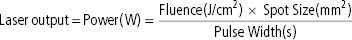
Power
W
Rate of energy delivery, laser output
Fluence
J/cm2
Amount of energy delivered per area
Pulse width
sec
Duration of laser exposure
Spot size
mm
Diameter of laser beam
Thermal relaxation time
sec
Time needed for the heated target to cool by 50% of its peak temperature through diffusion
Chromophore
Target of laser
Laser principles (LASER = Light Amplification by Stimulated Emission of Radiation)
Related posts:

Stay updated, free articles. Join our Telegram channel

Full access? Get Clinical Tree


