13. Dermatology


Few snippets


Extensor versus flexor skin surfaces: skin over the extensor surface is thicker and darker in color. This is designed to withstand trauma which is more likely to occur with extension movements than with flexion movements.


Skin in the back of your arm and upper body (extensor surface) is thicker and darker than skin in the front of your upper body or arm.


Skin in the front part of your leg is thicker and darker than in the back of your leg.


Intertriginous area (skin fold) is where skin touches skin frequently (e.g., groin and axilla). These areas are more likely to get infection with Candida, scabies, hidradenitis suppurativa, etc.


Rash variations: we included few classic pictures of various rashes, but in your free time try to look for other variations online. This can help you on exam. Just as humans, of the same Homo sapiens species, look different from one another, rashes of the same condition can look different too. If you Google search for atopic dermatitis or lichen planus, you will be amazed at how many variations you can find.


Terminology to describe skin lesions


Skin lesion type


Small (<1/2cm)


Big (> 1/2 cm)


Flat (non-elevated)


Macule


Patcha


No Image Available!

Elevated


Papule


Nodule


No Image Available!


No Image Available!

Fluid-filled blisters b


Vesicle


Bulla


No Image Available!

Red lesions


Petechiae


Purpura


No Image Available!


Source: James Heilman, MD, CC BY-SA 4.0, via Wikimedia Commons.


No Image Available!


Source: Coagulopathic Forms. In:Riede U, Werner M, ed.Color Atlas of Pathology: Pathologic Principles, Associated Diseases, Sequela. 1st ed. Thieme; 2004.


Loss of skin integrity


No Image Available!

aPatches that are palpable, or with corrugated, elevated surface are called plaques.


1


bIf the fluid inside blister/bullae is yellow, then it is called a pustule.


cSimilar to this principle, gastric erosion affects only mucosa, if there is involvement of submucosa, it is called an ulcer.


13.1 Skin Infections (Folliculitis, Furuncles, Carbuncles, Skin Abscess, Paronychia, and Hidradenitis Suppurativa)

2 Common risk factors of skin infections: diabetes, immunosuppression, obesity, infancy.


13.1.1 Folliculitis


Definition: Superficial infection around a hair follicle, most commonly due to Staphylococcus aureus.


Exam: Pinpoint erythema or pustules around follicles. It can be tender.


3


Rx: Depends on the area of involvement and severity.


No Image Available!

Differential dx: Bathtub folliculitis due to Pseudomonas. The lesions typically start below the neck (bathtub water submersion pattern). Treatment is avoidance or chlorination of hot tub. In severe cases use ciprofloxacin.


13.1.2 Furuncles, Carbuncles, Skin Abscess, and Paronychia


Definition:


Furuncles, carbuncles, skin abscess


Folliculitis can progress to small collection of infected material known as furuncle. When several furuncles come together to form a larger pus collection, it becomes a carbuncle, and a skin abscess forms.


No Image Available!


Furuncle on upper lip with edema.


Source: Furuncle. In: Sterry W, Paus R, Burgdorf W, eds. Thieme Clinical Companions—Dermatology. 1st ed. Thieme; 2006.


Paronychia


Localized redness, swelling, ± abscess in skin surrounding a nail area


No Image Available!


Source: Cgfalco, CC BY 4.0, via Wikimedia Commons.


Microbiology: Most common (MC) organism is S. aureus.


Physical Exam: As opposed to folliculitis which might not be tender, these are usually tender. A small collection or fluctuant mass is felt, with surrounding erythema and swelling.


Management:


No Image Available!


13.1.3 Hidradenitis Suppurativa


Predisposing factors: Obesity, smoking, genetic susceptibility, and postpuberty. Occurs in intertriginous areas (arm pits, inguinal, genital, and perianal area).


Pathophysiology


Presentation


Follicular occlusion → inflamed nodules


Recurrent painful inflammatory nodules in inner folds. As opposed to folliculitis, it is a recurrent chronic process that is potentially disfiguring and debilitating.


→ abscess → drainage


Purulent or serosanguinous discharge that might be foul-smelling


→ sinus tract and fibrous bands.
Multiple recurrences can lead to scarring and severe deformity.


No Image Available!


Right inguinal area. Source: Alharbi Z, Kauczok J, Pallua N. A review of wide surgical excision of hidradenitis suppurativa. BMC Dermatol. 2012; 12: 9.


Dx: Made clinically.


Rx: Lifestyle modification (e.g., maintaining hygiene) and, depending on severity, topical or systemic antibiotics and/or surgery (punch debridement or unroofing).


13.2 Other Bacterial Infections Involving Skin


Causative organism


Presentation


Management


Additional info


Leprosy


Mycobacterium leprae


Hypopigmented patch with anesthesia (i.e., loss of sensation in the lesion).


NSIDx: skin biopsy to demonstrate acid-fast bacilli.
Rx: dapsone


Disseminated leprosy infection can result in permanent damage to skin, nerves, limbs, eyes, etc.


Anthrax


Bacillus anthracis (cutaneous infection acquired from direct contact with infected livestock, e.g., in wool sorters).


A papule that progresses to ulcer with development of central necrosis and black eschar.


NSIDx: Gram stain and culture of ulcer.
Rx: ciprofloxacin or doxycyclinea


Bacillus can cause severe pulmonary disease with spore inhalation. As spores are very resistant (even to drying), powdered forms can be used as biological agents for terrorism.


MRS

ABCD.


13.3 Viral Skin Infections-HSV-1, HSV-2 and Herpes Zoster

4 The herpes group of viruses can remain dormant in a nerve ganglion for a long period of time. Certain precipitating factors such as stress (surgery, trauma, burn, etc.), or immunosuppression (e.g., advanced age or use of immunosuppressive medication) can lead to reactivation of the dormant virus.


13.3.1 Herpes Simplex Virus (HSV) Infection


HSV-1


HSV-2


Presentation


A single or multiple cluster of small vesicles, pustules, and/or multiple superficial ulcerations


Typical location


Oral area; rash most commonly occurs in vermilion borders of lip (known as herpes labialis or cold sores)


Genital area


Transmission


Infected oral secretions


Exposure to infected genital secretions during sex or child birth


Potential complication


  • Gingivostomatitis ± pharyngitis


  • Eczema herpeticum


  • Recurrent aseptic meningitis


  • Primary HSV-1 infection can cause meningoencephalitis (typically with temporal lobe involvement)


  • Usually does not cause encephalitis


  • Can cause recurrent aseptic meningitis


Rx


  • Consider antiviral valacyclovir, famciclovir, or acyclovir (+ clovir) during an episode.


  • In patients with frequent infections (4-6 episodes/year), consider chronic suppressive therapy with antivirals.


  • Dx is usually made clinically, but when in doubt we can do viral culture, viral polymerase chain reaction (PCR), direct fluorescent testing on sample, or Tzanck smear (to look for multinucleated giant cells).


  • Both can have dormant stage with recurrent reactivation and infection.


13.3.2 Herpetic Whitlow


Background: Cutaneous herpes simplex virus (HSV-1 or -2) can be acquired through contact with infected herpes genitalia or cold sores (e.g., dentist gets exposed to patient’s oral lesions). It can occur as a result of self-infection, too.


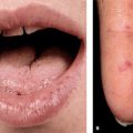
Presentation: Clear fluid-filled vesicles or pustules in fingers or hands. It is usually tender and can be recurrent.


Dx: Clinical, but when in doubt, we can do viral culture, viral PCR, direct fluorescent testing on sample, or Tzanck smear (to look for multinucleated giant cells).


Rx: Conservative management in most patients. In significant cases, consider antiviral valacyclovir, famciclovir, or acyclovir.


13.3.3 Herpes Zoster a.k.a. Shingles


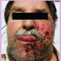
Background: After getting chicken pox (primary varicella zoster virus infection), the virus can remain dormant in sensory nerve ganglia for years. Precipitating factors such as immunosuppression can lead to reactivation of virus and eruption of rash along a sensory nerve root distribution.


Presentation:


  • Vesicles are in classical dermatomal distribution and unilateral (i.e., does not cross the mid-line). The rash is painful and itchy.


  • Some patient can present only with neuropathic pain in dermatomal distribution (preherpetic neuralgia). Pain can precede rash for days to weeks!


  • Severe postherpetic neuralgia (after onset of rash) can also occur.


No Image Available!


This 19-year-old immunocompetent patient had recurrent episodes.


Source: Brkljac M, Bitar S, Naqui Z. A Case Report of Herpetic Whitlow with Positive Kanavel’s Cardinal Signs: A Diagnostic and Treatment Difficulty. Case Rep Orthop. 2014; 2014: 906487.


No Image Available!


(a) Early zoster with grouped vesicles on an erythematous base. (b) More severe zoster, clearly showing dermatomal limitation.


Source: Zoster (Herpes Zoster, Shingles). In: Sterry W, Paus R, Burgdorf W, eds. Thieme Clinical Companions — Dermatology. 1st ed. Thieme; 2006.


Rx: Antiviral valacyclovir, acyclovir, or famciclovir (+clovir). In immunocompromised patients consider varicella zoster immune globulin.


Preventive measures:


Situation


Type of precaution in a health care setting (e.g., hospitals)


Indication for postexposure prophylaxis in patients who lack immunitya


Primary infection (chicken pox), disseminated zoster (when zoster involves more than 1 dermatome), and immunocompromised patient with dermatomal zoster


Airborneb + contact precautions


Yes


Uncomplicated herpes zoster


Standard precaution, including covering active lesions.


Only for close contacts of patients with open, weepy lesions. NOTE: crusted zoster lesions are not infectious.


aIndividuals with no immunity to VZV (varicella zoster virus) are:


  • Those who have not had two doses of zoster vaccine, or


  • Who have no laboratory evidence of immunity, or


  • Who have no verified chicken pox infection.


bRequires negative-pressure room. Same type of precaution is used for active tuberculosis.


Postexposure prophylaxis is recommended with two doses of zoster vaccine. Since it is a live vaccine, it is contraindicated in newborns, pregnant, or immunocompromised individuals. In these patients, use varicella zoster immune globulin instead.


Unique presentations of Varicella Zoster (aka Herpes Zoster) virus reactivation


When herpes zoster reactivates in particular distribution of cranial nerves


Presentation


Herpes zoster oticus (Ramsay Hunt syndrome)


Reactivation in geniculate ganglion (facial nerve ganglion)


Unilateral facial nerve paralysis with painful vesicles in auditory canal and/or external ear
Note: Due to the ganglion’s proximity to other cranial nerves, it can also affect cranial nerves 8 (hearing loss, tinnitus), 9, 10, etc.


Herpes zoster ophthalmicus


Reactivation in ophthalmic division of trigeminal nerve


Conjunctivitis, keratitis (corneal involvement), and iritis.
This can cause corneal scar or blindness, if severe.
Topical ocular steroid + antiviral therapy is recommended.


In a nutshell


Condition


Rx


Herpes simplex virus


Any of “ + clovir”.a If resistant, use foscarnet.


Herpes zoster or chicken pox


In immunocompetent host


Any of +clovira


In immunocompromised host


Any of +clovira
+
Varicella zoster immune globulin


aAcyclovir, famciclovir, or valacyclovir (except ganciclovir, which is used for cytomegalovirus).


13.4 Parasitic Skin Infections


13.4.1 Scabies


Background: Skin infection due to Sarcoptes scabiei, which is transmitted by close contact.


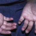
Presentation: Scattered papules and/or pustules ± underlying erythema found particularly in digital webs (interdigital space of hands or feet), palms, and intertriginous areas. These are very itchy. Look for history of exposure to patients with similar symptoms. Patients with HIV can develop extensive scabies with severe crusting, known as Norwegian scabies.


NSIDx: Find parasite in skin scrapings.


Rx: Topical permethrin or oral ivermectin. With crusted scabies, combine both oral and topical treatment.


Complication: Secondary bacterial skin infection can develop (e.g., impetigo, cellulitis).


13.4.2 Pediculosis (Head Lice)


Background: Infestation by lice can occur in hair-bearing areas; usually in head, but can occur in genital or axillary area.


Presentation: Prominent itching and skin excoriations. Secondary bacterial infection can develop.


NSIDx: Direct examination of hair-bearing areas.


Rx: Permethrin or lindane solution.


13.5 Intertrigo


Definition: Intertrigo is an umbrella term for inflammatory conditions located in skinfolds (intertriginous areas). It can develop due to mechanical skin-on-skin friction/irritation alone or become complicated by superimposed fungal or bacterial infection.


Risk factor: Diabetes, obesity, advancing age, and other forms of immunosuppression.


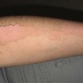
Presentation: Erythematous lesions that can be painful and pruritic. Secondary spongiosis can occur (formation of vesicles and pustules which can weep, ooze, and crust). Intertrigo can also present as diaper rash in children.


General treatment: Minimize friction (barrier creams) and prevent moisture buildup (absorptive powders such as corn starch).


Look for Secondary infection with dermatophytes, Candida, Corynebacterium (erythrasma), staphylococcus and streptococcus.


Erythrasma due to Corynebacterium infection


Candidal intertrigo


Differentiating feature


Sharply demarcated red to brown patches ± scales


Can have whitish film.
Look for satellite lesions (red papules or pustules) around the rash area.


No Image Available!


Source: Erythrasma. In: Sterry W, Paus R, Burgdorf W, eds. Thieme Clinical Companions-Dermatology. 1st ed. Thieme; 2006.


Intertriginous candidal infection, with typical satellite lesions


No Image Available!


Source: Oral Candidiasis. In: Sterry W, Paus R, Burgdorf W, eds. Thieme Clinical Companions—Dermatology. 1st ed. Thieme; 2006.


NSIDx


Wood lamp light typically reveals coral-red fluorescence.


Usually a clinical dx. Skin KOH preparation can be used.


Rx


Localized disease


Topical erythromycin, clindamycin, or fusidic acid


Topical azoles (e.g., clotrimazole, ketoconazole) or nystatin


Widespread disease


Oral erythromycin or clarithromycin


Oral fluconazole


No Image Available!


Itchy papules and burrows.


Source: Scabies. In: Sterry W, Paus R, Burgdorf W, eds. Thieme Clinical Companions—Dermatology. 1st ed. Thieme; 2006.


Primary skin disorders


Presentation


Condition


Additional points


Rx


Papules or plaques that can be of various colors. Rash can have overlying white lacy to net-like lines.
Flat-topped papules to plaques


No Image Available!


Source: Lichen Planus. In: Sterry W, Paus R, Burgdorf W, eds. Thieme Clinical Companions—Dermatology. 1st ed. Thieme; 2006.


Lichen planusa
Oral lichen planus can present with white lacy or net-like reticular lesions inside oral cavity.


No Image Available!


Source: Lichen Planus. In: Laskaris G, ed. Color Atlas of Oral Diseases. Diagnosis and Treatment. 4th ed. Thieme; 2017.


  • Associated with Hep C infection


  • Can also be drug-induced


  • Localized form: topical steroids


  • Generalized form:
    phototherapy, acitretin, or oral steroids.


Initial oval or round herald patch (looks like rash of tinea corporis), followed by development of maculopapular eruptions that occur in a Christmas-tree-pattern


  • Can be pruritic


Pityriasis rosea


If rash is atypical do VRDL or RPR test to screen for syphilis (palms and soles’ involvement points toward secondary syphilis)


Benign condition that usually resolves on its own.
Topical steroids can be used for very itchy lesions.


No Image Available!


Typical herald patch.


Source: Pityriasis Rosea. In: Sterry W, Paus R, Burgdorf W, eds. Thieme Clinical Companions—Dermatology. 1st ed. Thieme; 2006.


No Image Available!


Source: James Heilman, MD, CC BY-SA 3.0, via Wikimedia Commons.


Name giveth all.


  • Rosacea = facial redness, pronounced flushing reaction, and/ or telangiectasias (visible superficial vessels)


  • Acne = superficial papules and/ or pustules that look like pustular acne.


Acne rosacea


Get Clinical Tree app for offline access