1 Structure and Function of the Skin
Introduction
The skin is a complex organ and serves many important functions. It allows for interaction with the environment, while also protecting from injury. It is essential for wound healing, provides sensation, helps regulate body temperature, is a permeability barrier, protects from ultraviolet (UV) radiation and infection, and is responsible for much of an individual’s physical appearance. Because the skin is transected and manipulated in essentially every surgical procedure, it is important for the surgeon to understand its structure and various functions.
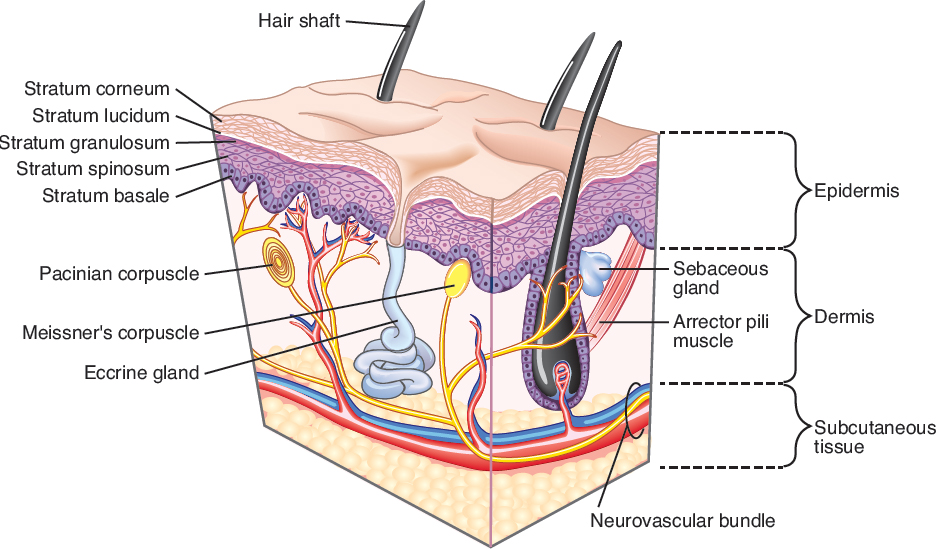
The skin is composed of three major layers: the epidermis, dermis, and subcutaneous fat. In an adult human, the skin weighs more than 5 kg and covers a surface area of approximately 2 m2. The epidermis is quite thin, ranging from 0.05–0.1 mm in depth. The dermis varies greatly in thickness, from 0.5 mm on thinskinned areas such as the eyelid or scrotum to more than 5 mm on the back. 1 , 2
The basement membrane separates the epidermis from the dermis at the dermal–epidermal junction (DEJ). Downward projections of epidermis known as rete ridges intercalate with upward-facing dermal papillae, helping solidify the connection between the skin layers. The subcutaneous fat lies beneath the dermis and contains blood vessels, lymphatics, and nerves which send branches into the dermis. Appendages, including hair follicles and eccrine and apocrine sweat glands, course through the dermis and open either on the epidermal surface (hair follicles and eccrine ducts) or into hair follicles (apocrine ducts) ( Fig. 1.1 ).
The Epidermis
The epidermis is the most superficial layer of the skin, and is composed of stratified squamous epithelium. It contains four cell types: keratinocytes, melanocytes, Langerhans cells, and Merkel cells. Keratinocytes are the most abundant cell and make up approximately 80% of cells in the epidermis. The layers of the epidermis, listed from superficial to deep, include the cornified layer (stratum corneum), the granular layer (stratum granulosum), the spinous layer (stratum spinosum), and the basal layer (stratum basale).
The stratum corneum (also known as the horny layer, keratin layer, or cornified layer) is composed of terminally differentiated, anucleate, flattened keratinocytes known as corneocytes, which are surrounded by an extracellular lipid matrix. 1 , 2 , 3 The stratum corneum varies in thickness from a few layers of cells on thinskinned surfaces like the periocular skin to many layers thick on acral surfaces such as the palms and soles. It accounts for the “barrier activity” of the epidermis, providing mechanical protection and preventing water loss and permeation of environmental substances. 1 , 4 , 5 Keratinocyte-derived antimicrobial peptides, such as defensins and cathelicidins, offer an innate immune defense against pathogens such as bacteria, viruses, and fungi. 1
An eosinophilic acellular layer, known as the stratum lucida or lamina lucida, can sometimes be seen beneath the stratum corneum on acral skin, such as the palms and soles. The granular layer (also known as the stratum granulosum) lies immediately beneath the stratum corneum and is named for its basophilic keratohyalin granules. These granules contain proteins important for the cornification and barrier function of the stratum corneum. One of these proteins, profilaggrin, is enzymatically cleaved to its mature form, filaggrin, following release from the keratohyalin granule. Filaggrin facilitates the aggregation of keratin filaments and is thus essential for formation of the stratum corneum. 1 , 2 Mutations in filaggrin cause ichthyosis vulgaris, a condition characterized by very dry, flaky skin due to increased transepidermal water loss in these patients. 1
The spinous layer (also known as the prickle cell layer or stratum spinosum) is located beneath the granular layer and is named for the spinelike appearance of the intercellular desmosomal connections when viewed microscopically. The spinous layer keratinocytes are polygonal in shape with abundant eosinophilic cytoplasm and oval vesicular nuclei, often with prominent nucleoli. 1 The spinous cells become progressively flatter as they differentiate and move toward the surface of the epidermis. The spinous cells contain lamellar granules, which contain lipids important for the integrity of the epidermis. Once released from the lamellar granules, these lipids act as “mortar,” helping the corneocyte “bricks” in the stratum corneum adhere to one another. 6
Beneath the spinous layer lies the basal layer (also known as the stratum basale) of the epidermis. It is composed of a single layer of cuboidal to columnar basophilic cells with a large central nucleus and prominent nucleolus. 1 The basal keratinocytes are mitotically active and give rise to the cells in the overlying layers of the epidermis. The basal layer is attached to the underlying basement membrane via hemidesmosomes. Melanocytes are interspersed between the basal cells, with approximately 1 melanocyte per every 10 basal keratinocytes.
Epidermal Cell Types
Keratinocytes
The ectodermally derived keratinocyte is by far the most abundant cell in the epidermis, accounting for approximately 80% of all epidermal cells. Each keratinocyte originates in the basal layer and throughout its 28 day life cycle will eventually reach the stratum corneum and subsequently desquamate. Keratinocytes contain keratin, a type of intermediate filament, which is important for the structure and function of keratinocytes. Humans possess 54 functional keratin genes, and there are two keratin gene families: type I (basic) and type II (acidic). 7 Keratin genes are coexpressed in pairs, with one acidic and one basic keratin filament in each pair. The specific keratin pairs vary based on cell and tissue type, developmental stage, disease conditions. and other factors. Each of the specialized epithelial tissues (skin, hair, nails, etc.) has its own keratin profile. 1 , 2
Keratins primarily play structural roles in keratinocytes. They are important components of the keratinocyte cytoskeleton and of desmosomes and hemidesmosomes, which serve to connect keratinocytes to one another and to the basement membrane, respectively. They also aid in cell signaling, the stress response, and apoptosis. 1 , 8 , 9 , 10
Related posts:


Stay updated, free articles. Join our Telegram channel

Full access? Get Clinical Tree


