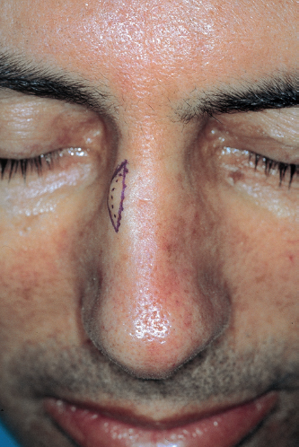
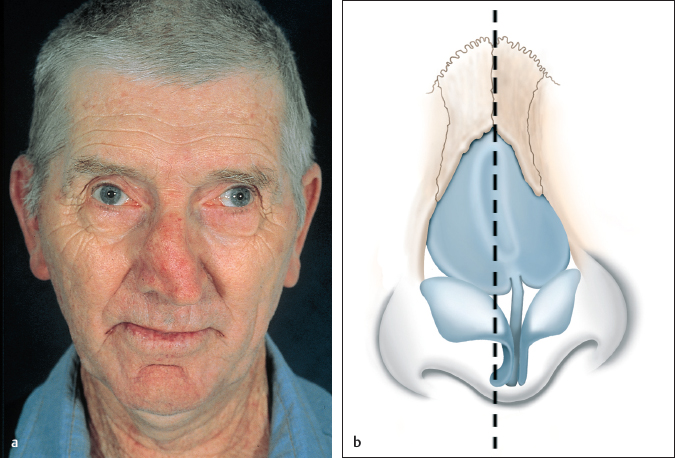
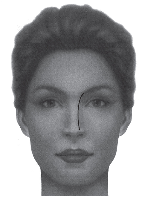
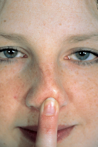
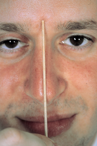
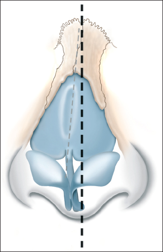
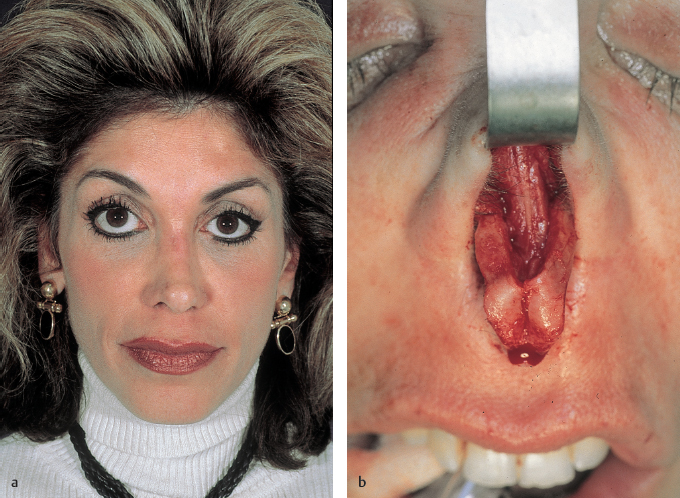
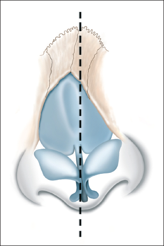
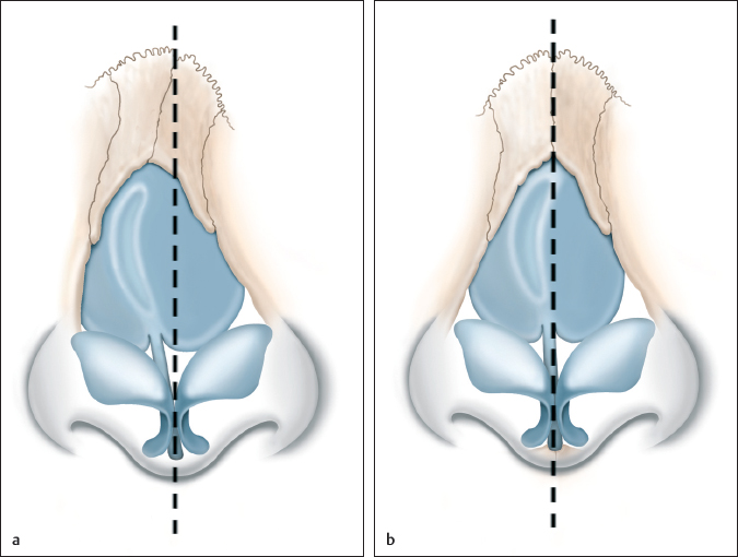
Chapter 10 10.4 Preoperative Considerations 10.5 Preoperative Analysis and Diagnosis 10.7 Principles of Postoperative Care There is no universally ideal nose, especially as one crosses ethnic and gender boundaries, although one consistent aesthetic trait found across all cultures is a straight dorsum. Correcting the deviated nose is a formidable rhinoplastic challenge for many reasons. The frontal view is often seen in photographs but is not otherwise a common perspective during normal interpersonal encounters. For this reason, many individuals will notice small dorsal deviations in photographs, which then prompt the surgical consultation. This true frontal view is the most challenging to perfect because even a subtle, focal area of fullness or depression can be conspicuous and readily detected as a dorsal asymmetry. Furthermore, correcting the twisted nose can be unpredictable because it relies on both sides of the nose healing in an identical way, with the same degree of swelling, scarring, and contracture. This may not always be the case. In some ways, a rhinoplasty can be viewed as two operations: one on the left side and the other on the right. Although the same technical maneuvers may be performed on each side, the rate of healing and degree of scar contracture can vary and lead to unpredictability. Rhinoplasty is a four-dimensional operation. Manipulating the bony and cartilaginous framework in a three-dimensional space is the first challenge. The fourth dimension is time. This powerful force is an essential consideration in surgical planning and its recognition and appreciation has had a tremendous impact on contemporary rhinoplasty techniques. There is a marriage between the cutaneous nasal deformity and the underlying anatomical cause, one that must be studied as a routine part of all preoperative analyses. After successfully identifying the external nasal problems, it is imperative to go the next step and investigate what bony or cartilaginous deformity is causing those findings. Such an exercise allows one to approach the surgical plan in a precise and targeted manner. This chapter will highlight the preoperative analysis and surgical repair of the deviated nose. The correction can be approached through a graduated algorithm that begins with a simple, minimally invasive maneuver and progresses toward destabilization and reconstruction. Representative cases will be used to demonstrate the analysis and rhinoplasty techniques. The indications for repairing a twisted nose can fall under two general categories, i.e., cosmetic and functional. Naturally, the cosmetic group is integrally involved with the patient’s perspective and complaints. Functionally, the twisted dorsum can be an active contributor to the cause of nasal obstruction, and correcting this deformity is often an important part of restoring nasal patency. Most twisted noses are the result of blunt trauma, where the nasal skeleton is displaced and immediately apparent. While we often think of bony injuries with blunt trauma, significant distortion to the cartilaginous framework can also occur and may be amenable to immediate repair. Most long-standing nasal deformities can be related to a history of nasal trauma, albeit remote or minor, and occasionally forgotten by the patient. Relatively small injuries to the lower two-thirds of the nose can disrupt the balance of intrinsic cartilaginous forces, which, over time, may result in progressive distortion and nasal twisting. Moreover, injuries at a younger age may influence the nasal growth centers and lead to asymmetrical development. Iatrogenic dorsal deformities can occur during a dorsal hump reduction, which inadvertently unmasks a midseptal deviation. Other rhinoplasty procedures can also heal and contract in an asymmetrical way and give rise to the twisted nose. The indications for a specific rhinoplasty maneuver to correct a dorsal deviation are dependent on the causes, which may be traumatic, iatrogenic, or idiopathic. A careful preoperative analysis of the structural aberrancy often dictates the approach and optimal surgical plan. At times, one must apply a stepwise approach such that a series of maneuvers are applied in a sequential fashion. For this reason, it is important to be facile with a host of surgical maneuvers before embarking on the repair of a twisted nose. There are relatively few contraindications to performing this type of rhinoplasty. One may encounter philosophical contraindications to repairing the crooked nose, such as an individual who will be exposed to repeated trauma (e.g., a boxer or rugby player). Under these circumstances, the timing of surgery is more at issue than the surgery itself. There are circumstances where straightening the deviated nose may compromise the nasal lumen and be relatively contraindicated. This could occur with a patient who desires a straighter nose with the collapsed side being considered more aesthetically pleasing. To create a symmetrical dorsum, the patient may request to have the normal side pinched medially, potentially giving rise to valve narrowing and obstruction. Identifying the good rhinoplasty candidate is as important as the surgery itself. A thorough history should include a commitment to get to know the patient as a person, seeking to understand a few specific personality traits. The motivation of individuals seeking a rhinoplasty can be diverse and some are considered healthy, while others are felt to be unstable. Ideally, a patient should pursue a rhinoplasty only after adequate contemplation and understanding of the procedure. The motivating force should be a personal wish to correct some specific deformity that is bothersome. After proper patient selection, a successful outcome can have far-reaching effects on self-image and self-esteem. Poor motivational factors include seeking cosmetic surgery to please others, correcting problems in their personal or professional lives, or in response to exogenous stresses in their lives. The physical expectations must also be carefully evaluated to ensure that they are realistic and within the realm of surgical possibility. The single most essential step toward realistic expectations is clear communication between the surgeon and the patient. There are physical limitations to some rhinoplasties, such as those relating to skin thickness or dramatic deviations to the dorsum, and these must be clearly defined preoperatively. It is also important to explain the balance between the nose and the face, such as the twisted nose on a person with preexisting facial asymmetry. Psychological factors and personality traits can influence the outcomes and final patient satisfaction. A history of psychiatric illness, impulsive behavior, and use of mood-influencing drugs should prompt further investigation to determine psychological candidacy. Some traits interfere with the ability to accept one’s body image, while others cannot tolerate the change. The following are some common personality types that should alert the surgeon preoperatively: • The dependent personality: overly compliant and leads the patient to interact in a subservient fashion to the surgeon. • The passive–aggressive personality: nonconfrontational but may display self-deprecating behaviors. • Obsessive–compulsive personalities: questions every detail yet remains indecisive, making effective communication difficult. • Histrionic personality: charming and dramatic, but insists on special attention and responds in an exaggerated and inappropriate way. • Paranoid personality: secretive, distrusting, and less tolerant of discomfort. Minors represent a special subset of patients as they may be brought to the surgeon by their parents. It is essential to determine who is seeking the cosmetic change and to ensure that the communication and instruction are mutual. The general teaching is that nasal cosmetic surgery should be delayed until the age of 15 for females and 17 for males. The two variables to consider before a pediatric rhinoplasty are emotional maturity and completed pubertal growth of the nasal skeleton. Both probably occur sooner in females than in males. Rhinoplasty in the older age group also involves unique emotional and anatomical factors. Older patients have lived with certain facial features for their entire lives and dramatic facial changes can be difficult to adjust to, occasionally having a negative impact on their self-image. As such, a conservative approach is further emphasized with this patient demographic. The older patient often has more brittle nasal bones, making osteotomies more challenging. Body habitus is worth noting, particularly any preexisting facial asymmetries that can occur. A perfectly straight nose on a crooked face may not appear balanced. Ethnicity and gender are important preoperative considerations for rhinoplasty but are less relevant in the management of the twisted nose because a straight dorsum is desirable in all cultures. An accurate preoperative analysis of the deviated nose goes beyond recognizing the external deformity; it requires a deliberate investigation into the underlying cartilaginous and bony anatomy, and the complexity with which it shapes the nasal dorsum. Each area of the nasal skeleton is responsible for a discrete cutaneous subunit of the nose, such as the nasal bones defining the upper third, the dorsal septum and upper lateral cartilages shaping the middle third, and tip being supported by the lower lateral cartilages and anterior septal angle. Cutaneous deviations, on the other hand, can be the result of more than one anatomical structure. The ideal nose blends into the face without calling attention to asymmetry, imbalance, or disproportion, allowing the casual observer to be drawn to other areas that typically define facial beauty, such as the eyes and lips. The aesthetic dorsum is straight, remains in the midline of the face, and may have a subtle concavity that reflects a narrower middle vault. The “brow-tip line” is a useful landmark that helps define an aesthetic dorsal contour. It begins from the medial brow, curving inferiorly along the dorsal border, gently blending with the tip-defining point. These lines should remain parallel and uninterrupted (Fig. 10.1). It is often useful to evaluate the nasal dorsum in segments rather than just a gestalt from the frontal view. Dividing the nose into an upper, middle, and lower third can help with delineating discrete aberrancies. The upper third of the nose is formed by the paired nasal bones and the frontal processes of the maxilla. The skin along the caudal border of the nasal bone is characteristically thin and allows small irregularities of bone, cartilage, or scar to be readily evident. Conversely, the skin and soft tissue at the nasion is much thicker and includes subcutaneous fat and the procerus muscle. Changes in the bony skeleton along this area tend to be camouflaged by the thicker overlying soft tissue, which drapes between the higher riding glabella and rhinion. The nasal bones also define the appropriate width and dorsal projection to the upper nose. The bony septum is a minor contributor to the dorsum in normal circumstances, but deviations to the upper third can involve the septum and must be considered. More will be discussed on this later in the chapter. Fig. 10.1 The brow-tip aesthetic line extending from the brow to the nasal tip seen in the aesthetic dorsum. The nasal tip should appear elegant, indiscrete, and in the midline. While the tip is not often discussed with the twisted nose, it too can be deviated and contribute to dorsal deviations. The midline position of the tip is dependent on both the lower lateral cartilages and caudal septum, especially the anterior septal angle. Like the middle vault, the culprit for a deviated tip may lie with either anatomical structure (or both) and a preoperative distinction is needed in order to develop a focused surgical repair. The anterior septal angle is often camouflaged by the thick tip skin and lower lateral cartilages, and palpation may be necessary to identify its position. When the caudal septum is deviated, it can bring the lower lateral cartilages with it and cause a passive tip deformity. Evaluation of the twisted nose is a challenging aspect of rhinoplasty and is best done in a methodical and systematic manner. An accurate diagnosis is a prerequisite for developing a preoperative surgical plan that is direct and target oriented. For these reasons, it is useful to evaluate the rhinoplasty patient with an algorithm that highlights some nuances that might otherwise go unnoticed. Preoperative nasal analysis should be organized and repeated several times. First, multiple views of the nose are useful. Clearly, the frontal perspective is most revealing of dorsal deviations (and the most difficult to perfect surgically), but different abnormalities can be appreciated from the submental, oblique, and lateral views. Photography serves two important functions in terms of analysis. It allows for repetitive, preoperative analysis, including within the operating room. Second, the physical act of taking photographs allows a unique perspective of the patient not typically achieved during normal consultations. The camera’s view finder, cropping, standardizing views, and the Frankfurt horizontal all contribute to a form of tunnel vision that leads to an objective analysis of the nose, apart from room décor, attire, and emotional expression. For example, one often asks the patient to lower his/her chin and refrain from smiling, two interpersonal habits that are prevalent during the consultations but influence the analysis. A hypoplastic chin or preexisting alar-columellar disproportion can be easily overlooked during an informal encounter but become readily apparent through a camera lens. During the physical examination of a rhinoplasty patient, there are two useful tools that are occasionally omitted. First, palpation of the nose is an invaluable asset that can reveal much in terms of bony and cartilaginous framework, stability, resilience, and soft-tissue problems. There are aspects of the nose that are best evaluated through careful palpation, such as the location of the anterior septal angle, the contour of the dorsal septum, tip support, and skin thickness (Fig. 10.2). During the examination, one must make it a point to investigate these areas and become familiar with the unique anatomy of each patient. A second useful tool when analyzing a twisted nose is to use a straight reference, such as a stick or cotton-tip applicator, in the precise midline of the face. With the midline clearly defined, one can inspect each third of the nose independently and better determine which component of the bony/cartilaginous framework is creating the cutaneous deformity (Fig. 10.3). Fig. 10.2 Depressing the nasal tip helps identify the position of the anterior septal angle. Fig. 10.3 An objective midline reference can assist in identifying which areas of the nose are deviated. The deviated dorsum comes in many forms and in no way can a single operation be universally applied to all patients. It is a critical to separate the cutaneous deformity of the nose from the underlying structural cause and anatomical pathogenesis. One must take a step backward from the cutaneous deformity to understand its anatomic etiology, and proceed forward with a surgical repair targeting this framework problem. The nose with a bony deviation is entirely different from the collapsed upper lateral cartilage or the dislocated caudal septum, although both may resemble a “twisted nose.” The nasal dorsum can be linear but deviated to one side, where all components of the nose are involved and need correction (Figs. 10.4 and 10.5). The bony dorsum can have a solitary irregularity, usually in the paramedian position, that creates an asymmetry and the appearance of a twisted nose. The causes of such isolated lesions can be related to old bony fractures, including where small osteophytes have grown from the disrupted periosteum (Fig. 10.6). The remedy for such problems may be as simple as sharp excision or rasping of the lesion. Some deviated noses are limited to the upper third and are the result of displaced nasal bones, while the dorsal septum and lower two-thirds are normal, as seen with nasal fractures (Fig. 10.7). Deflections of the bony pyramid, however, are not all identical and it is important to delineate (1) which side is aberrant, (2) the contour of the bone itself, and (3) potential involvement of the posterior bony septum. Bony deviations may involve only one side where an isolated segment is medially displaced. One must be alerted to the depressed nasal bone segment, as its repair may be unstable and require unilateral intranasal packing. Alternatively, these circumstances may be more efficiently managed with camouflage, onlay grafts. Careful palpation of the bony pyramid is done to identify any intrinsic concavity or marked asymmetry in width to the nasal bones. This analysis will influence the location of osteotomies as well as the need for intermediate osteotomies. It is difficult to predict which bony septal deflections will hinder realignment of the nasal pyramid and it is not uncommon to see significant endonasal deviations that do not interfere with dorsal repair. When the septal deviation is significant, it can be addressed either with endonasal closed reductions or percutaneous osteotomies. The twisted middle vault is possibly the most complex in terms of preoperative assessment. The majority of cases involve a primary deviation to the dorsal septum and passive distortion to the upper lateral cartilages (Fig. 10.8). This anatomy is best appreciated with direct palpation of the dorsum as well as by tightening the nasal skin across the lateral nasal walls, which can outline the dorsal septum. Because the upper lateral cartilages are firmly adherent to the dorsal septum (in the primary nose), they are equally distorted and contributing to the external concavity. Releasing the fibrous attachments between these two structures will typically reveal a persistent defection to the dorsal septal cartilage and a correction of the upper lateral deformity. This identifies the anatomical cause of the middle vault deviation and permits a targeted surgical correction. One should bear in mind that a twisted nose can be unmasked during a dorsal hump reduction when a preexisting midseptal deviation becomes the new dorsal strut. This iatrogenic twisted dorsum can be anticipated with careful intranasal inspection and corrected with traditional methods outlined below. A cartilaginous hump reduction also redefines the middle vault dorsum with an area of septal cartilage that is usually thinner, thus requiring prophylactic spreader grafts. There are two instances of middle vault deviations where the upper lateral cartilages are the primary culprit rather than the dorsal septum. The upper lateral cartilage may be disarticulated off its supporting structures (usually the dorsal septum but occasionally the nasal bone) and the result is a gradual medial displacement and depression that creates a unilateral concavity. There will be a disruption of the aesthetic brow-tip line on one side and the illusion of a twisted nose. When this occurs bilaterally and symmetrically, such as following a reduction rhinoplasty, it creates the typical “inverted-V” deformity encountered during many secondary rhinoplasties. The second scenario is when the primary causes are with the size, shape, or form of the upper lateral cartilages themselves; although less frequent, there are times where an intrinsic upper lateral deformity exists with a concavity or buckling of cartilage. This sidewall asymmetry will be interpreted as a twisted nose and can be addressed directly, through either repair or camouflage. Fig. 10.4 A linear deviation to the nasal dorsum. Fig. 10.5 (a) Nasal dorsum deviated the patient’s left. (b) Linear deviation of the nasal bones and dorsal septum. Lower lateral cartilages remain in the midline. Fig. 10.7 Dorsal deviation secondary to displaced nasal bones. Fig. 10.8 (a) Twisted dorsum involving the upper and middle thirds, with a deviation of the nasal bones and dorsal septum. (b) Twisted dorsum limited to the middle third and the dorsal septum. The lower third may also be twisted to one side and create the appearance of a twisted dorsum. Tip position is dependent on numerous forces that must work in concert in order to hold the tip in the midline; a disruption of this balance of forces can lead to marked asymmetry. Lateral displacement may be due to a malposition of the caudal septum (particularly the anterior septal angle) (Figs. 10.9 and 10.10). Tip structure and stability is best assessed with careful palpation because simple inspection can be misleading in terms of the causative agent. It is not uncommon for the anterior septal angle to be significantly deflected to one side and the cutaneous tip only slightly twisted. The caudal septum may be bowed or dislocated off the anterior nasal spine, causing deformities to both the columella and the nasal tip. Similarly, marked asymmetry of the paired lower lateral cartilages can be associated with enough twisting of the tip such that the entire dorsum appears irregular. The deviated nose can be associated with functional problems that are complex and related to pathology of the internal nasal valve, i.e., the area between the upper lateral cartilage and dorsal septum. Although this cross-sectional area represents a proportionally small amount of the intranasal lumen, it is responsible for a disproportionate amount of laminar airflow and small degrees of obstruction can quickly be symptomatic. Evaluating nasal obstruction related to the nasal valve must be done carefully and systematically. The goal is to identify exactly where the obstruction is and to distinguish a static narrowing from a dynamic collapse, as their respective treatments are inherently different. Examination of the nasal airway must be done with minimal distortion or artificial support to the nasal sidewalls, especially from the nasal speculum. Dynamic collapse of the nasal sidewall is generally repaired with cartilaginous batten grafts placed precisely along the epicenter of collapse with the intent to reinforce that area and resist the collapsing force during nasal inhalation. The static narrowing at the internal valve is usually thought of as a problem involving malpositioned upper lateral cartilages, typically remedied with spreader grafts, flaring sutures, or butterfly-type grafts.1–3 The dorsal septum is an important player in the anatomy of the internal nasal valve and can contribute significantly to the cause of valve obstruction. Clinical obstruction can occur on either the concave or the convex side of the septum, and it is difficult to predict without careful intranasal inspection. On the convex side of the nose, the dorsal septum deviates toward the airway and directly impinges on the internal valve from the medial side. Surgical correction is aimed at straightening the septum. When obstruction occurs on the concave side of the nose, the upper lateral cartilage is usually the culprit; it is usually medially displaced through either disarticulation off the septum or a buckling deformity. Correction involves lateralizing the upper lateral cartilage through spreader grafts. Similarly, a depressed nasal bone can cause both a cosmetic deformity to the dorsum and nasal obstruction at the level of the internal nasal valve. The displaced nasal bone brings with it the upper lateral cartilage and creates a secondary obstruction at the internal valve.
The Deviated Nose
10 The Deviated Nose
10.1 Introduction
10.2 Indications
10.3 Contraindications
10.4 Preoperative Considerations
10.4.1 Age
10.5 Preoperative Analysis and Diagnosis
10.5.1 Normal Anatomy and Diagnosis
10.5.2 Analysis of Aberrant Anatomy
Plastic Surgery Key
Fastest Plastic Surgery & Dermatology Insight Engine