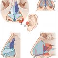
Chapter 8 Often, the most challenging part of rhinoplasty is modifying and refining the nasal tip. Endonasal delivery flap techniques have an extensive and successful history. This chapter will focus on the beauty, versatility, and the simplicity of endonasal tip surgery. Achieving tip definition has evolved since Joseph introduced cosmetic rhinoplasty in the late 1800s. This evolution is described well by Tebbetts.1 Initially, nasal tip-shaping techniques were destructive, consisting mostly of incising and resecting cartilage. Often the tip was approached in either a retrograde or a cartilage-splitting fashion. The limited visibility of these approaches increased the likelihood for possible asymmetry, with the destructive techniques that were implemented resulting in consistent loss of tip support and increased risk of secondary deformities. In contemporary rhinoplasty, primary outcomes such as solid structural support and unimpeded nasal airflow have taken on an equivalent and justified level of importance as the aesthetic outcomes and refinement. Focus has now turned towards cartilage preservation, and an emphasis has been placed on using various cartilage grafting techniques for support and contouring. With this shift in philosophy, there has been an increase in the amount of open structure rhinoplasty being performed. Supporters of the transcolumellar incision, and resulting broader exposure, maintain that there are more opportunities to use certain grafting techniques, and therefore more methods at their disposal to achieve more refined results and prevent late complications. Despite the approach, we now have evolved into an era of nondestructive tip-shaping techniques. These methods allow achievement of the desired aesthetic appearance while maintaining or recreating projection and functional tip support. This assures excellent results not just at 1 year, but also at 5 years, 10 years, and more. Our approach is based on the creation of the double-dome unit as described by McCollough and English.2 In addition, individual treatment of each dome to create the correct contour is further described. Long-term success using these techniques has been well described.3 We will first describe our basic surgical technique, followed by specific nasal tip deformities and the steps utilized to correct them. The ideal patient for these techniques has been described by Tardy et al.4 The ideal patient has a slightly bifid or broad tip with double-dome highlights. Thin to moderate thickness skin and sparse subcutaneous tissue allow for more refined results from these endonasal techniques. The alar cartilages themselves must be firm and strong. Finally, the alar sidewalls should be thin and delicate, yet resist collapse and recurvature. Most patients do not have these ideal features. Yet by using the endonasal approach and a progressive method with each tip, excellent aesthetic results can still be achieved. There are certain conditions in our experience that favor the use of the external columellar approach and allow accomplishing specific techniques much more easily. It is often difficult to deliver, in a safe and adequate manner, alar cartilages in a patient with scar tissue in the lobule from previous reconstructive nasal surgery or trauma. Additionally, in the setting of “cocaine nose” where there is extensive mucosal destruction, maintaining as much mucosal integrity is of utmost importance, and an endonasal approach would not be the ideal choice. Similarly, if one is trying to manipulate a cantilevered graft, place a caudal septal extension graft, or repair a markedly crooked or twisted nasal tip with discrepancies between the two medial crura, the external approach affords the surgeon a wider exposure to accomplish these goals. Again, while the endonasal approach is not contraindicated in these settings, some maneuvers are more easily performed with an open technique.5 Other indications for the external columellar approach include when a patient presents toward the extremes of overprojection, over-rotation, underprojection, and under-rotation of the lobule. All patients are initially seen in consultation with their selected surgeon. The consultation room is designed to put the patient at ease while still maintaining a professional environment. The nasal analysis begins with the patient on a comfortable swivel stool with the back in front of a three-way mirror with the physician directly behind him or her. Together they analyze the nose with the physician gently guiding the discussion. The three-way mirror offers a more three-dimensional conversation. An in-depth nasal history is taken during the consultation (Fig. 8.1a, b). Inquiries include any previous nasal trauma or surgery, difficulties breathing through the nose, any history of sinus disease or allergies, and current nasal medications. The physician reviews a more extensive medical history form, completed by the patient prior to consultation. Intranasal exam is also performed at this time to detect deformities of the septum, enlargement of the turbinates, or other intranasal pathology. The procedure should be thoroughly discussed at this time and goals summarized with the patient. The physician reviews with the patient what to expect on the day of surgery, including the length of surgery, anesthesia, recovery, and discharge. Initial postoperative care and restrictions regarding certain activities are also discussed. Finally, the limitations of the surgery as well as possible complications are given as part of obtaining informed consent. The consultation is then continued in the photography suite, where computer imaging is used to illustrate the physician’s goals for surgery. This allows for confirmation that both the patient and the surgeon agree on the desired aesthetic goals to be achieved. Following this, a full set of nasal images are taken for preoperative documentation. The last phase of the consultation is spent with the scheduling nurse, where questions can be answered in what often is a more comfortable setting for the patient. Fees are reviewed with the patient and signed copies of the procedures and fees are given to the patient. Any necessary lab work is arranged at this time. Prior to surgery, all patients receive folders with detailed instructions on surgery, prescriptions, and a booklet reviewing postoperative healing and expectations. All patients start an oral antibiotic the day prior to surgery, most often either oral cephalexin or azithromycin, and continue this for 5 days. The preoperative analysis is the foundation of any successful rhinoplasty operation but is even more crucial for tip reconstruction.6 The surgeon should assess the tip complex by inspecting the external contours, and interpret how the exam translates into the underlying nasal architecture. Both static and dynamic intranasal inspection of the external nasal valve, septum, turbinates, and internal nasal valve should be included and documented. A modified Cottle’s maneuver can be helpful in eliciting feedback from a patient presenting with obstructive symptoms. The tip shape should be described, for example, as bulbous, twisted, or infantile. Both the degree of rotation and the extent of projection should be evaluated. It is critical to assess skin thickness and this issue alone may dictate approach and/or procedure to be performed. Palpation is helpful in determining the nature, volume, strength, and resiliency of the lobular cartilages as well as in evaluating tip support. Finally, it is important to note columellar abnormalities and their relation to the alar cartilages. The patient is taken to the operating room and general anesthesia is induced prior to injecting local anesthesia. First pledgets soaked in 5% oxymetazoline are placed intranasally. After adequate time for decongestion, infiltration is started with 2% lidocaine with 1:50,000 dilution of epinephrine. No more than 7 to 8 mL is injected to avoid volume distortion of the nasal tissues. Individual variations in anatomy and expectations preclude the rote use of a single technique for each patient, and the technique and approach should be tailored to meet the operative goals. Several approaches exist including the caudal approach, transcartilaginous incision, retrograde approach, or our most commonly used delivery flap technique, which offers visualization of the entirety of the alar cartilages. The delivery flap technique begins with a high septal transfixion incision, and depending on existing tip projection, the septal transfixion incision can be carried down inferiorly into a complete transfixion incision, releasing the medial crural feet from the septum if further deprojection is desired (Fig. 8.2a, b). This is then connected with bilateral intercartilaginous incisions (Fig. 8.3). A plane of dissection is created at the superior septal angle over the dorsal aspect of the cartilaginous septum and upper lateral cartilages up to the bony cartilaginous junction when indicated. When further tip refinement or grafting is necessary, bilateral marginal incisions are made, and the lower lateral cartilages (LLC) are delivered. The delivery technique makes use of thin, outwardly beveled, Metzenbaum scissors to elevate the skin–soft tissue envelope off the underlying LLC, and the alar domes are individually delivered with a single hook and supported with the Metzenbaum scissors (Fig. 8.4). In this fashion, each dome is assessed and recontoured separately. The authors have found that the endonasal approach (incorporating bilateral marginal and intercartilaginous incisions) allows for excellent visualization of tip anatomy by presenting the alar cartilages as bipedicled chondrocutaneous flaps as previously described by the senior author.3
Endonasal Tip Rhinoplasty Approaches and Techniques
8 Endonasal Tip Rhinoplasty Approaches and Techniques
8.1 Introduction
8.2 Indications
8.3 Contraindications
8.4 Preoperative Considerations
8.5 Preoperative Analysis
8.6 Surgical Technique
Plastic Surgery Key
Fastest Plastic Surgery & Dermatology Insight Engine