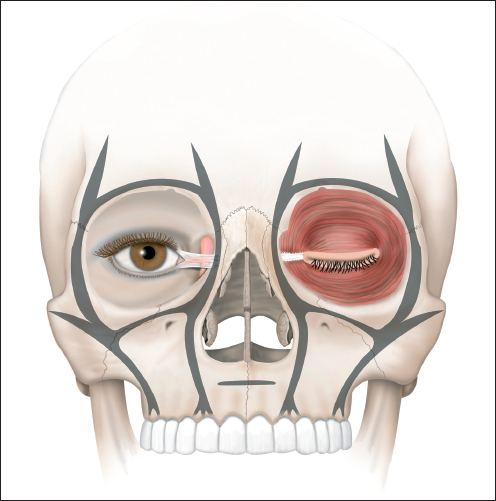
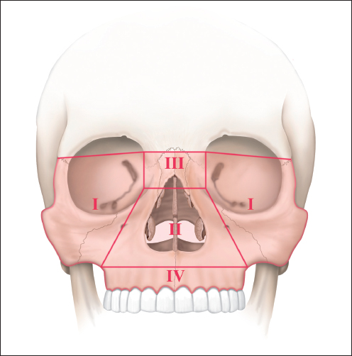
Chapter 14 14.2 Trauma-Relevant Anatomy of the Nose 14.3 Classification of Nasal Trauma 14.5 Management of Nasal Traumas The nose is the most prominent facial element. The fracture of the nasal pyramid is one of the most frequent bone fractures of the human body. The energy required to cause a fracture is lower than for other facial bone fractures. More than 50% of all facial fractures are injuries to the nose. In the course of increasing incidents of injuries to the facial area, the resulting mostly complex consequences pose great challenges for the trauma specialist, who, with his assessment and treatment, is responsible for the reconstruction of form and function.1–3 For nasal injuries, one can differentiate based on the type, direction, and energy volume of the impinging trauma between superficial soft-tissue injuries with lacerations of the skin and soft tissue, burns and frostbite, and fractures of the cartilage and bony framework and structure. High levels of energy striking the face often result in extensive and combined injuries. Not infrequently, injuries and especially fractures of the nose are considered minor injuries in an average clinical day and often treated with insufficient diagnostics as well as inadequate care. The incidence of posttraumatic deformities that have not only unaesthetic but also functionally unacceptable consequences is high. In many cases, the necessary revision septorhinoplasty has proved to be difficult. Therefore, practicable guidelines for the optimal medical care of acute nasal trauma are necessary. Currently, there are still discrepancies with regard to the timing and methodology involved in posttraumatic management. Posttraumatic repositioning of nasal bone fractures implemented early are generally carried out as simple, contained manipulations, resulting in cases requiring the corrective medical care of either rhinoplasty or septorhinoplasty. The data for frequency vary between 14 and 50%.2,4,5 A detailed anamnesis, in particular of the trauma event, as well as an exact clinical examination are especially important for the assessment of the injury. In doing so, precise knowledge of the fundamental anatomy is virtually essential for the surgeon. The osseous architecture of this compact region includes the twin nasal bones, front process of the maxilla, the maxillary process of the frontal bone, the lacrimal bone, the lamina papyracea of the ethmoid bone, the sphenoid bone, and the vomer. Fitted into this structure are the cartilage elements of the quadrangular cartilage of the septum and the upper and lower lateral cartilages of the external nose. The midfacial bony structures are reinforced by vertical and horizontal buttresses. The upper horizontal buttress is formed by the lower anterior rim of the sinus and the upper orbital rim, while the lower orbital rim functions together with the zygomatic bone as the lower horizontal buttress (Fig. 14.1). The twin naso-ethmoidal complex functions as a “central element” and forms the vertical buttress together with the frontal process of the maxillary bones and the lateral interior angle of the frontal bone. Only the thickened posterior edges of the nasal bones are components of the buttresses, but they protect the further dorsally located thin bones of the medial orbital wall. The central element is also the fixation point for the medial canthal tendon, which guarantees support for the bulb and the eyelids (Fig. 14.2). Tears to this support signify a traumatic telecanthus and a rounding of the medial canthus. However, the function of the musculus orbicularis oculi is not influenced by a mobile canthal tendon. In contrast, impairment of the lacrimal sac drainage can result, because this is surrounded by portions of the canthal tendon.6 Fig. 14.1 Horizontal and vertical columns constitute a static, supportive function in the midface. The vertical supporting column forms the central element; the upper horizontal column is formed by the frontal bone and the upper margin of the orbita; the lower horizontal column is formed by the lower orbital margins. The medial canthal tendon enters the bone of the medial canthus region that is part of the central element. An external portion of the tendon extends to the surface of the nasal bone. Fig. 14.2 Classification of midface fractures: I = zygomatico-orbital complex II = nasomaxillary complex III = naso-ethmoidal complex IV = dentoalveolar complex The nose as a central and prominent facial element can function as an energy absorber and thus as protective buffer of the viscerocranium. The cartilage portions have a high level of flexibility, and traumata with a low amount of energy can be partially absorbed without permanent damage. The variously thick bone structures determine the predilection sites of fractures, but the different bone thickness also has an influence on the extent of the fracture. Thus, older people with osteoporotic bones have comminuted fractures more frequently, whereas in children dislocative fractures are rare, but here greenstick fractures predominantly occur.7 The anatomical relations are significantly different in children in comparison to adults. The bones are shorter and the cartilage portion is larger. Additional protection is given because the bones are embedded in thicker soft tissue. Also, the nose is less prominent than in adults, which reduces the trauma consequences as the striking energy is distributed across a larger surface.8 On the other hand, various anatomical growth zones in the child’s nasal skeleton are strongly influenced. Consequently, the potential for growth impairment and problems with the development of the nasal framework and septum exist following trauma.9 The embedding of the nose in the midface requires that nasal fractures must be considered in the classification of midfacial fractures. In the classification according to Le Fort, bony injuries of the nose exist in types II and III (Fig. 14.3). The classification according to Becker and Austermann is divided into central, lateral, and centrolateral midfacial fractures (Table 14.1). Isolated nasal bone fractures are included in the midfacial fractures, whereas the fractures of the naso-orbito-ethmoid complex are synonymous with the centrolateral fractures. For isolated central nasal fractures, the categorization according to Simmen has been well established, divided into types I to IV. This categorization takes the direction of the trauma into consideration and specifies the trauma consequences on the osseous and cartilaginous system.10 In the classification according to Becker and Austermann, these fracture types are included in the category of central midfacial fractures of nasomaxillary and naso-ethmoidal types.11 A classification of the viscerocranium fractures with regard to the supporting structure mechanism seems sensible from a functional perspective but has generally not yet been accepted.7 Table 14.1 Classification of midfacial fractures
Nasal Trauma
14 Nasal Trauma
14.1 Introduction
14.2 Trauma-Relevant Anatomy of the Nose
14.3 Classification of Nasal Trauma
Central midfacial fractures | Lateral midfacial fractures | Centro-lateral midfacial fractures |
—Fractures of the nasal pyramid Type I—lateral trauma Type II—frontal trauma Type III—fronto-lateral trauma Type IV—caudal-frontal trauma —Fractures of the alveolar process Le Fort I Le Fort II | —Zygoma complex fracture —Zygoma arch fracture —Orbital fracture Orbital floor
Orbital wall | —Naso-orbitoethmoidal fracture —Le Fort III fracture |
14.3.1 Isolated Central Nasal Fractures
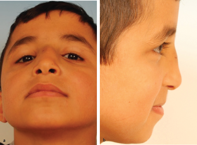
Type I corresponds to the unilateral depression of the nasal bone (Fig. 14.4). Fractures of this type are caused by the effect of a lateral impact with only low or moderate energy. An untreated fracture is apparent by an asymmetrical nasal pyramid, a damaged aesthetic eyebrow line, and the potential presence of a low level of protuberance formation on the rhinion (Fig. 14.5). The lamina perpendicularis and the septum cartilage remain intact in this type of fracture. The osseous-cartilaginous connections of the nasal bone and the upper lateral cartilages remain intact as well.
Type II is the multiple fracture of the nasal pyramid as a consequence of a frontolateral blunt trauma. The nasal bones and the lamina perpendicularis are fractured and the external fragments dislocate laterally. This fracture type results in a destruction of the central buttress with fracture and dislocation of the septum, whereby the osseous-cartilage connections are predominantly separated. The dislocation of the septum structures can occur along the entire length of the nose (Fig. 14.6). The long-term consequences are osseous-cartilaginous slanted noses with an occasional severely deviated and frequently also subluxated septum cartilage (Fig. 14.7). Intranasal avulsions of the mucosa and dislocation of cartilage fragments are very frequently observed. In the late phase, pronounced deformations and deviations are visible.
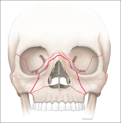
Fig. 14.3 Fracture lines of midface fractures according to LeFort types I–III.
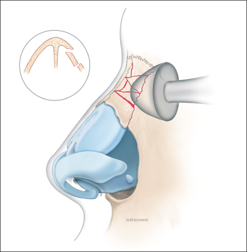
Fig. 14.4 Nasal fracture type I. Impression of the lateral bony nasal wall caused by a lateral impact.

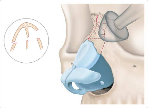
Fig. 14.6 Nasal fracture type II. Slanted nose with lateral displacement of the osseous nasal pyramid and fracture of the septum caused by a frontal–lateral impact.
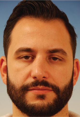
Fig. 14.7 Pronounced osseous–cartilaginous slanted nose following frontal–lateral trauma. The nasal pyramid is severely deviated and asymmetrically fixed. The consequence of the septum fracture is significant tension formation of the cartilaginous septum toward the right and subluxation of the anterior margin of the septum.
Type III is the consequence of direct frontal traumas, in which bilateral fractures and depressions or dislocations of the nasal bone occur. The lamina perpendicularis and the septum cartilage also frequently fracture as a result of the usually severe depressions. A separation of the connection between the nasal bones and the cephalic rim of the upper lateral cartilages often results as well. For this degree of injury, a relatively high level of energy is necessary (Fig. 14.8). The long-term consequences of an untreated fracture is expressed by a lowering and widening of the nasal pyramid, usually with a palpable protuberance formation on the bridge of the nose but also a saddle formation due to the lack of anchoring of the septum in the K-region (Fig. 14.9). Often a concha head avulsion at the height of the piriform aperture and mucous membrane avulsions with exposure of the cartilage is apparent endonasally. In addition, a deviation of the septum usually forms in the dorsal section. In the case of low trauma energy, only a marginal depression or an isolated avulsion of the nasal bones from the frontal bone may result. In this case, small step formations form on the nasal dorsum or on the nasion.
Fracture type IV is the result of a trauma striking in the direction of either caudal to cranial or dorsal to the tip of the nose. This causes a compression of the septum cartilage and the surrounding soft-tissue structures. The septum cartilage thus fractures and the osseous-cartilaginous connection to the lamina perpendicularis tears and results in a concomitant septum hematoma. The caudal fixation of the septum cartilage and the connection of the cranial septum rim to the cephalic rim of the lower lateral cartilages separate so that a complete or fragmented dislocation of the septum results (Fig. 14.10). An indirect sign for this fracture type is a hematoma in the upper lip at the height of the anterior nasal spine. This can be recognized by a cartilaginous saddle formation and rotation of the tip area with a reduction of projection (Fig. 14.11).
14.3.2 Naso-orbito-ethmoid Fractures
Classifications for the fractures of the naso-orbito-ethmoid complex were suggested by Markowitz et al12 and Jackson.13 Fracture type I consists of a one-sided noncomminuted fracture of the central segment. Two subtypes can be differentiated: (1) avulsion of the medial canthus ligament together with a piece of the lacrimal bone and (2) complete separation of the medial canthus ligament from the medial orbital wall. The consequences are a telecanthus with elapse of the medial palpebral commissure, narrowing of the palpebral fissure, limpness of the lids, and epiphora. The injuries of type II show one-sided comminutions and dislocations of the medial orbital wall, which, with more severe trauma, can also extend to the orbital roof or floor. The nasomaxillary columns and the maxilla are often affected, but the central segment remains. The clinical signs are similar to those of type I.
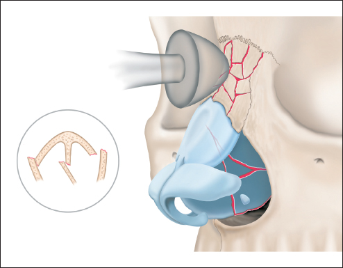
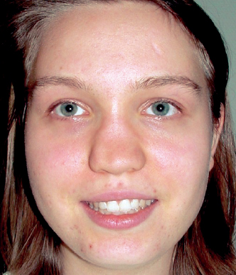
Fig. 14.9 Saddle nose following frontal trauma. The dissolution of the supporting function of the osseous pyramid and the septum has caused the entire nasal bridge to sink in, resulting in broadening of the nose and deformation of the nose tip. The lateral crus of the lower lateral cartilage are sunk in and the projection of the tip is reduced significantly and at the same time the nostrils clearly appear broadened.
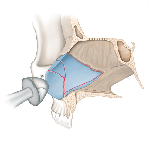
Fig. 14.10 Nasal fracture type IV. Compression and fracture of the septum resulting from a caudal-cranial impact.
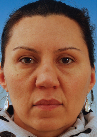
Fig. 14.11 Substantial septum deviation and cartilaginous slanted nose as a late effect following a septum fracture caused by caudal–cranial trauma.
In type III, there is such extreme comminution that the central element can no longer be identified and the septum, the nasal bones, and the frontal sinus are affected by the fracture and dislocation. Pronounced flattening and widening of the nasal dorsum and orbital displacement occur (Fig. 14.12).
14.4 Diagnostics
The clinical examination for nasal trauma with a suspicion of a nasal fracture should be conducted systematically. Because a nasal trauma can also be accompanied by craniofacial and cerebral injuries, the examination must also focus on cranial nerves, the cerebrum, and the eyes. The anamnesis for manifestations of allergic dispositions and chronically infected sinus illnesses is significant.
14.4.1 Inspection and Palpation
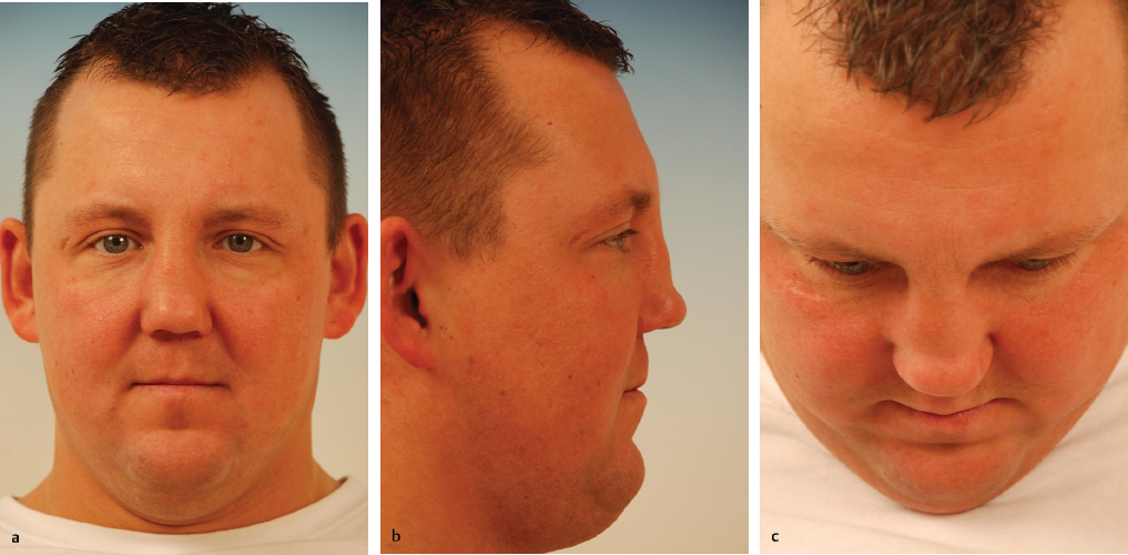
The external examination includes the inspection of the soft-tissue injuries, swellings, and deviations, as well as palpation of the nasal skeleton for abnormal movement, crepitation, depressions, a shortening of the nose, and also a possible widening of the nasal base (Fig. 14.13). In doing so, in particular, the intercanthal distance should be assessed. If the thumb and forefinger are each placed directly on the fixation point of the medial canthal tendon, the instability of the central fragment can be determined based on the extent of movement. A more sensitive estimation can be made according to the recommendations of Paskert and Manson14 by means of bimanual examination. An instrument inserted in the nose moves the mobile bone fragment against the externally palpating finger. In addition, the “traction test” can be executed by laterally pulling on the external edge of the lower lid. Asymmetries or abnormal movements indicate an avulsion of the medial canthal tendon. The integrity of the nasal framework can be checked through palpation of the nasal dorsum. A lack of resistance indicates a loss of osseous or cartilaginous buttresses in the central element. In cases of combined fractures of the nose and midface, the orbital rims should be palpated carefully. Steps and dehiscences in the bony edges of the orbital rims are suspect to fractures of the maxilla and orbit. An enophthalmus can immediately occur when the orbital floor developed large gaps after injury and content of the orbit moves downward into the maxillary sinus cavity. This clinical sign can be masked in the acute posttraumatic situation because of soft-tissue edema, hematoma, and ecchymosis.
14.4.2 Intranasal Diagnostics
Particular attention should be paid to the intranasal examination, for which an endoscope should always be used. It is the most important examination that can ensure a certain determination of the functional and aesthetic consequences of the nasal fracture. Verwoerd describes the pathogenesis of septum fractures of three septum zones with thicker cartilage as dorsoposterior, basal, and caudal.15 In contrast, the central section of the septum cartilage is thin. The thick posterior section of the septum cartilage supports the nasal dorsum. Therefore, trauma in the nasal dorsum area can cause caudal–basal to cephalo–dorsal lesions and horizontal fractures of the thin central regions. Fry presents the clear displacement of fractured septum cartilage fragments based on the separation of internal osseous cartilaginous connections16 (Fig. 14.14). Gunter and Rohrich show that the septum has a key function in the optimal care of nasal trauma and of the minimization of secondary deformities.17 All deformities and obstructions can be estimated with a rigid endoscope with a 4-mm optic (0 or 30 degrees). In doing so, one must pay particular attention in the cases of types II and III nasal bone fractures and naso-orbito-ethmoid fractures to the posterior osseous septum sections and to the vomer. A topical local anesthetic with 4% pantocaine and an additional reduction of the swelling with naphazoline is necessary in order to carry out a nasal endoscopy on conscious patients. It has been found that the endoscope should first be led along the nasal floor along the lower nasal concha to the posterior end of the septum. In addition to assessing the septum anomalies, the mucous membranes can be investigated for injuries and hematomas. These can occur on one or both sides. The disturbance of circulation to the septum cartilage resulting from the hematoma, which is provided by the perichondrium, can lead to irreversible damage after only 3 to 4 days (Fig. 14.15). Early recognition of these problems prevents the development of fibroses with ensuing septum displacement, abscess formations, and successive complete necrosis with nasal saddle formations. Pulling back the septum can allow for the recognition of possible injuries to the nasal concha and anything conspicuous, in particular bleeding, in the middle nasal passage.