, Shimin Chang2, Jian Lin3 and Dajiang Song1
(1)
Department of Orthopedic Surgery, Changzheng Hospital Second Military Medical University, Shanghai, China
(2)
Department of Orthopedic Surgery, Yangpu Hospital Tongji University School of Medicine, Shanghai, China
(3)
Department of Microsurgery, Xinhu Hospital Shanghai Jiao Tong University, Shanghai, China
Since the deep inferior epigastric artery perforator (DIEP) flap was first described in 1989 by Koshima et al. [1], the DIEP flap preserves abdominal wall function without sacrificing the functional muscles, has been proved to be a reliable option for reconstructive field, and becomes very popular in breast, head, and neck, as well as vaginal reconstruction in the field of plastic surgery [2–9].
Indications of this perforator flap with a short pedicle were superficial skin defects of the lower extremities, scalp, abdomen, or head and neck [10–13].
18.1 Vascular Anatomy
The deep inferior epigastric artery (DIEA) branches off from the external iliac artery opposite the deep circumflex iliac artery (DCIA). After crossing behind the lateral border, it enters the rectus sheath by passing anterior to the arcuate line midway between the pubis and the umbilicus. Generally, the deep inferior epigastric artery divides into two large branches (sometimes three), below the level of the umbilicus, that give off perforating vessels to the skin and muscle branches. The largest perforators (with diameters of 0.6 mm) are located within 6 cm of the umbilicus, in the medial one-third of the rectus muscle. The original free paraumbilical perforator flap described by Koshima et al. was based on a single paraumbilical perforator vessel that exhibits a suprafascial course after emerging from the anterior rectus fascia (Fig. 18.1).

Fig. 18.1
Vascular anatomy of the deep inferior epigastric artery
18.2 Illustrative Case
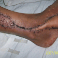
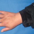
An 8-year-old boy was a victim of a motorcycle accident, which resulted in a skin defect on the dorsum of his right foot, exposing the metatarsal bones, and a defect of the extensor tendons. The wound was first managed with surgical debridement and VSD. Reconstructive surgery was performed 7 days after the injury (Fig. 18.2).

Fig. 18.2
Preoperative view (The case is offered by Professor Jun Liu and Dr. Xiaodan Xia, Hand Surgical Centre, Nanhua Hospital, Nanhua University, Henyang, China)
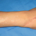
Flap Design
After thorough debridement of the necrotic tissue, the extensor tendons were fixed and the skin defect was treated with a 15 × 9 cm oblique DIEP flap which was based on one perforator (Fig. 18.3).

Fig. 18.3
Flap design
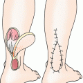
The location of perforators around the abdomen was preoperatively determined by an ultrasound Doppler, and the design of the flap was based on the size and three-dimensional features of the wound. The flap was designed including the point where the cutaneous perforators were located (Fig. 18.4).

Fig. 18.4
Schematic drawing of the flap design
Flap Elevation
Schematic drawing of the flap
Related posts:

Stay updated, free articles. Join our Telegram channel

Full access? Get Clinical Tree


