Hyperpigmentation disorders and skin lightening treatments have a significant impact on the dermatologic, physiologic, psychologic, economic, social, and cultural aspects of life. Skin lightening compounds, such as hydroquinone and topical corticosteroids, are often used to treat hyperpigmentation disorders, such as melasma, or lighten skin for cosmetic purposes. Despite their established effectiveness, a multitude of dermatologic and systemic complications have been associated with these agents. Regulatory agencies have also recognized the adverse effects of skin lighteners and many countries around the world now forbid the production and sale of these compounds, although this prohibition has not significantly curtailed distribution. Dermatologists and users of cosmetic products should be aware of the various components in bleaching compounds, their potential adverse effects, and alternative options for skin lightening.
Disorders of hyperpigmentation and skin lightening treatments have a significant impact on the dermatologic, physiologic, psychologic, economic, social, and cultural aspects of life. Skin lightening compounds or bleaching agents are chemicals used to achieve a lighter skin tone or whiten skin. These compounds are commonly used by individuals with hyperpigmentation disorders, such as melasma and postinflammatory hyperpigmentation (PIH), or those that desire lighter skin for cosmetic reasons. The most commonly used skin lightening products contain hydroquinone, topical corticosteroids (TCs), or mercury. Despite their apparent effectiveness, numerous cutaneous and systemic complications have been associated with these agents ( Table 1 ), resulting in more stringent regulations regarding the preparation and distribution of skin lightening products. In addition to the medical implications and patient safety concerns, the psychosocial aspects of hyperpigmentation disorders are important to consider, particularly their impact on quality of life, to further elucidate the motivations for skin bleaching.
| Hydroquinone | Mercurials | Topical Corticosteroids |
|---|---|---|
| Allergic contact dermatitis | Allergic contact dermatitis | Allergic contact dermatitis |
| Hyperpigmentation | Hyperpigmentation | Skin atrophy/striae atrophica |
| Corneal melanosis/degeneration | Anxiety/depression/psychosis | Acne vulgaris/periooral dermatitis |
| Exogenous ochronosis | Erythroderma | Cellulitis/dermatophytosis |
| Conjunctival pigmentation | Acute tubular necrosis | Cataracts/glaucoma |
| Trimethylaminuria | Membranous nephropathy | Hypertrichosis |
| Impaired wound healing | Peripheral neuropathy | Rosacea/telangiectasia |
| Nail discoloration | Positive antinuclear antibody test | Adrenal suppression/Cushing syndrome |
| Squamous cell carcinoma? | Tremor | Squamous cell carcinoma? |
Medical use of topical skin lightening compounds
Hydroquinone
The most commonly used over-the-counter (OTC) and prescription skin lightening preparation is a ubiquitous phenol compound known as hydroquinone or 1,4-dihydroxybenzene. Topical application of hydroquinone competitively inhibits melanogenesis through suppression of tyrosinase and subsequent release of semiquinone free radicals, which are toxic to melanosomes. In rat and human skin in vitro studies, hydroquinone has been shown to penetrate the epidermis and continue into the dermis, subcutaneous tissue, and circulation.
Hydroquinone’s ability to lighten skin was first reported by Oettel in 1936, when hydroquinone ingestion caused pigmentation changes in black-haired cats, which was reversible after hydroquinone discontinuation. Shortly after, in 1941, Martin and Ansbacher showed that hydroquinone ingestion induced graying of hair in mice. In the 1950s, hydroquinone was used as a sunscreen in the Southern United States, and users reported skin lightening as a complication. Since 1956, hydroquinone has been available in various OTC formulations for skin lightening purposes in the United States. In 1961, Spencer reported the first clinical trial evaluating hydroquinone as a skin lightener, and since then hydroquinone has remained the preferred treatment for various hyperpigmentation disorders, such as melasma, PIH, and lentigines.
Topical Corticosteroids
TCs are subdivided according to strength (class I–VII), with class I (ie, clobetasol) being the most potent and class VII (ie, hydrocortisone) the least potent. The strength of TCs is determined by the vasoconstriction assay, in which potency is associated with the degree of blood vessel blanching in the upper dermis. TCs have long been used for their skin lightening properties and are often the most commonly used skin lighteners in Africa. TCs are believed to bleach the skin through inhibiting pro-opiomelanocortin (POMC), the precursor protein for α-melanocyte-stimulating hormone (α-MSH), which is produced in the intermediate lobe of the pituitary to stimulate epidermal melanin production.
Mercurials
Mercury exists in three forms: organic, inorganic, and elemental. Mercury-containing creams and ointments have been used for centuries to treat infections (ie, syphilis), impetigo, phthiriasis (lice), and inflammatory skin diseases (ie, psoriasis), but more recently they have been used as skin lighteners. Mercurials exert their skin lightening actions through inhibition of sulfhydryl enzymes or mercaptans, ultimately resulting in suppression of tyrosinase (the rate-limiting enzyme in the melanin pathway) and decreased melanogenesis.
Adverse effects of topical skin lightening compounds
Hydroquinone
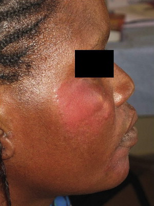
Although hydroquinone is one of the most effective and popular skin lightening compounds, it has been shown to cause multiple cutaneous and systemic side effects. The most common acute complication of hydroquinone use is irritant contact dermatitis (up to 70% of patients) ( Fig. 1 ), followed by PIH, hypopigmentation, and allergic contact dermatitis. Chronic complications of hydroquinone exposure include nail discoloration or “pseudo yellow-nail syndrome,” conjunctival pigmentation, corneal melanosis and degeneration, peripheral neuropathy, decreased skin elasticity, impaired wound healing, and wound dehiscence, particularly after abdominal procedures such as caesarian section or hysterectomy. Another unique complication of chronic hydroquinone use is trimethylaminuria or “fish odor syndrome,” characterized by a rotten fish body odor caused by excretion of trimethylamine in the saliva, sweat, urine, and vagina. An association between hydroquinone and squamous cell carcinoma has also been suggested, although all reported cases had a history of prior or concomitant TC use. Further studies are needed to properly determine the risk of skin cancer in hydroquinone users.
The most severe and widely recognized complication of chronic hydroquinone use is exogenous ochronosis, with at least 789 reported cases, 756 of which occurred in Africa. Ochronosis can exist in both endogenous and exogenous form, the former associated with alkaptonuria, an autosomal recessive disorder (1:25,000) characterized by the absence of homogentisic acid oxidase (HGOA), and the latter typically attributed to hydroquinone-containing compounds. Exogenous ochronosis has also been associated with antimalarials, ; carbolic acid (phenol) leg ulcer compresses ; resorcinol ; mercury ; levodopa ; and picric acid. In the inherited form, HGOA enzyme deficiency leads to homogentisic acid accumulation, which polymerizes to form ochre-colored pigments that deposit in the skin, cartilage, and tendons. Alkaptonuria is classically characterized by the triad of painless cutaneous hyperpigmentation (ochronosis), arthritis, and homogentisic aciduria, in which urine turns black when left standing or on contact with air or an alkali. Thickening of the cartilage of the pinnae, calcification of the aortic valve and prostate gland, and dark cerumen are also characteristic. Although exogenous ochronosis is clinically and histologically similar to its endogenous form, it is not inherited and no systemic symptoms are observed.
The term ochronosis (“yellow disease” in Greek) was coined by Virchow in 1866, when he described a patient whose cartilage appeared blue-black grossly, but was yellow-brown when viewed microscopically. In 1901, Pick reported the first exogenous form of ochronosis in a patient with prolonged exposure to phenols. In 1975, Findlay and colleagues described the first cases of hydroquinone-induced exogenous ochronosis in 35 South African Bantu women that used high-concentration (3.5%–7%) hydroquinone for several years. In 1979, Dogliotte and Liebowitz classified exogenous ochronosis into three different stages: (1) erythema and mild pigmentation, (2) hyperpigmentation, black colloid milia, and scanty atrophy, and (3) papulonodules with or without surrounding inflammation. The first case in the United States of exogenous ochronosis caused by hydroquinone use was reported in 1983 by Cullison and colleagues, which was followed with reports by Hoshaw and colleagues, Connor and Braunstein, and Lawrence and colleagues, all in patients using low-concentration (≤3%) hydroquinone for a short duration (≤1 year). Although various theories exist regarding the pathogenesis of exogenous ochronosis, the most accepted is that of Penneys’ which attributes the condition to hydroquinone’s inhibition of the enzyme HGOA, leading to the accumulation of homogentisic acid, which then polymerizes to form ochre pigments in the papillary dermis.
Exogenous ochronosis is characterized by gray-brown or blue-black macules coalescing into patches, which are occasionally accompanied by pinpoint, dark brown, caviar-like papules. Exogenous ochronosis is typically symmetrically distributed in photoexposed areas, particularly over osseous surfaces in the infraorbital and zygomatic regions, leading some to speculate that ultraviolet exposure might be a risk factor. Histologically, exogenous ochronosis is characterized by normal epidermis, curved ochre-colored banana-shaped structures in the papillary dermis, dermal solar elastosis, and degeneration of collagen and elastic fibers. Occasionally, sarcoid-like granulomas with multinucleated giant cells are seen surrounding the ochronotic particles. On dermoscopy, blue-gray amorphous areas can be seen obliterating follicular openings. The ochronotic pigment stains blue-black with methylene blue and black with Fontana stain; they do not stain with Prussian blue for iron.
Several modalities have been experimentally used to treat exogenous ochronosis, but results have not been reassuring. Some reports have noted improvement with carbon dioxide laser, dermabrasion, Q-switched ruby laser, Q-switched alexandrite laser (755 nm), tetracycline and retinoic acid, whereas cryotherapy and trichloroacetic acid have not shown efficacy. Occasionally, hydroquinone discontinuation leads to reversal of the hyperpigmentation, but this can take up to several years.
Topical Corticosteroids
Although TCs are frequently used as skin lightening agents, they are associated with multiple dermatologic and systemic side effects. Common cutaneous complications associated with TC application include acne vulgaris, allergic contact dermatitis, skin atrophy, perioral dermatitis, hypertrichosis, rosacea, striae atrophica, and telangiectasias. The skin on the neck seems particularly prone to atrophy, sometimes producing a rippled, “plucked chicken” appearance, otherwise known as pseudo-pseudoxanthoma elasticum. Chronic periocular TC use can cause ophthalmologic complications, such as cataracts and glaucoma. TC users are also particularly prone to developing infections such as cellulitis, dermatophytosis, erysipelas, folliculitis, and scabies. Dermatophyte infections are especially common and include tinea incognito, widespread tinea corporis and tinea faciei, which can mimic roseacea or lupus erythematosus. Furthermore, chronic users are more likely to develop viral warts, which tend to appear simultaneously on the neck and upper trunk, a finding that Olumide and colleagues nicknamed the “pseudo Leser-Trélat sign.”
TC use is also associated with various systemic complications such as hypothalamic-pituitary-adrenal (HPA) axis suppression, Cushing syndrome, diabetes mellitus, and hypertension. The most worrisome of these complications is HPA dysfunction causing adrenal insufficiency, which can be life-threatening. Adrenal suppression has long been regarded as a complication of high-dose TCs (ie, >50 g/wk of clobetasol propionate), but patients taking low doses (ie, 7.5 g/wk) have also experienced adrenal suppression. In 2001, Perret and colleagues assessed the functionality of the HPA axis among decade-long TC users in Sénégal and reported significantly lower plasma cortisol levels in response to cosyntropin stimulation compared with controls.
Another potential adverse effect from chronic daily use of a potent TC is “steroid addiction syndrome,” which is characterized by intense burning and potentially permanent erythema due to withdrawal vasodilatation. The irreversible form of pronounced erythema, often referred to as homme rouge , occurs more commonly in male users.
Other less-common reactions reported with TC use include avascular necrosis of the femoral head and squamous cell carcinoma. Additionally, chronic use of TCs may mask other pathologic conditions, as was observed in a patient with leprosy. The risk of these adverse reactions is potentiated when high-potency formulations are used on sites with fragile, thin skin (ie, face, armpit, groin) for prolonged periods or under occlusion, which promotes penetration. Individuals particularly at risk for developing adrenal suppression include infants and patients with damaged skin barriers.
Mercurials
Mercury-containing skin lightening agents or mercurials usually contain either mercury chloride or calomel and ammoniated mercury chloride, which are inorganic salts. Mercury, as thimerosal, is also commonly used in the manufacturing of mascara and other cosmetics. Acute or chronic exposure to topical mercury-containing compounds can cause dermatologic, renal, and neurologic toxicity. Classically, mercury poisoning was associated with felt hat manufacturers, hence the name “mad hatters disease,” or in patients treated for cutaneous disorders such as syphilis or impetigo. However, skin lightening products have also emerged as a major cause of mercury toxicity.
Common cutaneous complications of mercury use include allergic contact dermatitis, flushing, erythroderma, purpura, gingivostomatitis, and nail discoloration. Chronic use of mercurial skin lighteners can cause a paradoxic hyperpigmentation, which might be caused by dermal deposition of mercury-containing granules. Mercury-induced neuropsychiatric toxicity includes metallic taste, tremor, peripheral neuropathy, erethrism, memory loss, anxiety, depression, and psychosis. Other complications include positive antinuclear antibody titers and a suggested association with systemic lupus erythematosus (SLE), as metallic mercury exposure has been shown to accelerate SLE development in lupus-prone mice. Maternal use of mercury-containing soap during pregnancy has resulted in prenatal and postnatal intoxication, as mercury can cross the placenta. In children, inorganic mercury exposure has also been associated with acrodynia.
Nephrotoxicity is another potential complication related to mercury use. In one report, 50% of young African women in Kenya who used mercury-containing skin lightening creams developed glomerular lesions. The type of mercury-associated kidney injury depends on the form of mercury and the rate of administration. Organic and metallic mercury are lipophilic and typically cause neurotoxicity, whereas inorganic mercury typically causes nephrotoxicity. Still, any form of mercury can cause tubular or glomerular renal disease depending on the length of contact, with acute exposure usually causing tubular injury (acute tubular necrosis) and chronic exposure usually causing glomerular injury (membranous nephropathy, immune complex–mediated glomerulonephritis, minimal change disease). Cole and colleagues showed that the amount of mercury applied to the skin is proportional to the amount excreted by the kidneys. Still, mercury-induced membranous nephropathy typically resolves spontaneously after exposure cessation.
Adverse effects of topical skin lightening compounds
Hydroquinone
Although hydroquinone is one of the most effective and popular skin lightening compounds, it has been shown to cause multiple cutaneous and systemic side effects. The most common acute complication of hydroquinone use is irritant contact dermatitis (up to 70% of patients) ( Fig. 1 ), followed by PIH, hypopigmentation, and allergic contact dermatitis. Chronic complications of hydroquinone exposure include nail discoloration or “pseudo yellow-nail syndrome,” conjunctival pigmentation, corneal melanosis and degeneration, peripheral neuropathy, decreased skin elasticity, impaired wound healing, and wound dehiscence, particularly after abdominal procedures such as caesarian section or hysterectomy. Another unique complication of chronic hydroquinone use is trimethylaminuria or “fish odor syndrome,” characterized by a rotten fish body odor caused by excretion of trimethylamine in the saliva, sweat, urine, and vagina. An association between hydroquinone and squamous cell carcinoma has also been suggested, although all reported cases had a history of prior or concomitant TC use. Further studies are needed to properly determine the risk of skin cancer in hydroquinone users.
The most severe and widely recognized complication of chronic hydroquinone use is exogenous ochronosis, with at least 789 reported cases, 756 of which occurred in Africa. Ochronosis can exist in both endogenous and exogenous form, the former associated with alkaptonuria, an autosomal recessive disorder (1:25,000) characterized by the absence of homogentisic acid oxidase (HGOA), and the latter typically attributed to hydroquinone-containing compounds. Exogenous ochronosis has also been associated with antimalarials, ; carbolic acid (phenol) leg ulcer compresses ; resorcinol ; mercury ; levodopa ; and picric acid. In the inherited form, HGOA enzyme deficiency leads to homogentisic acid accumulation, which polymerizes to form ochre-colored pigments that deposit in the skin, cartilage, and tendons. Alkaptonuria is classically characterized by the triad of painless cutaneous hyperpigmentation (ochronosis), arthritis, and homogentisic aciduria, in which urine turns black when left standing or on contact with air or an alkali. Thickening of the cartilage of the pinnae, calcification of the aortic valve and prostate gland, and dark cerumen are also characteristic. Although exogenous ochronosis is clinically and histologically similar to its endogenous form, it is not inherited and no systemic symptoms are observed.
The term ochronosis (“yellow disease” in Greek) was coined by Virchow in 1866, when he described a patient whose cartilage appeared blue-black grossly, but was yellow-brown when viewed microscopically. In 1901, Pick reported the first exogenous form of ochronosis in a patient with prolonged exposure to phenols. In 1975, Findlay and colleagues described the first cases of hydroquinone-induced exogenous ochronosis in 35 South African Bantu women that used high-concentration (3.5%–7%) hydroquinone for several years. In 1979, Dogliotte and Liebowitz classified exogenous ochronosis into three different stages: (1) erythema and mild pigmentation, (2) hyperpigmentation, black colloid milia, and scanty atrophy, and (3) papulonodules with or without surrounding inflammation. The first case in the United States of exogenous ochronosis caused by hydroquinone use was reported in 1983 by Cullison and colleagues, which was followed with reports by Hoshaw and colleagues, Connor and Braunstein, and Lawrence and colleagues, all in patients using low-concentration (≤3%) hydroquinone for a short duration (≤1 year). Although various theories exist regarding the pathogenesis of exogenous ochronosis, the most accepted is that of Penneys’ which attributes the condition to hydroquinone’s inhibition of the enzyme HGOA, leading to the accumulation of homogentisic acid, which then polymerizes to form ochre pigments in the papillary dermis.
Exogenous ochronosis is characterized by gray-brown or blue-black macules coalescing into patches, which are occasionally accompanied by pinpoint, dark brown, caviar-like papules. Exogenous ochronosis is typically symmetrically distributed in photoexposed areas, particularly over osseous surfaces in the infraorbital and zygomatic regions, leading some to speculate that ultraviolet exposure might be a risk factor. Histologically, exogenous ochronosis is characterized by normal epidermis, curved ochre-colored banana-shaped structures in the papillary dermis, dermal solar elastosis, and degeneration of collagen and elastic fibers. Occasionally, sarcoid-like granulomas with multinucleated giant cells are seen surrounding the ochronotic particles. On dermoscopy, blue-gray amorphous areas can be seen obliterating follicular openings. The ochronotic pigment stains blue-black with methylene blue and black with Fontana stain; they do not stain with Prussian blue for iron.
Several modalities have been experimentally used to treat exogenous ochronosis, but results have not been reassuring. Some reports have noted improvement with carbon dioxide laser, dermabrasion, Q-switched ruby laser, Q-switched alexandrite laser (755 nm), tetracycline and retinoic acid, whereas cryotherapy and trichloroacetic acid have not shown efficacy. Occasionally, hydroquinone discontinuation leads to reversal of the hyperpigmentation, but this can take up to several years.
Topical Corticosteroids
Although TCs are frequently used as skin lightening agents, they are associated with multiple dermatologic and systemic side effects. Common cutaneous complications associated with TC application include acne vulgaris, allergic contact dermatitis, skin atrophy, perioral dermatitis, hypertrichosis, rosacea, striae atrophica, and telangiectasias. The skin on the neck seems particularly prone to atrophy, sometimes producing a rippled, “plucked chicken” appearance, otherwise known as pseudo-pseudoxanthoma elasticum. Chronic periocular TC use can cause ophthalmologic complications, such as cataracts and glaucoma. TC users are also particularly prone to developing infections such as cellulitis, dermatophytosis, erysipelas, folliculitis, and scabies. Dermatophyte infections are especially common and include tinea incognito, widespread tinea corporis and tinea faciei, which can mimic roseacea or lupus erythematosus. Furthermore, chronic users are more likely to develop viral warts, which tend to appear simultaneously on the neck and upper trunk, a finding that Olumide and colleagues nicknamed the “pseudo Leser-Trélat sign.”
TC use is also associated with various systemic complications such as hypothalamic-pituitary-adrenal (HPA) axis suppression, Cushing syndrome, diabetes mellitus, and hypertension. The most worrisome of these complications is HPA dysfunction causing adrenal insufficiency, which can be life-threatening. Adrenal suppression has long been regarded as a complication of high-dose TCs (ie, >50 g/wk of clobetasol propionate), but patients taking low doses (ie, 7.5 g/wk) have also experienced adrenal suppression. In 2001, Perret and colleagues assessed the functionality of the HPA axis among decade-long TC users in Sénégal and reported significantly lower plasma cortisol levels in response to cosyntropin stimulation compared with controls.
Another potential adverse effect from chronic daily use of a potent TC is “steroid addiction syndrome,” which is characterized by intense burning and potentially permanent erythema due to withdrawal vasodilatation. The irreversible form of pronounced erythema, often referred to as homme rouge , occurs more commonly in male users.
Other less-common reactions reported with TC use include avascular necrosis of the femoral head and squamous cell carcinoma. Additionally, chronic use of TCs may mask other pathologic conditions, as was observed in a patient with leprosy. The risk of these adverse reactions is potentiated when high-potency formulations are used on sites with fragile, thin skin (ie, face, armpit, groin) for prolonged periods or under occlusion, which promotes penetration. Individuals particularly at risk for developing adrenal suppression include infants and patients with damaged skin barriers.
Mercurials
Mercury-containing skin lightening agents or mercurials usually contain either mercury chloride or calomel and ammoniated mercury chloride, which are inorganic salts. Mercury, as thimerosal, is also commonly used in the manufacturing of mascara and other cosmetics. Acute or chronic exposure to topical mercury-containing compounds can cause dermatologic, renal, and neurologic toxicity. Classically, mercury poisoning was associated with felt hat manufacturers, hence the name “mad hatters disease,” or in patients treated for cutaneous disorders such as syphilis or impetigo. However, skin lightening products have also emerged as a major cause of mercury toxicity.
Common cutaneous complications of mercury use include allergic contact dermatitis, flushing, erythroderma, purpura, gingivostomatitis, and nail discoloration. Chronic use of mercurial skin lighteners can cause a paradoxic hyperpigmentation, which might be caused by dermal deposition of mercury-containing granules. Mercury-induced neuropsychiatric toxicity includes metallic taste, tremor, peripheral neuropathy, erethrism, memory loss, anxiety, depression, and psychosis. Other complications include positive antinuclear antibody titers and a suggested association with systemic lupus erythematosus (SLE), as metallic mercury exposure has been shown to accelerate SLE development in lupus-prone mice. Maternal use of mercury-containing soap during pregnancy has resulted in prenatal and postnatal intoxication, as mercury can cross the placenta. In children, inorganic mercury exposure has also been associated with acrodynia.
Nephrotoxicity is another potential complication related to mercury use. In one report, 50% of young African women in Kenya who used mercury-containing skin lightening creams developed glomerular lesions. The type of mercury-associated kidney injury depends on the form of mercury and the rate of administration. Organic and metallic mercury are lipophilic and typically cause neurotoxicity, whereas inorganic mercury typically causes nephrotoxicity. Still, any form of mercury can cause tubular or glomerular renal disease depending on the length of contact, with acute exposure usually causing tubular injury (acute tubular necrosis) and chronic exposure usually causing glomerular injury (membranous nephropathy, immune complex–mediated glomerulonephritis, minimal change disease). Cole and colleagues showed that the amount of mercury applied to the skin is proportional to the amount excreted by the kidneys. Still, mercury-induced membranous nephropathy typically resolves spontaneously after exposure cessation.
Psychosocial aspect
Although dermatologic disorders are not generally considered to be life-threatening, suicide has been reported in patients with “skin failure.” Psychodermatologic problems are more common in women, and facial symptomatology seems to be a particular risk factor for depression and suicidal ideations. Disorders of pigmentation, such as melasma, vitiligo, PIH, lentigo, and idiopathic guttate hypomelanosis, are prevalent and have been shown to significantly impact health-related quality of life (HRQOL) in a deleterious manner. A recent United States study showed that 80% of randomly selected patients at a private dermatology clinic had pigmentation disorders that significantly affected their quality of life. Approximately 47.3% of patients felt self-conscious about their skin, 32.7% felt unattractive, and 23.6% felt their disorder affected their day-to-day activities, although few patients sought or received treatment for these conditions. Treatment with skin lightening agents has been shown to improve these unfavorable psychosocial and HRQOL issues.
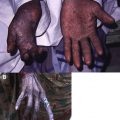
The habitual use of skin lightening compounds has long been commonplace in Africa, particularly Ghana ( Fig. 2 ), Kenya, Nigeria, Sénégal, and Zimbabwe, and in India, but has recently become recognized in other parts of the world, including North America, South America, Central America, Great Britain, Europe, Japan, Southeast Asia ( Fig. 3 ), and the Middle East. For more than 50 years, the mainstay of therapy for Black, White, Asian, Indian, and Hispanic individuals with hyperpigmentation disorders has been hydroquinone. Although traditionally regarded as a female practice, men in Africa commonly use skin lighteners, and recent articles suggest that they are becoming popular among men in India, North America, and Central America.
Related posts:
 Buruli Ulcer: Advances in Understanding Mycobacterium ulceransInfection
 Outbreak of Nontuberculous Mycobacterial Disease in the Central Pacific
 Arsenical Keratoses in Bangladesh—Update and Prevention Strategies
 Chagas Disease: Coming to a Place Near You
 Female Genital Mutilation: What Every American Dermatologist Needs to Know
Human Immunodeficiency Virus and Leprosy: An Update
Female Genital Mutilation: What Every American Dermatologist Needs to Know
Human Immunodeficiency Virus and Leprosy: An Update
Stay updated, free articles. Join our Telegram channel

Full access? Get Clinical Tree


