The aim of the current review is to provide an overview of the use of reflectance confocal microscopy to detect early skin aging signs. This new imaging tool holds the promise to morphologically explore the epidermis and upper dermis at nearly histologic resolution and over time. The main confocal findings of aged skin include the presence of irregular honeycombed pattern, linear skin furrows, mottled pigmentation, and distinct collagen types (coarse and huddled).
Key points
- •
Skin aging is a complex and multifactorial biological process that involves a multistep pathway in which chronologic and photo aging are closely entangled.
- •
Reflectance confocal microscopy (RCM) can perform early detection of specific skin aging signs.
- •
Epidermal and papillary dermal changes can be morphologically assessed and readily monitored over time and over treatment with nearly histologic resolution and in a noninvasive manner.
- •
With RCM, aged skin typically displays an irregular honeycombed pattern with variable mottled pigmentation and the presence of flattened rete-ridges that coexists with polycyclic papillary contours. The papillary dermis shows a variable degree of changes of collagen with coarse collagen and huddled collagen. Finally, the presence of curled elastotic fibers, referred to solar elastosis , is observable in elderly skin.
Introduction
“Age has no reality except in the physical world” Gabriel García Márquez ( Love in the Time of Cholera ). With this said, it is clear that aging is an ineluctable process that affects humans beings. Unlike the aging signs of internal organs, the skin demonstrates the first obvious signs of the passage of time with the consequent impact on a patient’s social life.
Skin aging can be formally conceptualized into intrinsic and extrinsic aging, the latter not being easily disentangled from the former. The importance of aging lies in the enormous consumer demand for agents or treatments that can prevent or reverse its stigmata, its strong association with skin tumors, and the clues it provides regarding the nature of aging itself. In light of this, it is mandatory to detect early skin aging signs when the process can be readily reversed or, at least, minimized.
As a direct consequence, a precise and real-time quantification of aging is of outmost importance for in vivo staging of the dynamic process. Several bioengineering methods have been proposed to extensively and noninvasively assess skin aging in its early phase of development.
The current review focuses on the use of reflectance confocal microscopy (RCM) for the early detection of skin aging and for treatment monitoring.
Reflectance Confocal Microscopy
Minsky developed RCM in 1957; since then, it has gained clinical and research popularity in the last decades, faster than any other devices. The reasons rely on the fact that RCM is a totally noninvasive technique that permits us to get optical en face sectioning of the skin with good contrast and high resolution, providing cytologic and architectural details. Furthermore, the examination of a given skin lesion can be repeated over time, rendering this method extremely useful for treatment monitoring or dynamic evaluation of biological phenomena (ie, growing melanocytic nevi).
Technical notes
Briefly, a confocal microscope consists of a point source of light, condenser, objective lenses, and a point detector. The pinhole collects light emanating only from the in-focus plane. The mechanism of bright contrast in RCM is backscattering. In gray-scale confocal images, structures that appear reflective have components with high refractive index compared with their surroundings and are similar in size to the wavelength of light. Backscattering is primarily governed by the structures’ refractive index compared with surrounding medium. Highly reflective skin components include melanin, collagen, keratin, and other elements, such as cytoplasmic organelles. The confocal scanning produces high-resolution black and white horizontal images (0.5 × 0.5 mm) with a lateral resolution of 1.0 μm and axial resolution of 3 to 5 μm. A sequence of full-resolution individual images at a given depth is acquired and combined together to create a mosaic ranging in size from 2 × 2 mm to 8 × 8 mm. A VivaCube (Mavig, Munich, Germany) composed of 3 to 4 mosaics with a 25-μm step is usually acquired for facial skin. Furthermore, a vertical VivaStack (Mavig, Munich, Germany) can be imaged. It consists of single high-resolution images acquired from the top skin surface up to 200 μm to obtain a sort of optic biopsy. The VivaStack modality is useful for the assessment of the epidermal thickness. Recently, a handheld RCM has been introduced on the market. This version is a smaller and flexible device that is quite useful in difficult-to-access areas (skin folds, ears). Unlike the wide-probe version, it has on-instrument control for laser power, imaging depth, and capture; but it does not allow scanning a large field of view.
Morphologic Aspects of Skin over the Age
Skin aging affects both epidermis and dermis; the process involves keratinocytes (KCs), melanocytes, and all cells that are present in the skin. In light of the nearly histologic resolution of RCM, it is readily feasible to detect morphologically the changes occurring over time. To start with, it is mandatory to know the overall picture of normal and young skin and then to progressively go through the changes observable in elderly skin.
Healthy Young Skin
In healthy young skin, the epidermis appears as a multilayer tissue with paradigmatic confocal findings depending on the skin level. The stratum corneum appears as a highly refractive surface surrounded by darker skin furrows. Corneocytes are large, ranging from 10 to 30 μm, polygonal, and enucleated. Skin furrows appear as dark folds between islands of KCs that are typically arranged in a rhomboidal pattern formed by intersecting skin furrows ( Fig. 1 ). Of note, the shape and arrangement of the skin folds strongly depends on the body site (being almost absent on the forehead and well represented on the abdomen) and the individual’s age.
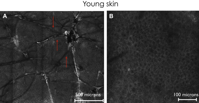
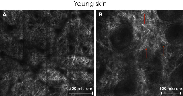
Going deeper, the stratum granulosum is composed of polygonal KCs presenting a grainy cytoplasm because of the presence of organelles. The KCs cohesively assemble, forming a honeycombed pattern because of its similarity with the honeycomb of bees (see Fig. 1 ). The contour of the cell is usually brighter than the cytoplasm and perfectly outlined. This pattern is commonly observed in light skin types. In contradistinction, dark skin types show pigmented KCs that are usually bright cells, small in size and always polygonal, separated by a darker contour (cobblestone pattern), resembling the negative of the honeycombed pattern. On the face, a peculiar pattern is caused by the presence of numerous hair follicles that appear as dark round areas with a central roundish area from which arises the hair shaft. At the stratum spinosum level, the size of KCs tends to decrease but the cells still have a polygonal shape. The honeycombed pattern is easily observable. Below the spinous layer, there is a single layer of basal cells at the dermoepidermal junction (DEJ) that are uniform in size and shape but are smaller and more refractive than spinous KCs because of the melanin caps forming bright disks on top of the nuclei. It is intuitively obvious that the brightness of KCs depends strongly on the skin photo-type: the darker the skin photo-type, the brighter are the basal KCs. Conversely, skin photo-type I-II is characterized by basal KCs with low refractivity that constitutes barely visible dermal papillae. At the dermoepidermal level, in the presence of regular rete-ridges, basal cells form round or oval rings of bright cells (KCs) surrounding dark dermal papillae. On the face, rings are not usually observed. In young subjects, the dermis shows the presence of thin reflective reticulated fibers that form a weblike structure ( Fig. 2 ).
Aged Skin
Regarding the epidermis, characteristic changes have been found in aged skin; the main findings have been reported for facial skin. There is a decreased epidermal thickness with aging. Of note, a slight increase in epidermal thickness was found in middle-aged subjects whereby the presence of polycyclic papillary contours was simultaneously present. It is well known that polycyclic papillary contours correspond histopathologically to the presence of solar-lentigo features with bulbous and fused epidermal cristae. Thus, it could be speculated that the skin might became hyperplastic in response to solar insult and, later, severely atrophic with progressive thinning.
The overall epidermal surface analysis reveals a predominantly linear furrow pattern with the rhomboidal pattern becoming progressively larger and then completely linear without intersecting lines.
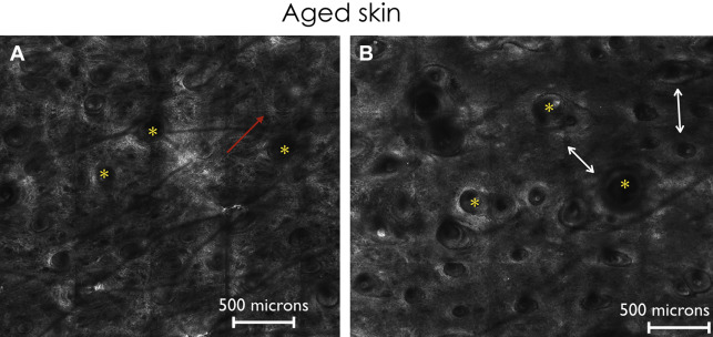
Unlike young skin, KCs in the elderly shows a wider variation in terms of shape, cellular outlines, and architecture. Those morphologies mirror a distinct epidermal pattern named irregular honeycombed pattern ( Fig. 3 ); the changes occurring at different ages account for a variable degree of deviation from the regular honeycombed pattern until the complete disarray of the epidermis that occurs in presence of an actinic keratosis.
When examining the changes occurring on melanocytes with age, it has to be clarified that in normal conditions melanocytes cannot be distinguished from KCs; they are detectable in tumors (ie, melanocytic nevi and melanoma) or in inflammatory skin conditions (ie, melasma). With this said, the indirect effect on pigmentation can be observed with RCM, even in the preclinical stage.
More specifically, the presence of mottled pigmentation, defined as small clusters of bright KCs amid a honeycombed pattern, can be variably observed in different age groups. The late sign of aging or photoaging, clinically visible as age spots, is seen on confocal microscopy as changes of a solar lentigo.
At the dermal level, RCM provides an overview of the collagen and elastic tissue that might present different degrees of degeneration over time. In young subjects, the dermis shows the presence of thin reticulated fibers that are progressively replaced by thick coarse fibers or even amorphous huddles in the elderly ( Fig. 4 ). Furthermore, when severe solar elastosis is present, RCM highlights the presence of curled wavy fibers corresponding to elastotic changes.