45
Volar Dislocations of the Proximal Interphalangeal Joint
Lisa L. Lattanza and Steven Z. Glickel
History and Clinical Presentation
A 20-year-old woman sustained an injury to her right ring finger during a tackle while playing rugby. She was not able to describe the exact mechanism, as it happened in a “pile-up.” She was seen in the emergency department that day.
Physical Examination
A swollen and tender proximal interphalangeal (PIP) joint of the right ring finger was noted, with slight rotational deformity of the digit. The patient was unable to actively extend the PIP joint. There was brisk capillary refill of the digit and the skin was intact.
Diagnostic Studies
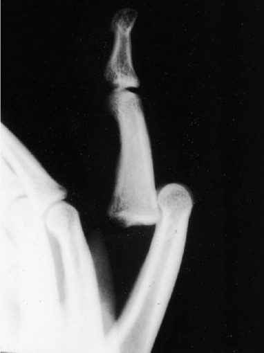
Anteroposterior and lateral radiographs of the right ring finger showed a volar PIP joint dislocation without fracture (Fig. 45-1).
PEARLS
- Flex the PIP and MP joints when reducing the dislocation.
- Determine if the dislocation is straight volar or volar rotatory.
- Mobilize early if stable.
PITFALLS
- Most volar dislocations do not require aggressive operative intervention.
- Beware of late PIP joint stiffness or boutonniere deformity.

Figure 45-1. Volar proximal interphalangeal (PIP) joint dislocation.
Differential Diagnosis
PIP joint sprain
Dorsal PIP joint dislocation
Volar PIP joint dislocation
Middle phalanx fracture
Proximal phalanx fracture
PIP joint fracture/dislocation
Diagnosis
Volar PIP Dislocation
Unlike dorsal PIP joint dislocations, volar dislocations are rare. It is important to understand the mechanism of injury and the resultant damage to the underlying structures to devise a sensible treatment plan. It is also necessary to differentiate the straight volar dislocation from the volar rotatory dislocation as the structures damaged, and therefore the treatment, are different.
Nonsurgical Management
There are two types of volar PIP joint dislocations: a straight volar dislocation and the volar rotatory dislocation. To fully understand the management of these two distinct entities, it is essential to understand the different injuries.
Stay updated, free articles. Join our Telegram channel

Full access? Get Clinical Tree


