1. Vitiligo/non-segmental vitiligo (NSV)
1.1 Generalized
1.2 Acrofacial
1.3 Universal
1.4 Mucosal
1.5 Mixed
2. Segmental vitiligo (SV)
21.2.1 Vitiligo/NSV
The term vitiligo includes different usually multifocal clinical subtypes that are all clearly distinct from SV. The depigmented macules and patches vary in size from a few to several centimeters in diameter and involve both sides of the body with a tendency toward symmetrical distribution. Vitiligo/NSV can be generalized, acrofacial, universal, mucosal, and mixed (associated with SV). Rare types of vitiligo/NSV include vitiligo punctata and hypochromic vitiligo/vitiligo minor.
21.2.1.1 Generalized Vitiligo
This is the most common form of the disease. It is characterized by lesions involving the face, trunk, and extremities, usually in a symmetrical pattern (Fig. 21.1).
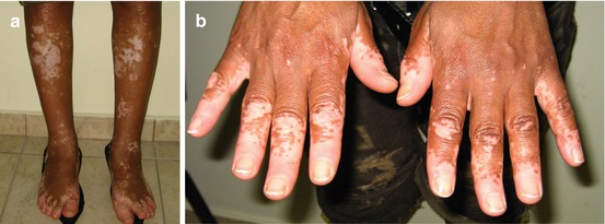
Fig. 21.1
Generalized vitiligo: symmetrical lesions on the shins and feet (a) and hands (b)
21.2.1.2 Acrofacial Vitiligo
Acrofacial vitiligo is limited to the face, head, hands, and feet (Fig. 21.2). Depigmentation of the distal fingers and facial orifices is usually present. Acrofacial vitiligo may evolve into typical generalized vitiligo.
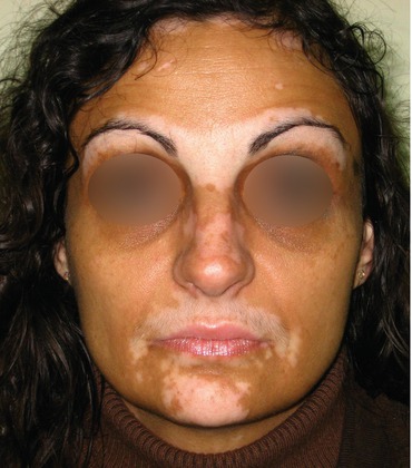
Fig. 21.2
Acrofacial vitiligo: lesions on the face (lesions on the hands were also present)
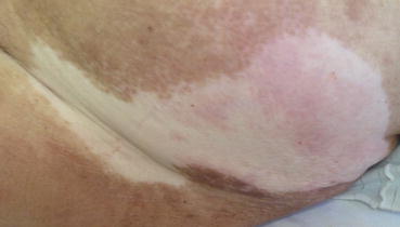
21.2.1.3 Universal Vitiligo (Vitiligo Universalis)
In universal vitiligo, more than 80 % of the body surface area is depigmented (Fig. 21.3). It is the most extensive form of the disease, and hair involvement is also common. Some pigmentation may be still present, especially in sun-exposed areas. Universal vitiligo can be the end point of a progressive generalized vitiligo that has evolved to nearly complete depigmentation of the skin and hair.
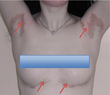
Fig. 21.3
Universal vitiligo: note the few remaining islands with normal pigmentation (arrows)
21.2.1.4 Mucosal Vitiligo
Mucosal vitiligo is characterized by involvement of the oral and/or genital mucosae (Fig. 21.4). It is included in vitiligo/NSV when it is associated with skin involvement. If the skin lesions are not present, mucosal vitiligo is classified as undetermined/unclassified vitiligo.
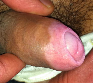
Fig. 21.4
Mucosal vitiligo: lesions on the prepuce and glans penis
21.2.1.5 Mixed Vitiligo
Mixed vitiligo refers to segmental vitiligo which has been followed by vitiligo/NSV with a delay of at least 6 months [4]. In patients with mixed vitiligo, the “segmental” part of the disease is usually more resistant to treatment. Risk factors for the progression of segmental to mixed vitiligo include the presence of halo nevi and leukotrichia [5].
21.2.1.6 Rare Types
Vitiligo punctata refers to multiple, small (confetti-like), sharply demarcated depigmented macules (Fig. 21.5).
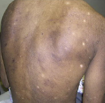
Fig. 21.5
Vitiligo punctate: confetti-like lesions
Hypochromic vitiligo/vitiligo minor is characterized by the presence of hypopigmented lesions alone or in association with completely depigmented lesions [6]. This type of vitiligo/NSV has been very sparsely reported, and it seems to be limited to dark-skinned individuals. Histological examination should be performed to rule out hypopigmented mycosis fungoides (Fig. 21.6).
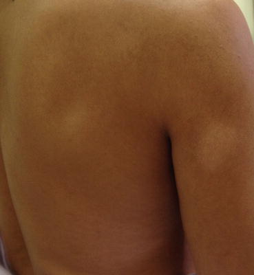
Fig. 21.6
Mycosis fungoides lesions resembling hypochromic vitiligo
21.2.2 Segmental Vitiligo
In segmental vitiligo, one or more vitiligo lesions are distributed on a unilateral segment of the body. The lesions usually (but not always) respect the midline (Fig. 21.7).