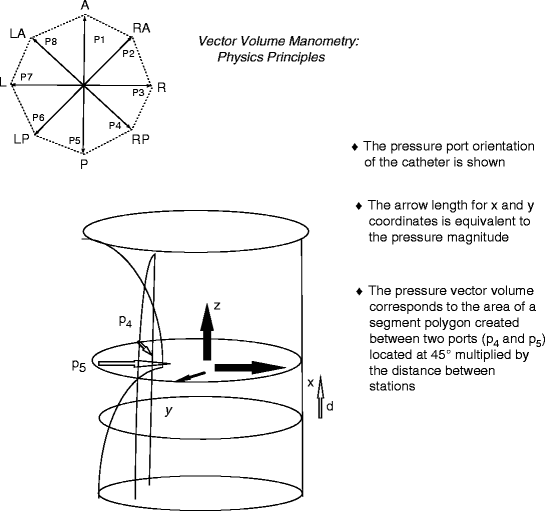
where 1 is the level above the anal verge with recordable pressures, d is the distance between station measurements, and P doublets of pressure are the pressure vectors for each sector wedge. The sine is 45 given the eight different channels used, but this would vary if more channels were employed. As such, the volume has no parameter representing the total integration of vector polygons at a given speed of catheter withdrawal. The PC is interfaced with a high-resolution color monitor, which has a graded palette to create a color-coded vectorgram that can be rotated to assess graphic indentations representing increases in overall sector pressure [7, 8]. For an average sphincter length at a withdrawal speed of 1 cm/s, there are an average of 15,000 individual data points with smoothing vectography provided by spline-curve interpolation to create the vectorgram. For this reason, an increase in the number of channels would result in less data interpolation and a smoother image. The vectorgram may be obtained either as an open mesh-net or as a solid-state design. Figure 7.1 shows the physical principles of vector volume construction.

Fig. 7.1
Physical principles of vector volume construction/manometry. An eight-channel, radially disposed polyethylene catheter is used. At a constant rate of withdrawal, this provides a sector polygon of summated pressures at 45° angles that are interpreted to create a vector volume measurement and vectorgram. L left, LA left anterior, LP left posterior, A anterior, RA right anterior, R right, RP right posterior, P posterior (Reproduced with permission from Zbar et al. [9])
There are few comparisons of conventional ARM and VVM in either health [9] or disease [10]. Work comparing conventional ARM with VVM has been conducted in normal patients and those with passive fecal incontinence, full-thickness rectal prolapse, and chronic anal fissure, measuring the mean resting vector volume (MRVV), mean squeeze vector volume (MSVV), HPZ lengths (at rest and during squeeze), maximal averaged pressure at rest (MR), maximal averaged pressure during sustained squeeze (MPS), and the percentage asymmetry as the percent deviation of the integrated cross-sections from a perfect circle. Sectorial pressures have been pooled for analysis, creating right, left, anterior, and posterior mean pressures. As expected, significant differences at rest and during squeeze are demonstrable for MRVV, MSVV, MPR, and MPS that mirror those obtained with conventional ARM, whereas, in general, MPR values tend to exceed mean resting anal pressure, and MPS values tend to be lower, on average, than mean squeeze pressure values. It may be that during automated catheter withdrawal, there is some voluntary sphincter contraction that triggers the MPR recording to be higher than the mean resting anal pressure value obtained with conventional ARM. This also may result from perfusate leakage. The lower value of MPS over mean squeeze pressure (in conventional ARM) may reflect a greater difficulty in sustaining an adequate squeeze contraction during the vector volume technique (Table 7.1) [11].
Table 7.1
Correlation coefficients and P values at rest and on maximal squeeze between vector volume manometry and conventional manometric measurements
MRAP | MRVV | HPZ | MPR | |
|---|---|---|---|---|
MRVV | 0.51 | |||
(<0.001) | ||||
HPZ (length) | 0.17 | 0.43 | ||
(0.07) | (<0.001) | |||
MPR | 0.79 | 0.70 | 0.19 | |
(<0.001) | (<0.001) | (0.05) | ||
Asymmetry (%) | −0.21 | −0.24 | −0.07 | −0.29 |
(0.027) | (0.009) | (0.45) | (0.003) | |
MSP | MSVV | HPZ | % asymmetry | |
MSVV | 0.59 | |||
(<0.001) | ||||
Asymmetry (%) | −0.13 | −0.24 | ||
(0.18) | (0.01) | |||
HPZ (length) | −0.14 | 0.26 | −0.04 | |
(0.14) | (0.006) | (0.72) | ||
MPS | 0.86 | 0.78 | −0.11 | −0.19 |
(<0.001) | (<0.001) | (0.26) | (0.05) |
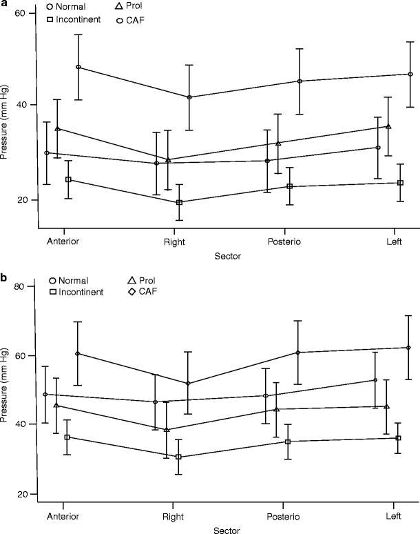
Although there is an expected sectorial ordering from patients with incontinence through to hypertonic anal fissure, there is no evidence of inherent sectorial differences for the different anorectal conditions, although there is a trend toward higher anterior sector pressures in patients with fissure. This sectorial variation has been identified in some other studies [12–14] and is shown in Fig. 7.2a, b. The correlation coefficients between conventional and vector volumetric variables for rest and squeeze confirm a strong correlation for HPZ length measurement with both techniques (see Table 7.1). There is, however, no correlation between sectorial asymmetry and demonstrable EAS defects [15] or in patients after internal anal sphincter (IAS) division for chronic anal fissure [16], although recent data has suggested that VVM may assist in defining those cases with an EAS defect in the last centimeter of the anal canal [17, 18]. It would seem, however, that when initial ultrasound is inconclusive in the diagnosis of a reparable sphincter defect, an ultrasonographically defined use of VVM would somewhat defeat its purpose [19, 20]. Further recent data have shown significant differences in all vector resting parameters, HPZ length at rest, and percentage asymmetry at rest, along with changes in most squeeze variables, after IAS division for topically resistant chronic anal fissure [16], with marked differences between postoperative continent and incontinent cohorts, particularly in resting HPZ length. This latter finding may suggest an overly zealous internal anal sphincterotomy that has been previously endosonographically recorded [21] and during which the extent of IAS division often can be far greater than intended. In continent postoperative patients, percentage resting asymmetry tends to increase (by about 6.7 %), whereas in incontinent postoperative cases it tends to fall (by about 3.1 %). The explanations for these changes in squeeze parameters are not understood, but it is conceivable that there is excessive voluntary sphincter fatigue after surgery between groups (even in continent cohorts), where impending leakage occurs because contents entering the anal canal after rectal motor activity are poorly discriminated. This latter phenomenon, known as “anorectal sampling,” permitting the distinction between flatus and feces, is discussed in the section in Chap. 6 on the rectoanal inhibitory reflex and is believed to represent one of the functions of the IAS [22]. It also may be that in some patients there is a constitutively deficient subcutaneous, overlapping segment of the EAS (as has been shown using endoanal magnetic resonance imaging preoperatively in some patients with fissure [23]) so that distal IAS division will render the distal anal canal unsupported and lead to incontinence and possible attendant weakness in postoperative squeeze function [24]. At this time, the role of VVM must still be regarded as experimental, although it has provided an interesting tool for the study of sectorial sphincter asymmetry. The equipment and software is expensive and not widely available, but as a manometric instrument, VVM has been validated sufficiently [25]. It is conceivable that, with its use in prospective trials of patients with fissure, it could identify those patients who are likely to function poorly after IAS division and assist in the decision making for sphincter-sparing surgical alternatives [26, 27]. Further specific parameter assessment may delineate subtle dysfunctions that may predictably respond to directed biofeedback therapies in some forms of incontinence after anorectal surgery and that may better identify patients more suited to neorectal reservoir reconstruction or who are precluded from perineal rectosigmoidectomy for rectal prolapse [28–30].


Fig. 7.2
(a) Sectorial pressures at rest (means and 95 % confidence intervals) as derived from vector volumetry for different anorectal conditions. (b) Sectorial pressures during sustained squeeze (means plus 95 % confidence intrervals) as derived from vectorvoumetry for different anorectal conditions. Prol prolapse, CAF chronic anal fissure, (Reprinted with permission from Zbar et al. [9])
Neurophysiologic Testing
Traditional neurophysiologic testing (NPT) in the anal canal consisted of the use of painful concentric needle electromyography (CNEMG) and single-fiber electromyography (SFEMG); techniques that were designed to aid operative decision making for EAS sphincter repair. This was coupled with the use of the endoanal of pudendal nerve terminal motor latency (PNTML) assessment using contact electromyography (EMG) where it had been deemed that extensive (particularly bilateral) pudendal neuropathy was a negative prognostic variable for the prolonged successful outcome of sphincteroplasty in incontinence. In the first case, the advent of validated endoanal ultrasonography has obviated the need for CNEMG and SFEMG [31, 32], whereas PNTML assessment has not precluded the successful use of sphincter repair [33]. NPT has taken on a resurgence, initially with an improved understanding of the mechanism of action of dynamic graciloplasty in fecal incontinence (covered elsewhere in this book), where there was a need to correlate the changes in muscle physiology predictive of a successful outcome. There also has been an increase in the use of sacral neuromodulation and peripheral nerve stimulation techniques (largely for incontinence but also in some forms of slow- and normal-transit severe constipation), where the assessment of the central mechanisms using somatosensory evoked potentials has proven to be of value in the prediction of longer-term success. Much of this work has come about as a translation of neurophysiologic and neuroanatomic understanding in urodynamics. Standard testing using NPT technology that was part of routine anorectal practice today has only a specialized place in the treatment of those with incontinence and in the assessment of reoperative cases [34].
Nerve conduction and EMG studies measure the efferent (motor) innervations; afferent fiber injury is more difficult to quantify. Traditional EMG was first described by Beck [35] in 1930, [35] with the design of a concentric needle by Adrian and Bronck [36] in 1929 adapted for its use; the basic technique differs little from these initial descriptions. Within this estimation, a motor unit consists of a single anterior horn cell, all its peripheral nerve fibers, motor end plates, and the muscle fibers it innervates. One muscle fiber (MF) is innervated by a single motor neuron (MN), but one MN can innervate many MFs. The composition of an MF depends on its functional demands, where striated MFs typically are divided into two main types, namely, type I and type II. Type I muscle units are slow tonic fibers, whereas type II fibers are fast phasic fibers [37]. In this context, the MFs of the levator ani are mostly type I maintaining constant tone and type II MFs are more widely distributed in perianal and periurethral sites [38]. The use of dynamic graciloplasty as a stimulated technique of the anal sphincter aims to convert predominantly type II into type I fibers by conditioning, an effect that has been shown to occur in successful cases using immunohistopathology.
For the purposes of recording, during voluntary contraction of individual units within a given muscle, these units combine into a motor unit potential (MUP), where the amplitude of the signal obtained contains each single fiber potential, and the shape of the signal depends upon the number of fibers discharging simultaneously. The duration of the signal is the time between the first recorded deflection and its return to baseline. For the anus, EMG can be performed by surface, concentric needle (CN), single fiber (SF) and wire electrodes. EMG mapping of the anal sphincters has largely become unnecessarily invasive, and although puborectalis EMG may be more accurate, particularly during provocative maneuvers, dynamic magnetic resonance imaging (MRI) has largely replaced its use. If EMG is to be used at all, it has a specialized role in an ever-diminishing number of laboratories where the overall experience of its use is now restricted. It will define MF denervation and reinnervation and some cases of sphincter integrity where there is doubt based on ultrasonography or MRI doubt. The latter is of little consequence in some cases of failed sphincteroplasty because many of these patients will be considered for sacral nerve stimulation, regardless of the status of their sphincter integrity. The medium-term data in this area, however, are still not available and often includes an eclectic group of patients that may not be strictly comparable [39]. It is potentially possible that EMG recordings may have clinical benefit in some cases of poor functional outcome after the repair of anorectal malformations [40].
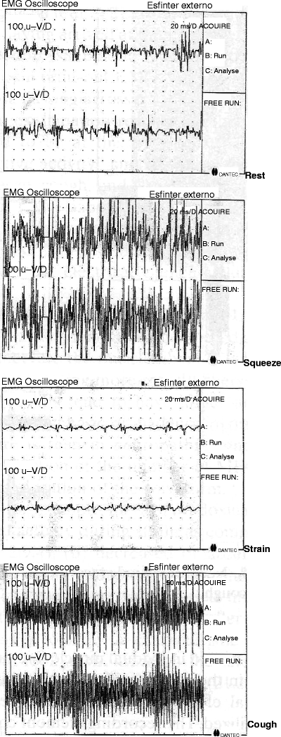
CNEMG of the EAS (Fig. 7.3) was the first technique used, and it evaluates spontaneous activity, recruitment patterns, and MUP waveforms. The concentric needle consists of a fine (0.7 mm) platinum wire mounted inside a metal cannula with a larger diameter, about 65 mm wide, so that the inner wire is fully insulated. Optimal recording occurs with a frequency range of 10 Hz–10 KHz and a sensitivity of 100–500 mV, with a sweep speed of 20 ms per record at rest and during squeeze, strain, and cough. The introduction of the CN electrode on to the left and right sides of the sphincter is accompanied by a reactive MUP discharge that is separable from an initial insertion discharge, an effect that disappears rapidly when the patient relaxes during the procedure. In those cases of denervation, normal MUP activity is replaced by fibrillation denervation potentials with separable EAS and puborectalis recordings. Each of these activities are separated during provocative maneuvers as the needle is both advanced and withdrawn. Abnormal waveforms will be evident with a reduction in the number of MFs or in denervation. During the reinnervation process that occurs from preserved axons, motor units tend to have larger amplitudes, have longer durations, and become polyphasic in character. In myopathic states, the motor units tend to have lower overall amplitudes and be low duration waveforms. The technique also can better define paradoxical puborectalis syndrome with increased activity during strain [41]. Overall decreased amplitudes, and activity is seen in postobstetric or traumatic EAS damage. In some cases, spontaneous fibrillation and myoclonia in underlying neurological disorders [42].


Fig. 7.3
Concentric needle electromyographic recording of the external anal sphincter (Reprinted with permission from Rosato and Lumi [46])
SFEMG was originally described by Stälberg and Trontelj [43] to record individual MF action potentials as a complement to CNEMG [43]. An electrode with a smaller needle and a surface of only 25 μm is used and is capable of detecting electrical signals over a recording surface of only 0.0003 mm2


Stay updated, free articles. Join our Telegram channel

Full access? Get Clinical Tree


