Fig. 19.1
Vaser selectivity: breast tissue after Vaser action. Fat cells have been emulsified and removed. All the vertical supporting collagen and elastic fibers have been spared, as it is clearly visible, with the attachment with underlying tissues
19.2 Lipoabdominoplasty
The main advantages of the lipoabdominoplasty are:
1.
Minimal undermining of the abdominal wall
2.
Superficial liposuction of the lower part of the abdominal wall down to the fascia lata fat
3.
Preservation of the vascularization coming from big perforators of the abdomen
Those three main innovations have decreased the complication rate in abdominoplasty such as bleeding and possibility of hematoma formation; necrosis of the skin and the subcutaneous tissue, due to diminishing vascularization from a section of the perforator vessels; and seromas.
In lipoabdominoplasty liposuction of the upper abdomen is carefully performed in a conservative way and affects mostly the intermediate and deep layers. This is understandable, as to diminish the risk of necrosis of the abdominal wall from a too aggressive liposuction. When lipoabdominoplasty reaches its limits, then Vaser lipoabdominoplasty starts.
19.3 Vaser Lipoabdominoplasty Technique
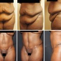
Preparation for Vaser lipoabdominoplasty is similar to the drawings and measurements of a standard abdominoplasty. Drawings of center line from the xiphoid to pubis and of the apron to be removed are marked in standing position of the patient (Fig. 19.2). The full area of tissue removal from the lower abdomen is marked, drawn, and double-checked with the patient lying down. Measurement of the length of the entire apron (upper and lower portion of skin resection) is checked to have two similar length flaps to join after resection and approximation.
Fig. 19.2
Preoperative markings
Lateral side of drawings is double-checked to prevent dog-ears, due to impairment of two different flaps (in length and thickness). The umbilicus is marked, and preoperative position is checked and outlined. An area of 5 cm width by 5 cm length is marked on top of the patient’s navel position. This represents the expected area of undermining of the upper flap of the abdomen, down to the rectus abdominis fascia. The drawings must be modified according to patient needs. Moreover, the upper abdomen and lower abdomen and lateral flanks are marked (Fig. 19.2, vertical red lines). These areas are treated with Vaser liposelection before the surgical resection of the planned skin of the lower abdomen.
The intended benefits of this surgical Vaser liposelection are:
1.
Completely remove the unwanted areas, by thinning the fatty component, and allow a better mobilization of the areas. Once the treated flaps are thinned, they are easily mobilized and moved down to reach the pubis.
2.
Sculpture the upper abdomen and flanks, in which the full shape of the body is increased, with a more natural shape and contouring.
Vaser lipoabdominoplasty starts with the emulsification with tumescent solution of the apron which is due to be removed (Fig. 19.3). Tumescent solution contains:
Fig. 19.3
Tumescent infiltration of the lower abdomen panniculus (apron)
1.
1000 mL of saline solution
2.
1 mL of adrenaline
3.
300 mg of lidocaine
Adrenaline helps diminish blood loss. Lidocaine enzymes prolong postoperative anesthesia of the treated areas. The full infiltration of the lower abdomen apron requires from 800 mL to 2000 mL, depending on the size of the apron to be resected. Infiltration starts in the superficial layers and then affects the deeper layers (Fig. 19.4).
Fig. 19.4
Infiltration starts in the superficial layer and then goes on the deep layer
The reason is that Vaser will start in the superficial layers first, which will require adequate fluids in the area. To help emulsification of fat with the effects of cavitation determined by ultrasound energy, infiltration is then extended to the full abdomen and flanks, depending on the areas treated. The full upper abdomen required 800–2000 mL of infiltration of the tumescent solution, depending on the size of the patient.
At this stage, Vaser is started (Fig. 19.5). A 3.7 mm, two-ring probe is normally chosen, with power set at 90 % of the total. The surgical layers are treated first since tumescence is a temporary state and the tissues tend to lose firmness as fluids go in the deeper layers by gravity (Fig. 19.6). The Vaser probe affects the deeper layers of the lower abdomen. Full time of action of Vaser in the entire lower abdomen varies from 7 to 12 min.
Fig. 19.5
Vaser 3.7 mm two rings to the superficial fat layer of the lower abdomen
Fig. 19.6
Vaser to the deeper layer of the lower abdomen
For the full abdomen and flanks, total Vaser time ranges 12–20 min.
Once the lower abdomen is fully treated, Vaser is extended to the upper abdomen, starting with the umbilicus region (Figs. 19.7 and 19.8). Vaser probes act in the superficial layers first to free and mobilize the upper abdominal flap. The probe works 1 cm deep to the skin layer, thus freeing adhesion of skin superficial layer with deeper connective lax tissues (Fig. 19.9). The free mobilization of the upper abdomen includes the free areas up to the xiphoid and flanks as well (Figs. 19.10 and 19.11).
Fig. 19.7
Vaser to the upper abdominal wall
Fig. 19.8
Vaser of the upper abdomen
Fig. 19.9
Vaser of the upper abdomen
Fig. 19.10
Vaser of the upper abdomen
Fig. 19.11
Vaser of the upper abdomen
Vaser liposelection is selective and affects only the adipocytes, sparing vessels and nerves, and collagen fibers, with elastic tissues. This property enhances the safety of the technique which can easily be combined with abdominoplasty. Vaser liposelection can be more extended, involving all the areas of the body at the same stage.
There is no fear of damage to the vascularization of the tissue, despite an extended treatment of the superficial layers of the subcutaneous tissue, as well as the deeper layers of fat.
1.
Superficial layers allow an easier mobilization of flaps (abdomen, flanks).
2.
Deep layers allow a defatting of the treated area.
19.3.1 Liposuction
Liposuction of the treated areas starts from the lower abdomen. The VentX cannulas, 3.7 mm diameter, are first used for the superficial layer (Fig. 19.12). Superficial fat aspiration is then started in the contralateral side (Fig. 19.13
 Abdominoplasty Combined with Cesarean Section: Discussion of the Evidence
Retrospective Analysis of Never Events in Panniculectomy and Abdominoplasty Patients and Their Financial Implications
Abdominoplasty Combined with Cesarean Section: Discussion of the Evidence
Retrospective Analysis of Never Events in Panniculectomy and Abdominoplasty Patients and Their Financial Implications
 No Drain Abdominoplasty with Progressive Tension Suture: The Logic, Simplicity, and Aesthetics
 Evolution of the High-Superior-Tension Lipoabdominoplasty: The High-Tension Abdominoplasty
 Tension Suture Technique Combined with Lidocaine-Adrenaline-Saline Infiltration Decreases Complications and Promotes Recovery in Abdominoplasty
 A Study of Postural Changes After Abdominal Rectus Plication Abdominoplasty


Related posts:
Abdominoplasty Combined with Cesarean Section: Discussion of the Evidence
Retrospective Analysis of Never Events in Panniculectomy and Abdominoplasty Patients and Their Financial Implications
No Drain Abdominoplasty with Progressive Tension Suture: The Logic, Simplicity, and Aesthetics
Evolution of the High-Superior-Tension Lipoabdominoplasty: The High-Tension Abdominoplasty
Tension Suture Technique Combined with Lidocaine-Adrenaline-Saline Infiltration Decreases Complications and Promotes Recovery in Abdominoplasty
A Study of Postural Changes After Abdominal Rectus Plication Abdominoplasty
Stay updated, free articles. Join our Telegram channel
Full access? Get Clinical Tree
