1. Physiological factors
Anatomic factors: integrity of musculoskeletal system; neural control; visual and kinesthetic awareness; fatigue; age
2. Psychological factors
Emotional or mental attitudes may influence the behavior of the muscle system and stances or movements adopted; demand of work place
3. Physical factors
Body physique; flexibility; strength
38.2 Anatomic Consideration in Posture Balance
Abdominoplasty is a surgical procedure of removing excess abdominal skin and fat and tightening of lax anterior abdominal wall muscle. A musculofascial plication is a major component of abdominoplasty in a patient with divarication of the rectus muscles. During plication the surgeon approximates toward the midline the junction of the lateral abdominal musculofascial complex and lateral rectus sheaths on both sides [4]. This approximation causes a tightening of the muscles of the lateral abdominal complex and increasing of intra-abdominal pressure. Therefore, the muscles are placed in a more efficient point to increase their force-generating capacity while the intra-abdominal pressure provides more stability and stiffness of the spine without concurrent activity of the abdominal and back extensor muscles. These two phenomena greatly increase the effectiveness of the lateral muscle complex as a spine stabilizer [5].
Abdominal rectus plication abdominoplasty causes great changes in body mass and significantly improves body image and self-esteem. By providing physical benefits, it improves spine stability and body posture. Indeed, redundant abdominal skin and weakness of the anterior abdominal wall are often associated with kyphosis which the patients try to hide because they consider it to be a source of embarrassment. The reduction of body masses placed on the anterior part of the body provides postural changes, leading to the retro positioning of the pelvis with compensation of the head and shoulders (Table 38.2).
Table 38.2
Key points
Proper evaluation of the postural changes after abdominoplasty includes the following key components |
Calculating BMI at time of presentation and assessing stability of weight |
Analyzing medical history especially those associated with obesity and bariatric surgery |
Analyzing the actual body posture and factor effecting posture. Diagnosing the body contour deformities |
Evaluation of psychosocial considerations and the motivation of patients. Analyzing his or her ideal self-image form |
Understanding his or her goals and expectations |
Formulating a complete treatment plan and possible complications of the surgery |
Few medical scientific data are available on the effect of abdominal rectus plication abdominoplasty on the human posture. A study by Mazzocchi et al. [3] suggests that abdominal rectus plication abdominoplasty makes changes in body mass and its distribution which significantly improve the body posture and stabilization of the spine. These psychological components are particularly evident in the first postoperative period, while in the late follow-up period, a complete balance of psychological and emotional components of postural control is obtained.
38.3 Body Image, Self-Esteem, and Quality of Life
Body image, more specifically body image dissatisfaction, plays a central factor in motivating a number of appearance-enhancing behaviors, including weight loss, cosmetic and fashion purchases, and aesthetic surgery [6]. In general, “body image” refers to an individual’s experiences of embodiment, especially self-perceptions and self-attitude toward one’s appearance. Body image is typically viewed as a multidimensional construction, consisting of perceptual, attitudinal, and behavioral components. The two most central body image dimensions are body image evaluation (satisfaction and dissatisfaction) and body image investment (psychological importance of one’s appearance to his or her sense of self or self-worth). Dissatisfaction with one’s body image can cause anxiety and reduce self-esteem as well as alter interpersonal relationship. Indeed, excess abdominal skin is thought to create a functional disability that adversely affects a person’s quality of life because of the disproportionate lower body weight. The discovery of a new body image eliminates dissatisfaction, reduces anxiety, and increases self-esteem, which provides psychological and physical benefits that improve the quality of life [6].
A number of studies have found that body image dissatisfaction is associated with more favorable attitudes toward cosmetic surgery [5]. Other investigations have suggested that cosmetic surgery patients typically report increased investment in and dissatisfaction with their body image before cosmetic surgery, as compared to a person not interested in aesthetic procedures [6]. Body image has long been discussed in the context of plastic surgery and first appeared in the work of Edgerton et al. in the 1950s and 1960s [7].
Body image dissatisfaction can also be a failure of several psychiatric disorders which include eating disorder, depression, gender identity disorder, and schizophrenia. The psychiatric disorder of perhaps greatest relevance to cosmetic surgery is body dysmorphic disorder (BDD). BDD is characterized as a preoccupation with slight or imagined defect in appearance that leads to significant disruption in daily functioning, and they can report low levels of satisfaction with cosmetic surgery [8].
As has been suggested by many plastic surgeons over the years, the greatest benefits of plastic surgery are psychological in nature. Abdominoplasty has a positive impact by reducing the level of concern and dissatisfaction with body image. A new body image discovered by the patient eliminates dissatisfaction, reduces anxiety, and increases self-esteem, which provides psychological and physical benefits that improve the quality of life. In some cases we can see a significantly improved self-esteem and mental health even when final aesthetic outcomes are not yet visible, which highlights the psychological impact of aesthetic plastic surgery [9].
38.4 Anatomic Consideration in Abdominoplasty
The knowledge of the anatomy of the abdominal wall is key to achieving optimal result in abdominoplasty. The surgeon must know the anatomic landmarks of the abdominal wall, muscle and fascia anatomy, zone of adhesions, innervation and nerve territory, blood supply of the skin flap, adipose tissue, and umbilical zone as well as lymphatic supply.
In the anterior abdominal wall from inferior costal margin to the pubic symphysis, the rectus sheath around the rectus abdominis muscle forms an anchor for the attachment of the lateral abdominal muscle complex (the external oblique, internal oblique, transverse abdominal muscles, quadratus abdominis). In the lumbar spine, the tendon of origin of the lateral abdominal muscle complex fuses with the middle and posterior layers of the thoracolumbar fascia. The middle and posterior layers of the thoracolumbar fascia then attach to the transverse processes and spinous processes of the lumbar vertebrae. According to this the rectus abdominis muscle and its sheath form a structure that biomechanically influences the mechanics and stability of the lumbar spine, particularly on the lumbar lordosis angle and sacral inclination angle [2, 10].
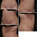
The patterns of the fat distribution in the body vary with sex, race, heredity, and age group. The subcutaneous tissue according to histologic distinctions is composed of two different layers, the superficial and the deep. The superficial adipose layer is made up of compact, dense pockets within well-organized fibrous septa, while the deep adipose layer consists of looser, more areolar fat bound by a haphazard network of partitions [11]. This classification is of less clinically significant. The concept of “surgical fat layers” divides the subcutaneous fat into three layers according to depth. Each layer represents a zone of relative safety or caution during suction lipectomy. The deep and intermediate layers are always safe to treat, while the superficial layer of densely compacted fat should be suctioned with great caution because of the great risk of deformities and skin irregularities afterwards (Figs. 38.1 and 38.2).
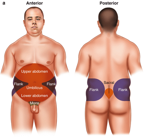
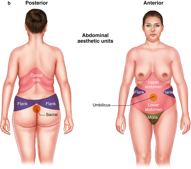
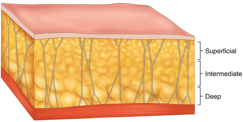
Fig. 38.2
Fat distribution in male (a) and female (b) patients (Reprinted with permission from Matarasso [43])
The total number of fat cells is unique to the individual and is genetically determined. It is slightly influenced by environmental and nutritional factors at an early age and remains stable in late life. Each body habitus depends on enlargement of the genetic number of adipocytes in both gender and fat-cell replication by post-adipocytes. Obese patients have larger adipose cells than do nonobese patients. Cell size also varies with location in the body (subcutaneous or deep layers) and the gender group. According to the distribution on the body regions, the adipose deformities are concentrated primarily above the umbilicus, below the umbilicus, or at the waistline. Elderly women usually present with a lower abdominal apron of skin and fat, and males have generalized obesity with increased intra-abdominal fat [12].
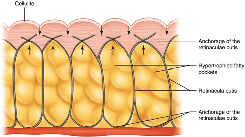
Cellulite is not a pathologic condition. It is the result of hypertrophy of the superficial fat cells and edema (fluid retention) within the subcutaneous tissue. Lockwood [13] describes two types of cellulite formation: a primary hypertrophied superficial fat cell and a secondary cellulite related to skin laxity. Although cellulite can affect both men and women, it is rarely found in men. A number of factors cause cellulite formation. The most prominent factor is estrogen and progesterone hormones which stimulate lipogenesis and inhibit lipolysis. There is also a difference between men and women in the way the connective tissue expands the adipocytes into the upper reticular dermis close to the epidermis. Additional contributing factors are water retention and poor circulation. According to that, conservative treatment includes lymphatic drainage, increasing microvascular circulation, reducing size and volume of the fat cell (decreasing tension on surrounding connective tissue), and promoting lipolysis (Fig. 38.3).
38.5 Abdominal Wall Deformity Classification
The abdominal wall may be affected by pregnancies, weight variation, and previous abdominal surgical scars. Some patients may present a weakening of the musculoaponeurotic layers attributable to aging or to a congenital condition. These factors alter the body contour of the abdominal wall and its function [14]. Surgical correction on the abdominal wall should restore function and body posture, providing a balance between anterior and posterior abdominal complex muscles and improving the cosmetic aspect.
In clinical situation with acquired deformity of the abdominal wall, the surgeon must first reconstruct these deformities which may include or not cosmetic correction of the abdominal wall.
Deformities of the musculoaponeurotic layers can be congenital and genetic or acquired. Collagen type I and III are in the composition of abdominal aponeuroses. A genetic condition is when aponeuroses with a high concentration of collagen type III in fibers are weaker than those with a higher rate of type I fibers [15]. Another factor that may play a role in correction of the musculoaponeurotic layer is the collagen type I, III, IV, and V deposition in muscles. With aging there is an increase in the total number of fibers and the muscles become less flexible and pliable [16]. Also, it is important to stress that there is a relationship between the musculoaponeurotic deformity and the skin excess of the abdomen. The quality of the extracellular matrix of the skin, aponeurosis, and muscles has similarities in the same individual. More complex skin deformity is related to more complex musculoaponeurotic deformities (Fig. 38.4).
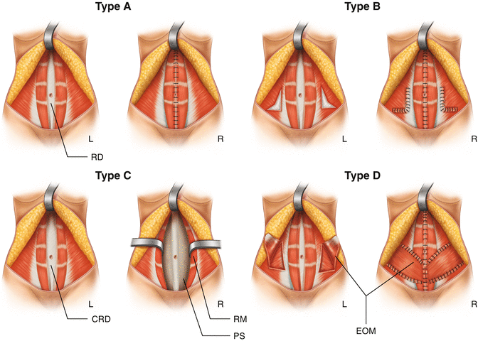
Fig. 38.4
Types of abdominal deformity and their correction. Type A, rectus diastasis secondary to pregnancy; correction is by plication of the anterior rectus sheath. Type B, rectus diastasis with laxity of the musculoaponeurotic layer; correction by plication of the anterior rectus sheath and external oblique aponeurosis. Type C, congenital lateral insertion of the rectus muscles at the costal margins and probable hernias. Correction by undermining the posterior rectus sheath, invagination of the linea alba, and anchoring the anterior rectus sheath to the midline. Type D, rectus diastasis and poor waistline definition. Correction is accomplished by anterior rectus sheath plication and medial advancement of the external oblique muscles (Reprinted with permission Nahas [22])
A common acquired deformity of the musculoaponeurotic layers is the diastasis of the rectus abdominis as a postpartum occurrence. During pregnancy the linea alba is particularly affected by the expansion of the abdomen. The width of the linea alba is known as the inter-recti distance and normally varies along its length from the xiphoid to the pubis symphysis. Beer et al. [17] measured the width of the linea alba with ultrasound imaging in nulliparous women aged between 20 and 46 years and found the mean width to be highly variable, reporting 7 mm ± 5 at the xiphoid, 13 mm ± 7.3 cm above umbilicus, and 8 mm ± 6.2 cm below the umbilicus.
Obesity is a worldwide health problem. The health consequences of obesity are manifested in an increased incidence of diabetes, hypertension, osteoarthritis, obstructive sleep apnea, deep venous thrombosis (DVT), and postoperative complications. Abdominal rectus plication abdominoplasty as a part of the post-bariatric body-contouring surgery causes a great change in body mass and significantly improves body image and self-esteem. In some time it is associated with high rates of patient satisfaction, although postoperative complications continue to have negative impact on the initial satisfaction. The changes in those parameters provide psychological and physical benefit that generally improves spine stability and body posture. Indeed, redundant abdominal skin and weakness of the anterior abdominal wall are often associated with kyphosis which the patients try to hide what they consider to be a source of new embarrassment. Posture subsequently reached a state of equilibrium when the emotional aspect and psychological component of posture control are stabilized [18, 19].
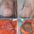
Another common acquired deformity is postoperative incisional hernia. The gradual enlargement of the hernia over time results in loss of abdominal wall strength with effects on postural maintenance, respiration, micturition, defecation, and biomechanical properties with an impact on the patient’s physical capacity and quality of life. In novel days many variations of ventral hernia repair exist with autologous tissue or prosthetic material (Fig. 38.5) [20].
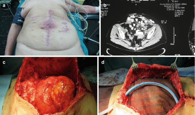
Fig. 38.5
(a) Ventral hernia. (b) MRI (magnetic resonance imaging) scan of the abdominal wall. (c) Intraoperative hernia. (d) Hernia repaired
38.5.1 Abdominal Wall Deformity Classification Based on Skin Type Excess Evaluation (Table 38.3) [21]
Table 38.3
Correlation between the classification that considers excess skin and the one that evaluates musculoaponeurotic laxity
Skin deformity | n (%) | Musculoaponeurotic deformity | n (%) |
|---|---|---|---|
Type I | 3(3.6) | Type A | 3 (3.6) |
Type II | 24 (28.9) | Type A | 15 (18.1) |
Type B | 6 (7.2) | ||
Type C | 3 (3.6) | ||
Type III | 56 (67.5) | Type A | 46 (55.5) |
Type B | 4 (4.8) | ||
Type C | 4 (4.8) | ||
Type D | 2 (2.4) | ||
Total | 83 (100) | 83 (100) |
1.
Type I patients present mild excess skin with a high-positioned umbilicus
2.
Type II patients have mild excess skin and a well-positioned umbilicus
3.
Type III patients present severe excess skin
38.5.2 Abdominal Wall Deformity Classification Based on Musculoaponeurotic Deformities
Divides patients into four groups (Table 38.3) [22]:
1.
Type A: Patients present fusiform rectus diastasis secondary to pregnancy. Both rectus muscles are inserted on the costal margin. The method of correction is plication of anterior rectus sheath.
2.
Type B: Patients present a bulging of the musculoaponeurotic layer after the correction of diastasis and have vertical elongation of the abdominal wall. To correct these deformities, an “L”-shaped plication of the external oblique aponeurosis should be done in addition to the plication of the anterior rectus sheath.
3.
Type C: Patients present congenital rectus diastasis, on which the insertion of the rectus muscle is lateral in the costal margin. To correct this deformity, an advancement of the rectus muscles is performed.
4.
Type D: Patients present poorly defined waistline and a strong aponeurosis of the external oblique muscle which will be advanced medially after the anterior rectus sheath plication.
38.6 The Body-Contouring Operations
These have been classified into two categories:
1.
Aesthetic body contouring
2.
Body contouring after massive weight loss
Evaluation and classification of the anatomic characteristics precede decision making and the selection of the appropriate operation. The excess skin, striae, fat, musculofascial laxity, and diastases of the rectus muscles with the assessment of the adjacent areas including the flanks, thighs, and back are the major components to be assessed in both operation groups (abdominoplasty and other body-contouring procedures).
Matarasso [23] classifies abdominal deformities based on severity of the skin, fat, and muscular flaccidity and selects the most appropriate surgical technique for abdominal contouring (Table 38.4).
Table 38.4
Abdominal deformity and recommended procedure for correction
Category | Skin | Fat | Musculofascial layer | Treatment |
|---|---|---|---|---|
Type I | Minimal laxity | Variable | Minimal flaccidity | Suction-assisted lipectomy |
Type II | Mild laxity | Variable | Mild lower abdominal flaccidity | Mini abdominoplasty |
Type III | Moderate laxity | Variable | Moderate lower and/or upper abdominal flaccidity | Modified abdominoplasty |
Type IV | Severe laxity | Variable | Significant lower and/or upper abdominal flaccidity | Standard abdominoplasty with suction lipectomy |
Rohrich et al. [24] use a modified Matarasso classification that is based on clinical assessment of the degree of skin redundancy, amount of abdominal flay, skin thickness and tone, and status of the abdominal musculature (Table 38.5).
Table 38.5
Abdominal deformity and recommended procedure for correction
Class | Skin/fat excess | Diastasis | Treatment |
|---|---|---|---|
Ia | No | No | UAL or SAL alone |
Ib | No | Yes | UAL or SAL alone ± endo |
IIa | Mild | No | Diastasis repair |
IIb | Mild | Yes | UAL or SAL alone |
IIIa | Moderate | No | UAL or SAL alone ± endo |
IIIb | Moderate | Yes | Diastasis repair UAL |
IVa | Significant | No | Infraumbilical mini abdominoplasty and diastasis repair + UAL epigastrium |
IVb | Significant | Yes | Traditional abdominoplasty + UAL of the flank |
Traditional abdominoplasty + diastasis repair + UAL of the flank |
Reprinted with permission from Rohrich et al. [24]
SAL suction-assisted lipoplasty, UAL
 Abdominoplasty Combined with Cesarean Section: Discussion of the Evidence
Retrospective Analysis of Never Events in Panniculectomy and Abdominoplasty Patients and Their Financial Implications
Abdominoplasty Combined with Cesarean Section: Discussion of the Evidence
Retrospective Analysis of Never Events in Panniculectomy and Abdominoplasty Patients and Their Financial Implications
 No Drain Abdominoplasty with Progressive Tension Suture: The Logic, Simplicity, and Aesthetics
 Evolution of the High-Superior-Tension Lipoabdominoplasty: The High-Tension Abdominoplasty
 Tension Suture Technique Combined with Lidocaine-Adrenaline-Saline Infiltration Decreases Complications and Promotes Recovery in Abdominoplasty
 Abdominal Wall Repair Post Hernia in Kidney and Liver Transplantation


Related posts:
Abdominoplasty Combined with Cesarean Section: Discussion of the Evidence
Retrospective Analysis of Never Events in Panniculectomy and Abdominoplasty Patients and Their Financial Implications
No Drain Abdominoplasty with Progressive Tension Suture: The Logic, Simplicity, and Aesthetics
Evolution of the High-Superior-Tension Lipoabdominoplasty: The High-Tension Abdominoplasty
Tension Suture Technique Combined with Lidocaine-Adrenaline-Saline Infiltration Decreases Complications and Promotes Recovery in Abdominoplasty
Abdominal Wall Repair Post Hernia in Kidney and Liver Transplantation
Stay updated, free articles. Join our Telegram channel
Full access? Get Clinical Tree
