94
Vascular Neoplasms and Reactive Proliferations
Lesions of vascular origin are broadly, and somewhat imperfectly, classified as neoplasms (tumors), malformations, telangiectasias, or reactive proliferations. The growth of a neoplasm is largely autonomous (i.e. not reactive). Malformations, in general, are not actively proliferating (see Chapter 85). Telangiectasias represent pre-existing capillaries that are persistently dilated but lack a proliferative component (see Chapter 87). Reactive proliferations represent endothelial cell proliferation that is in response to some factor (e.g. fibrin, hypoxia, trauma).
Neoplasms/Tumors
Cherry Angioma
• Bright red, 1- to 6-mm papule, commonly on the trunk or upper extremities (Fig. 94.1); appears during adulthood; early, tiny lesions can be macular.
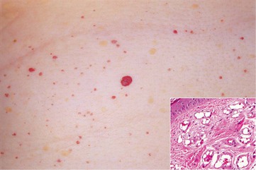
Fig. 94.1 Cherry angiomas. Multiple, slightly compressible red papules. Histologically, congested capillaries and postcapillary venules expand the papillary dermis (insert). Courtesy, Jean L. Bolognia, MD.
• Dermoscopy: red to red-blue lacunas (well-demarcated round to oval red to red-blue structures).
Glomus Tumor
Tufted Angioma
• Pink to dark red patches and plaques with superimposed papules (Fig. 94.3) that slowly enlarge and occasionally regress.
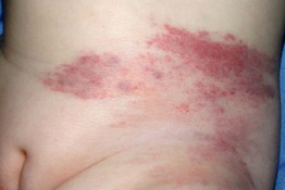
Fig. 94.3 Tufted angioma. Mottled red patches and superimposed papules are typical clinical features. Courtesy, Julie V. Schaffer, MD.
• Commonly found on the neck and trunk.
• Congenital or acquired during childhood or young adulthood.
• On a spectrum with kaposiform hemangioendothelioma.
Hobnail Hemangioma/Targetoid Hemosiderotic Hemangioma
• Uncommon red-purple papule on the trunk or extremities of children or young adults; papule may have a surrounding pale area and outer ecchymotic ring, thus creating a target lesion (Fig. 94.4).
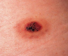
Fig. 94.4 Hobnail hemangioma (targetoid hemosiderotic hemangioma). Note the target-like appearance. Courtesy, Ronald P. Rapini, MD.

