(1)
Department of Medicine, College of Medicine and Health Sciences, Al Ain, United Arab Emirates
The upper third of the face is made up of the forehead , glabella, where one may argue are part of the forehead and the temples . Given the rule of thirds, the distance between the upper third should equal that of the middle and third. The area transcends the hairline superiorly to the root of the nose inferiorly. Changes that occur with time cause the upper third to appear more concave with the thinner skin leading to textural changes as well as the formation of lines that run perpendicular to the muscles underneath the skin.
The upper third ’s major function is to display emotions. Anger, happiness, and fear can easily be conveyed through seeing just the forehead of that person without the need to see the rest of the face. It is highly dynamic owing to the muscles that originate and insert themselves in the same area as compared to middle thirds where muscles, though originating from that location, insert themselves in structures in the lower face, another highly dynamic part.
Treatment of the upper third is essential when taking care of a person requesting volume rejuvenation. When combined with botulinum toxin , the result is highly satisfactory for both the patients and the injectors. The upper third , however, is recipient to the least amount of filler injections when all three domains are taken into consideration.
Forehead
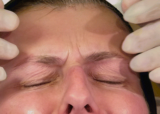
Augmentation of the forehead , albeit uncommonly performed is something worth looking into and understanding. It is not a region that is injected by the novice, as understanding the anatomy is key as well as realizing the right indication. Perhaps the most important part of the forehead that may be treated is the glabella . Although the part is fairly and readily injected with botulinum toxin , there are times that filler should be used to help remove etched lines caused by many years of frowning or non-treatment. In addition, fat compartments in the forehead do exist and those can atrophy with aging.
Patients do not generally present asking for augmentation of the forehead . Most will come to discuss the glabellar lines and the horizontal creases. The physician should inform patients especially those in their mid to late 40s that their forehead may benefit from fillers and the expected results will be softening of the lines as well as decreasing the concavity of the forehead head that occurs with time due to the loss of the fat compartments . The process also gives a youthful look and shine when performed, as the skin is brought back to its taut, original self. This is a holistic approach each and should be discussed during the consultation process. As a rule, patients will not come asking that their forehead be injected.
When treating the glabella , the injector should be aware of the major vasculature, pertinent to the area, most importantly, the supratrochlear artery . Damage to this artery can lead to neurovascular compromise affecting the forehead , with backwash embolism into the vessel, potentially also leading to the blindness . The vessel lies medial to the supraorbital foramen . Cadaveric studies show that the vessel lies in the general area of the vertical creases caused by contraction of the corrugator supercilli muscle. This is exactly the same area that is injected with fillers when treating the glabellar lines, meaning the injector is injecting right over the vessel. It is important to understand that though this may be the case, the supratrochlear artery originates deep from the bone, at roughly 1 o’clock in the right eye and 11 o’clock in the left. The vessel continues to lie below the corrugators till about 2 cm from of the superior orbital rim , where it then penetrates the frontalis and lies more superficial.
The other major vessel in the area is the supraorbital artery . This vessel lies more lateral to the suptratrochlear and can be readily palpated by feeling for the supraorbital foramen on the superior orbital rim . Damage to this vessel is uncommon as augmentation is not something that is readily performed in that area. Advanced injectors may use fillers and the best matter to avoid the penetrating the vessel and leading to a potential complication is to use a blunt tipped cannula, but the area is generally avoided. Damage to both vessels may occur to their branches higher up on the forehead when both vessels penetrate the frontalis muscle and are more superficial.
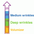
Other matters that need to be taken into consideration when injecting the area of the forehead are knowing the type of filler injected and knowing the level these fillers are injected. When injecting the forehead fat compartments and taking into consideration the stiffness and G′, fillers of low to medium G′ should be considered. This allows the fillers to be fairly distributed in the area without leaving any major lumps in the area. That being said, it is not recommended that the horizontal line be injected. Those lines tend to become less apparent with subsequent injections and not through injections of the lines themselves as the vessels are in the superficial plane in that area.
Procedure
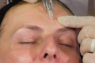
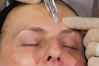
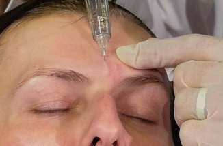
Glabella :
- 1.
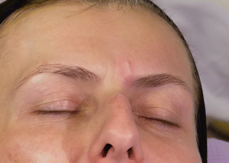
Identify the area to be injected by asking the patient to frown. If the patient is unable to frown, you may push the skin medially to elicit the movement and be able to visualize the lines.
- 2.
Discuss with the patient the need of neuromodulation. This will help in decreasing the line initially in addition to increasing the longevity of the filler itself.
- 3.
If the patient has had the area treated with a neuromodulator , assess the glabellar lines if they are actually apparent.
- 4.
Mark the areas to be treated.
- 5.
It is recommended that injection should be performed using a hyaluronic acid (HA) filler. HA fillers can be readily dissolved if a neurovascular complication occurs immediately or later.
- 6.
Needles (recommended to be 30G or smaller) are preferred over than cannulas.
- 7.
Injection should be performed superficially. This is done by introducing the needle parallel to the skin at about 10–15°. The injector is able to visualize the silhouette of the needle.
- 8.
It is recommended that the injections be performed away from the orbital rim towards the hairline as that also help ensure that the filler, if inadvertently injected into the blood vessel moves away from the eye and thus decreasing the chance of blindness .
- 9.
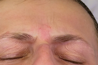
When pushing the filler in, the injector is able to see blanching of the area injected. This is not caused by the embolism of the major vessels, but due to the pressure of the filler on the tissue, mainly the capillaries of the dermis.
- 10.
Retrograde or anterograde injection may be performed.
- 11.
Aspiration may be performed, though unnecessary, as a lack of blood in the hub does not necessarily mean that the needle is in the vessel lumen.
- 12.
Massage the area after treatment.
Forehead Horizontal Lines:
- 1.
It is not recommended that these lines be treated as the vessels lie in the superficial plane.Related posts:

Stay updated, free articles. Join our Telegram channel

Full access? Get Clinical Tree