(1)
Department of Medicine, College of Medicine and Health Sciences, Al Ain, United Arab Emirates
If there ever was one area on the face that will give you the most when it comes to facial augmentation with fillers, it would have to be the middle third of the face. Fillers when injected in that area help showcase the most commonly visualized part, the eyes, and subsequently the area under them. Thus, it is important to understand the anatomy in addition to the safe zones where the material may be injected.
Areas of convexities that need to be enhanced during the process of soft tissue augmentation
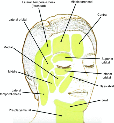
The middle third is made up of the peri-ocular area, the cheeks and the nose . Each of those areas is injected differently than the other with different fillers needed depending on the indication. The middle third of the face is also different in men and women. In men, due to the effect of testosterone, the mid cheek is slightly more depressed and volume enhancement happens in that area with lateral extension onto the zygomatic arch generally avoided. As a matter of fact, injecting that area in men can lead to feminizing features. By injecting the mid cheeks , the tear troughs may not necessarily require any enhancement, as most hollowness that are perceived as tear trough deformities are caused by atrophy of the mid cheeks . Augmentation of the tear troughs in men may lead to an increase in the convexity of the mid cheek , which is also a feminine feature. In women, there are three very important areas that are usually injected to enhance the middle third . These are the mid cheeks , the lateral cheeks (area of the zygomatic arch ), and the tear troughs with priority dependent on the individual indication.
Deep fat compartments of the face
Anatomically the layers of the middle third are comprised of the skin, fat compartments , both superficial and deep, muscles and bones of the skeleton. With aging many changes occur in those areas. Both superficial and deep fat compartments tend to undergo volumetric changes with a decrease in the size of those compartments. That being said, not all compartments undergo the same volume loss with time. The nasolabial fold (NLF) fat compartment , for example, does not decrease with time when compared to its neighboring compartments and may show a relative increase. This is important to realize as this negates the old age adage of injecting the NLFs. As a matter of fact, this proves that injecting the NLFs will cause a much more unnatural result.
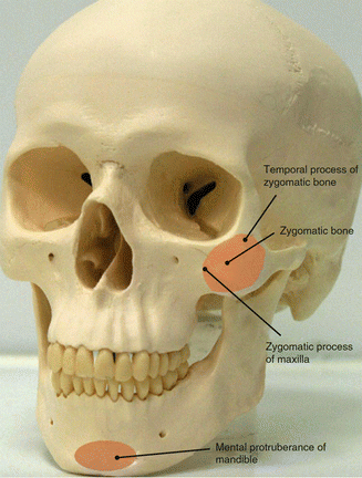
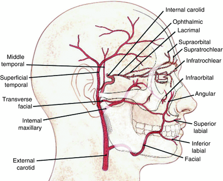
Blood circulation in the area is restricted to the middle part of the middle third and compromises of branches of the facial artery , mainly the angular which tends to branch off to feed the nose and the transverse facial artery . In addition the infraorbital artery also comes off of the infraorbital foramen . Though difficult to palpate, the foramen is found slightly medial the supraorbital foramen and opens at a downward angle from the maxilla about 1 cm from inferior orbital rim . This is important as injection in that area can be perpendicular at a 90° when placing a bolus, but an angled injection introduced from the lower part of the cheek upwards should be avoided as to not lacerate the artery. Thus, when it comes to filler injection and this artery being the most prominent in the mid cheek , it is important to inject from the lateral end to the medial with the injection point being on the zygoma and moving medially to the maxilla. This will insure that any potential damage to the vessel is avoided.
The angular artery passes by the pyriform fossa, a depression lateral to the nose . While this vessel is deep at first, it starts moving further superficial and in that area it is relatively found lying just in the subdermal plane. This is important to know because though the NLFs may be injected with fillers in the dermal/subdermal plane, once the superior part that is lateral to the nose is augmented, injection should be placed deeply onto bone to avoid any potential complication, as neurovascular compromise into the area will lead to potential necrosis across the side of the nose and above it. This may be avoided with the use of cannulas.
The vessels feeding the nose are branches of the angular artery and anastomose with those arising from the infraorbital and supratrochlear arteries. This makes this location the most sensitive in terms of potentially causing adverse events. The nasal arteries lie on the lateral walls and anastomose on the bridge of the nose and dorsum. This clinically is translated into injections needing to be made strictly on the midline and in the deeper plane. It is important to know that even when injecting with a cannula, injections should be made along that midline as much as possible to avoid possible damage to the vessels.
Anatomy of the blood circulation of the face
Cheeks
Injection of the cheeks has become the gold standard of soft tissue augmentation procedures. When performed, the cheeks , tear troughs , and also the nasolabial folds are corrected. Thus, it is a procedure that is not only desirable, but required when assessing and treating any patient with fillers. There are differences in the cheeks and bony prominences of both men and women and differences when it comes to injecting fillers do exist between the two sexes. In addition, there are also ethnic differences. The Asian skull has slightly more prominent zygomatic arches when compared to the Caucasian skull. This leads to an apparent flattening of the mid face in the former. Thus, injection of fillers is usually performed in the mid cheek in the Asian population and the zygomatic arch is not augmented to avoid further lateral projection. In the Caucasian population, both areas are readily injected. Injection of the mid cheek corrects the tear trough allowing for less filler to be used. Thus, it is important to inject the cheeks first and then inject the tear trough after reassessment. The zygomatic arch may also be injected to increase lateral projection. The arch should be slightly fuller than their area underneath it. This is observed as a shadow effect that gives the cheek a three-dimensional look. Makeup artists use blushers to enhance that effect and this can be performed with filler material to give a much more youthful appearance.
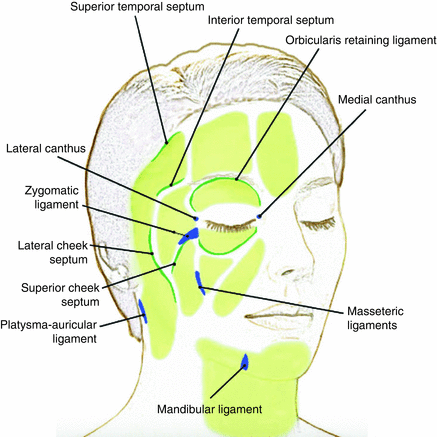
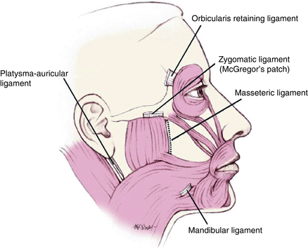
Retaining ligaments and septae
When injecting the cheeks , it is important to be aware of the correct level of placement of the filler. Thicker fillers may be injected in the deep fat compartment and that may be performed by injecting the filler using a cannula in a linear threading method or bolus. Needles may be used to do the same, though they are safer when injecting a bolus into the correct compartment. A filler once placed in the deep compartment will not migrate. In the midface, the suborbicularis oculi fat and deep cheek fat represent deeper fat compartments that provide volume and shape of the face. The deep fat compartments are divided by septae or an extensive network of retaining ligaments . It is these that prevent the filler from moving from one compartment to the other very similar to a partition in a room. Unlike other retaining ligaments , the zygomatic retaining ligament is a true ligament that connects the inferior border of the zygomatic arch to the dermis and is found just posterior to the origin of the zygomaticus minor muscle. This is clinically seen as a groove in the cheeks of some people. Augmentation of that groove may prove challenging for two reasons. The first is that injecting into it will not make it disappear given that injectors are injecting an area where the zygomatic ligament runs through. The second challenge is avoiding the facial vein , which also lies in that groove. This can cause for a significant hematoma if it bleeds. Another true ligament is the lateral orbital thickening that appears on the superolateral orbital rim and meets the orbital retaining ligament , which surrounds the orbit in a circular fashion. The ligament also acts as the superior border of the suborbicularis oculi fat compartment. Augmentation of that area underneath it helps decrease the tear trough deformity.
The superficial fat compartments , although separated by septae, allow for communication of the filler from one point to the other and it is important to realize that only thinner or more dynamic fillers, those that become readily integrated into the soft tissue, should be placed in that area as this plane is notorious in causing bumps.
With aging, the retaining ligaments under the eye become weaker. In addition to volume loss in the superficial and deep fat compartments , this results in visible folds and grooves in the cheeks and under the eye.
Retaining ligaments and septae1
Procedure:
- 1.
Identify the area to be injected (in most circumstances, it is the mid cheek ).
- 2.
There are many points that may be safely identified when it comes to injecting the cheeks and all depend on the region of augmentation. The mid cheek compartment is where the bulk of augmentation will happen and this may be accessed laterally (least chance of an adverse event occurring), inferiorly (risk of penetrating through the infraorbital foramen and vessels), or at a 90° angle with a needle.
Related posts:

Stay updated, free articles. Join our Telegram channel

Full access? Get Clinical Tree