34. Upper Blepharoplasty
Ashkan Ghavami, Foad Nahai
RELEVANT ANATOMY1–3 (Fig. 34-1)
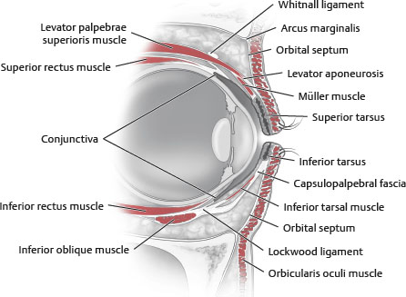
Fig. 34-1 Cross section of the upper and lower eyelids.
UPPER EYELID LAYERS
■ Anterior lamella: Skin, subcutaneous tissue (retroorbicularis oculi fat [ROOF] and suborbicularis oculi fat [SOOF]), and orbicularis oculi muscle (OOM)
■ Middle lamella: Orbital septum
NOTE: Problems in this layer most commonly lead to the continuum of cicatricial contraction (more common in the lower lid).
■ Posterior lamella: Tarsus and conjunctiva
ORBICULARIS OCULI MUSCLE
■ Innervation (Fig. 34-2)
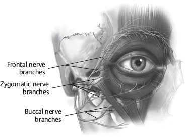
Fig. 34-2 Medial and lateral innervation points warrant consideration of orbicularis oculi muscle nerve preservation (particularly the pretarsal portion) to avoid postoperative adverse effects.
• Frontal, zygomatic, and buccal contributions from facial nerve (CN VII)
• Medial and lateral innervation points
■ Three portions (Fig. 34-3)
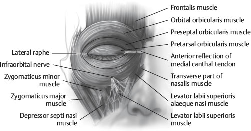
Fig. 34-3 Muscular anatomy of the periorbital region.
TIP: Considering the OOM as a sphincter muscle with three segments facilitates understanding the consequences of eyelid surgery, botulinum toxin administration, ligament release, and preservation of muscle innervation.
• Orbital
► Outermost portion
♦ Superficial to corrugator supercilii muscle (CSM) and procerus muscles
– Interdigitates laterally and medially with CSM under dermis and with frontalis muscle fibers
► Voluntary action
► Functions as tight closure of eye
• Preseptal
► Directly overlies septum
► Voluntary and involuntary components
► Assists with blinking mechanism
• Pretarsal
► Tightly adherent to tarsal plate
► Involuntary
► Responsible for blink mechanism
► Innervation from zygomatic branch of facial nerve
► Most involved in proper tear movement
TIP: The preseptal orbicularis is adherent to the septum, and careful dissection in the proper submuscular plane is required. No pinkish hue or transverse fibers should be left on the septum, which would indicate retained orbicularis fibers and an improper plane. At least a 6 mm strip of pretarsal orbicularis must be preserved for proper eyelid “sphincter” function.
TARSOLIGAMENTOUS COMPLEX
■ Upper tarsus
• 7-11 mm wide
• Müller muscle inserts onto superior border of tarsal plate.
• Anterior levator aponeuorosis fibers insert onto superior tarsal border.
■ Fascial insertions on the upper border of the tarsus1-4:
• Help to form shape, position, and magnitude of upper eyelid crease
• Levator aponeurosis
• Orbital septum
■ Orbicularis fascia: Firmly attached at posterior surface of orbicularis “sphincter”; fuses with levator aponeurosis at level of lid fold; offers mechanical and nutritional (possibly lymphatic) support4
• Fuses with orbicularis retaining ligament (ORL)5–9
■ Conjoined fascia: Present between the eyelid fold and lash line (deep to orbicularis and superficial to tarsal plate). This is an extension or fusion of the orbicularis fascia with the levator aponeurosis at a variable location superior to the tarsus.
■ Lateral raphe
• Lateral extension of the OOM along the lateral orbital rim and zygomatic complex
• Deep and superficial components of orbicularis insertion form lateral canthal tendon and lateral raphe.
• Contributes to “lateral orbital thickening” against the lateral orbital rim, where the ORL fuses5–7
• Acts as lateral anchor (fulcrum) for eyelids
■ Lateral canthal tendon (anterior and posterior limbs) (Fig. 34-4)
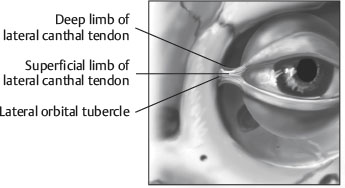
Fig. 34-4 The lateral canthal tendon has posterior and anterior limbs.
• Formed by:
► Lateral horn of levator palpebrae superioris
► Lockwood ligament
► Check ligament of the lateral rectus muscle
► Deep preseptal and pretarsal orbicularis muscle
MEDIAL CANTHAL TENDON
■ Tripartite structure (anterior horizontal, posterior horizontal, and vertical components)
■ Formed by:
• Deep head of pretarsal orbicularis
• Medial Lockwood ligament
• Check ligament of medial rectus muscle
• Whitnall ligament
ORBICULARIS RETAINING LIGAMENT6–9 (Fig. 34-5)
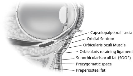
Fig. 34-5 Orbicularis retaining ligament (ORL). Orbicularis oculi muscle (OOM).
■ Also known as orbitomalar ligament8 or malar septum9 in lower periorbita
■ “Near-circumferential” retaining structure encircling upper and lower orbit7
■ Lax and longer laterally; more taut (short) medially
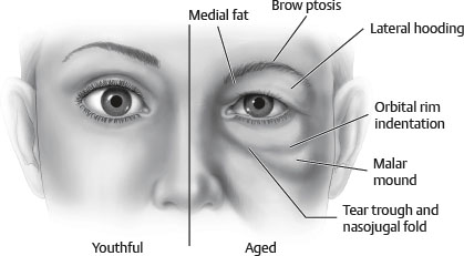
• Lateral laxity may partially explain lateral hooding.
• Medial tightness may be the reason for lack of medial hooding and tear trough phenomenon (medial depression/line at inferomedial periorbita).
■ Extends from OOM to the periosteum
■ True retaining ligament
■ May have lymphatic properties
■ Protects ocular contents: Semipermeable membrane
TIP: Blunt or sharp transection/disruption of ORL helps to smooth the tear trough and blend the lid-cheek junction. Release in the upper periorbita is required for effective browlifting. Medial preservation in the corrugator region may minimize medial brow splaying.
PRESEPTAL FAT
■ Between orbital septum and orbicularis muscle
■ Can contribute to upper eyelid lateral hooding
■ Upper lid: ROOF
■ Lower lid: SOOF
ORBITAL SEPTUM
■ Protective function
■ Extension of orbital periosteum
• Fuses with periosteum to form the arcus marginalis in upper and lower periorbita
■ Upper septum: Extends from superior orbital rim to insertion on levator aponeurosis at varying levels (10-15 mm above superior tarsal border)
■ Lower septum: Extends from inferior orbital rim to the capsulopalpebral fascia (5 mm below lower tarsal border)
■ Can have attachments with the ORL
LEVATOR PALPEBRAE MUSCLE
■ Muscle origin: Lesser wing of sphenoid
■ Insertion: Superior edge of tarsus (conjoined fascia)
■ Innervation: CN III
■ Action: 10-15 mm upper lid excursion and sustained lid elevation from contractile tone
NOTE: The amount of excursion and function is helpful in selecting an eyelid ptosis procedure.
■ Whitnall ligament: Fascial condensation 14-20 mm from superior edge of tarsus. translates posterior vector of pull into a superior direction
TIP: The main cause of postoperative upper eyelid ptosis is “unrecognized” preoperative ptosis. Examination for upper eyelid ptosis preoperatively is essential.
MÜLLER MUSCLE
■ Posterior lamella of levator palpebrae muscle
■ Origin: Levator muscle
■ Insertion: Superior edge of tarsus
■ Innervation: Sympathetic system
■ Action: 2-3 mm of upper lid lift
TIP: If inadvertent lid ptosis is caused by diffusion of botulinum toxin as a result of improper technique (i.e., violation of the ORL), then the use of pharmacologic eyedrops that stimulate the Müller muscle can help until full levator function returns.
ORBITAL FAT PADS
■ Distinct compartments (Fig. 34-6)
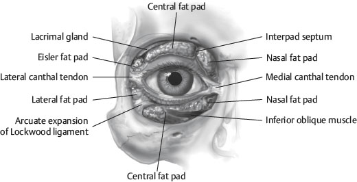
Fig. 34-6 Controversy exists regarding the true distinction of separate fat compartments; however, preservation of fat volume during blepharoplasty is a hallmark of the modern surgical approach.
• Two compartments in upper lid (medial and middle)
► Medial is more pale, vascular, and fibrous.
► Trochlea of superior oblique muscle separates the medial and middle compartments.
► Minor lateral fat pad (Eisler fat pad) is present in some.
• Three in lower lid (medial, central, and lateral)
► Inferior oblique muscle separates medial and central compartments.
LACRIMAL APPARATUS
■ Palpebral and orbital segments separated by levator aponeurosis
■ Located posterior to lateral portion of superior orbital rim
• Lacrimal drainage system
► Punctum drains to canaliculus, which drains to lacrimal sac, which drains to nasolacrimal duct.
► Active pump mechanism
♦ Blinking creates negative pressure in lacrimal sac, allowing tears to pass through the punctum and canaliculus into the sac.
♦ Eye opening increases sac pressure and passes tears into the nasolacrimal duct.
TEARS
■ Function
• Lubrication for lid excursion
• Antibacterial properties
• Oxygenation of corneal epithelium
• Smooth, refractive globe surface
■ Three layers
• Lipid layer: Superficial and thin; reduces evaporative loss; secreted by meibomian glands and accessory sebaceous glands of Zeiss and Moll
• Aqueous: Thicker, secreted from lacrimal gland and accessory glands of Wolfring and Krause
• Mucoid: Maintains lid contact with globe; produced by mucin goblet cells
■ Basic secretion
• Accessory lacrimal glands of Wolfring and Krause, mucin goblet cells, and meibomian glands
■ Reflex secretion
• Main lacrimal gland, parasympathetic
INDICATIONS AND CONTRAINDICATIONS
CLASSIC INDICATIONS (UPPER EYELID)
■ Excess upper eyelid skin
■ Upper eyelid fold excess
■ Lack of upper eyelid fold
• Asian ethnicity with indistinct lid crease (see Chapter 34)
NOTE: The fold may be present but masked by excess fat and poorly formed or positioned conjoined fascia.
■ Fine periorbital or eyelid rhytids
TIP: A brow evaluation is critical in all patients (see Chapter 33). Often, a browlift unmasks a poorly defined or visible upper lid crease.
EYELID PATHOLOGY AND DEFORMITIES
■ Dermatochalasis: True excess of upper eyelid skin
■ Steatoblepharon: Excess fat protruding through septum
■ Blepharochalasis
• Thin upper and lower lid skin allows presentation of cyclical lid edema (with or without erythema).
• IgE and histamine are released.
• In 80% of patients, onset is before 20 years of age.
• Edema is refractory to antihistamines and steroids.
■ Blepharoptosis
• Drooping of upper eyelid
• Measured by distance to light reflex of pupil (marginal reflex distance [MRD])
• See Chapter 39 for further details.
■ Pseudoblepharoptosis
• Eyelid margin is in normal position; however, excess upper lid and/or brow weight is ptotic (MRD is within normal limits).
• This may indicate blepharoplasty in conjunction with a browlift procedure.
■ Ptosis adipose: Excess attenuation of canthus and septum
TIP: Blepharoptosis is not a contraindication to blepharoplasty, but it must be fully evaluated, informed, discussed, and treated.
SENIOR AUTHOR TIP: Eyelid ptosis should be corrected during blepharoplasty.
PREOPERATIVE EVALUATION
HISTORY
■ Patient expectations
• Functional versus aesthetic
• Detailed discussion is needed to inform patients of the cause of the problem (with the aid of a handheld mirror) and what can be done to correct it.
• Unrealistic expectations are unmasked and discussed.
• Video imaging has been most helpful in discussing patients’ expectations and whether they are realistic.
SENIOR AUTHOR TIP: With the Internet and media as a prevailing “pseudoeducational” force, patients may come in telling the doctor what procedure they need or want, as if ordering at a restaurant (e.g., “I don’t want a browlift or anything fancy, just a little of this excess skin removed.”) Patient education is more and more critical in today’s practice environment. Surgeons should always recommend and do what they think is correct. Our job is to inform patients, make the recommendations that we think are best, and discuss the procedure or procedures, risks, and expected outcomes, including the quality of the result and the expected recovery. With this information, patients can make a truly informed decision.
PERTINENT MEDICAL CONDITIONS
■ Eyelid inflammatory conditions (Reiter syndrome)
■ Grave disease
■ Benign essential blepharospasm
■ Dry eye syndrome
• Ask about eyedrop use and probe for details about dry eye symptomalogy.
• Contact lens use
• Bell phenomenon test
• Consider Schirmer test
TIP: A history of dry eyes with decreased tear production (frequent use of eye lubricants), combined with postblepharoplasty tear film loss from lagophthalmos, can lead to corneal exposure (keratoconjunctivitis or ulceration). An abnormal Bell phenomenon increases the risk of corneal complications. A more conservative blepharoplasty with possible temporary tarsorrhaphy may be best versus no surgery at all for this group of patients.
■ History of LASIK surgery
• Best to wait 12 months after LASIK surgery to allow corneal incision time to heal
SENIOR AUTHOR TIP: Some ophthalmologists even recommend allowing 24 months to heal following LASIK surgery.
■ Epiphora: Excess tearing
• History of Bell palsy (crocodile tearing)
• Gustatory epiphora
■ Blepharoptosis
• Discussed previously and in Chapter 39.
■ General medical conditions
• Coagulopathies
► Anticoagulant/antiplatelet therapy or medication
CAUTION: Postoperative bleeding/hematoma after blepharoplasty is a serious complication that can lead to blindness. Early recognition is critical.
• Severe “periorbital” allergic symptomatology
• Thyroid dysfunction
• Hypertension
• Renal or cardiac abnormalities
• Neurologic
► Myasthenia gravis
► Horner syndrome
OCULAR EXAMINATION
■ The best visual acuity for each eye is recorded.
• May require a more accurate assessment by an ophthalmologist (especially with insurance-related cases)
► Patient referred to ophthalmologist if any abnormalities noted
■ Bell phenomenon: If lids are forcibly held open while patient attempts to close them, then globe should rotate upward.
• Built-in protective mechanism
• When not present, patient more susceptible to dry eye exacerbation with even minimal postoperative lagophthalmos
LACRIMAL FUNCTION TEST
■ Most important in elderly and patients with a history of dry eyes
• May consider ophthalmologic referral preoperatively
■ Schirmer test I: Basic and reflexive secretions
• Whatman filter paper (Whatman, Inc.): 5 by 35 mm, distal 5 mm folded; fold placed on lateral sclera
• More than 10 mm wetting after 5 minutes is normal.
■ Schirmer test II: Basic secretion
• Performed after topical anesthesia applied (eliminates reflex component)
• Usually <40% of Schirmer test I
■ More advanced tests: Tear film breakup, rose bengal staining, tear lysozyme electrophoresis