Modern cleft surgery requires four-dimensional and functional anatomic understanding of the cleft (and noncleft) lip, nose, and alveolus. Some techniques for nasolabial repair rely more on precise anatomic geometry, whereas others afford the surgeon a more flexible design. Consistent anthropometry enables accurate assessment and reporting of long-term outcomes; such reports are needed to guide perioperative care, delineate optimal repair principles, and resolve ongoing controversies.
Key points
- •
Modern cleft surgery requires four-dimensional and functional anatomic understanding of the cleft (and noncleft) lip, nose, and alveolus.
- •
Some techniques for nasolabial repair rely more on precise anatomic geometry, whereas others afford the surgeon a more flexible design.
- •
Consistent anthropometry enables accurate assessment and reporting of long-term outcomes; such reports are needed to guide perioperative care, delineate optimal repair principles, and resolve ongoing controversies.
Introduction
Since 390 bce , the treatment of upper lip clefts has challenged generations of surgeons. Cleft care is now concentrated in specialized, high-volume centers where multidisciplinary teams can best serve patients and their families. The American Cleft Palate–Craniofacial Association (ACPA) has established a framework for such centers that guides multidisciplinary team composition, interdisciplinary communication, cultural competence, psychosocial services, and outcomes assessment ( www.acpa-cpf.org ). Modern cleft reconstruction requires three-dimensional (3D) and functional anatomic understanding of the cleft (and noncleft) lip, nose, and alveolus. Accurate nomenclature defines shared challenges, improves classification schema, and builds consensus (where possible). Multiple repair techniques and modifications reflect the rich variety and continuous evolution of operative principles in cleft reconstruction. Diligent anthropometry enables accurate assessment of outcomes over time, the so-called fourth dimension.
Embryology and epidemiology
Lip development occurs from the fourth to the seventh weeks of gestation. The frontonasal prominence is formed from mesenchymal tissue ventral to the forebrain and leads to development of the medial and lateral nasal prominences. The paired maxillary and mandibular prominences are formed from neural crest cells migrating from the first pharyngeal arch. The primary palate, or premaxilla, similarly forms from the fusion of the medial palatine processes. A unilateral cleft lip occurs when there is failure of complete fusion between the advancing maxillary prominence and the fused medial nasal prominences on one side ( Fig. 1 ). The cleft of the lip extends through the maxillary dentoalveolus to the incisive foramen when the medial palatine processes fail to fuse. The secondary palate, posterior to the incisive foramen, develops from the 6th to the 12th weeks of gestation as the lateral palatine processes fuse. A cleft palate occurs when there is incomplete fusion of the lateral palatine processes with the medial palatine process and/or nasal septum ( Fig. 2 ). Various anomalous combinations in lip and palate development occur; it is more common to have cleft lip and palate (46%) than isolated cleft palate (33%) or isolated cleft lip (21%). Additional failures of mesenchymal penetration result in an array of facial clefts.
Cleft lip (with or without cleft palate) occurs most commonly in boys and has a 6:3:1 ratio of left/right/bilateral involvement. If a child is born with a cleft lip, the risk of having another child with cleft lip is 4% and 9% after 2 affected children. If a mother and child are affected, the risk to the second child is 15.3%. The condition occurs in approximately 2 of 1000 Asians, 1 of 1000 white people, and 0.5 of 1000 African Americans. Unlike isolated cleft palate, cleft lip is usually sporadic and associated with only a few syndromes: van der Woude (autosomal dominant, lip pits), 22q deletion (DiGeorge anomaly, conotruncal malformation, velocardiofacial syndrome), and Stickler (type 2 collagen mutation, retinal detachment).
Applied anatomy
Restoration of normal surface, muscle, and mucosal anatomy is paramount in patients with cleft lip. The upper lip is lined posteriorly with nonkeratinized oral mucosa and anteriorly by keratinized vermillion and hair-bearing skin. The red line divides labial mucosa from vermillion, whereas the vermillion border separates vermillion from white roll. The white roll is a shiny, hairless convexity lying just above the vermillion border. The normal philtrum is composed of a central depression between 2 ridges that emanate upward from the peaks of Cupid’s bow toward the medial footplates of the lower lateral nasal cartilages. Philtral ridges are normally formed by decussating fibers of orbicularis oris muscle that insert superficially into overlying dermis. The orbicularis oris has 2 functional components: lip retraction (fibers originating superficially from facial mimetic muscles) and oral sphincter closure (fibers originating deeper from modiolus). Within vermillion, orbicularis oris becomes superficial and concave. Known here as the pars marginalis, its shape contributes to the lip’s natural pout.
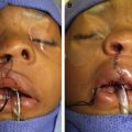
In a unilateral cleft, the medial lip is short with a flat philtral ridge and narrow vermillion. The lateral lip also has deficient mucosa, vermillion, and white roll medial to the Noordhoff point (of maximum vermillion height). The orbicularis oris is deficient, with aberrant muscle insertions into the alar base, nasal sill, columella, and dermal cleft margins. The cleft nasal deformity is variable and corresponds with severity of the cleft lip. The columella is short and the anterior nasal spine/caudal septum deviates away from the cleft. In complete unilateral clefts, the nasal sill, nasal floor, alveolus, and primary palate are also disrupted, allowing unrestricted growth of the greater (medial) maxillary segment, which splays the maxillary arch. The cleft-side ala and associated lower lateral cartilage are widened and distorted by the ala’s attachments to the collapsed lesser (lateral) maxillary segment. The primary palate (containing premaxilla, central alveolus, and philtrum) is anterior to the incisive foramen, whereas the secondary palate (containing maxilla, palatine bones, palatal musculature, uvula, and pterygoid plates) is posterior. The incisive foramen is important because it distinguishes the complete cleft lip defect from a cleft palate defect, and also because it contains the nasopalatine nerves and sphenopalatine artery.
Cleft classification
Numerous classification schemas have been proposed for cleft lip ; we think that a simple system with objective, clearly defined anatomic features is most useful for practitioners. A cleft lip is distinguished by 2 main characteristics: (1) unilateral versus bilateral deformity and (2) complete versus incomplete deformity. A complete cleft lip is characterized by through-and-through involvement of the lip, nasal sill, nasal floor, and alveolus, whereas incomplete clefts show partial penetrance through these structures. Thirty percent of patients with complete cleft lip still show a Simonart band.
Veau described the spectrum of incomplete unilateral clefts. Various colloquialisms such as minimal and occult have since been used to describe the array of incomplete labial, muscular, and dental cleft phenotypes. Yuzuriha and Mulliken describe lesser form labial clefts (those at the far end of the unilateral incomplete spectrum) as minor form (notched vermillion-cutaneous junction extending 3 mm or more above the normal Cupid’s bow peak), microform (notch less than 3 mm above the normal Cupid’s bow peak), or mini-microform (disrupted vermilion-cutaneous junction without elevation of Cupid’s bow peak) ( Fig. 3 ). For each, nasal severity reflects that of the lip. Yuzuriha and Mulliken’s classification is practical because it guides optimal operative technique by cleft severity.
Anthropometric analysis and planning
Anthropometry is the measurement of the human individual and permits comparison of anatomy under normal and abnormal conditions. Dr Leslie Farkas, father of medical anthropometry, and Farkas and colleagues, provided normative measurements of the lip and nose. Cleft surgeons use anthropometry to objectively measure aesthetic outcomes over time. With an applied understanding of the fourth dimension (growth), these data are retrospectively applied to improve the initial 3D design at primary nasolabial repair.
There are fundamental nasolabial anthropometric landmarks essential to cleft surgery ( Fig. 4 ): alare (ala) is the most lateral point of each ala, subalare (sbal) is the most inferior point of each alar base, the highest point of columella (c) lies atop each hemicolumella and is level with each nostril peak, subnasale (sn) defines the angle between columellar base and upper lip, crista philtri superior (cphs) is atop each philtral column at the same horizontal line drawn through sn, crista philtri inferior (cphi) lies at the base of each philtral column (each Cupid’s bow peak), labiale superius (Ls) lies at the midpoint of the upper vermilion (Cupid’s bow trough), stomion (sto) is the point along the vertical facial midline that bisects the free margin of the upper lip, and cheilion (ch) is located at each labial commissure.
Anthropometric measurements can be obtained by direct or indirect methods. Direct anthropometry is the gold standard, but requires experience and a cooperative patient. For young patients, this is most accurately obtained in the operating room under general anesthesia. Indirect anthropometry can be obtained by two-dimensional (2D) photography but requires image standardization and calibration for linear measurements; 2D photography is best for discerning proportions and angles. As an alternative, 3D photography allows photographs to be analyzed with software accompanying 3D camera systems. This technique has gained favor as an alternative to direct anthropometry in children because images are captured in as little as 3.5 milliseconds. Wong and colleagues evaluated the validity and reliability of nasolabial anthropometry using 3D photography compared with direct anthropometry and found that linear measurements were highly correlated and overall precision of 3D measurements were within 1 mm of direct measurements.
Relevant lip and nose measurements for unilateral cleft deformities include heminasal width (sn-al), nasal width (al-al), nasal tip projection (sn-prn), columellar length on each side (sn-c), columellar width (c-c), labial height (sn-cphi, sbal-cphi), and lip length (cphi-ch). Understanding future growth is crucial when performing primary cleft lip repair. The surgeon must normalize nasolabial measurements not at the time of repair but when the child reaches 5 to 10 years of age. Mulliken and LaBrie studied anthropometric changes over time after unilateral nasolabial repair and found that heminasal width (sn-al) increases more on the cleft side than noncleft side during the first 6 years of life. Lateral drift of the alar base was also described by Millard, who secured the alar base to the caudal septum to reduce alar creep. Byrd preserves a muscular roll below the alar base that he sutures to soft tissue adjacent to the anterior nasal spine and contralateral medial crural footplate. Slow-growing structures such as nasal tip projection (sn-prn) and columellar length (sn-c) should be made longer than those of age-matched, gender-matched, and ethnicity-matched subjects at the time of primary repair. Fast-growing features such as nasal width (al-al) and prolabial width (cphs-cphs and cphi-cphi in bilateral deformities) are intentionally made narrower than those of normative subjects. Vermilion-mucosal height is a fast-growing features that is intentionally made longer because of concern for deficiency of tissue in the median tubercle.
Goals of surgery
Nasolabial repair of the unilateral cleft deformity should restore normal midfacial anatomic relationships and obtain long-term symmetry. Regardless of the technique used, a primary goal is to achieve lip height (sn-cphi and sbal-cphi) on the cleft side that matches the noncleft side. An ideal technique allows for more adjustments and produces a favorable scar pattern. Blair delineated the stigmata of the unilateral cleft nasal deformity: deviation of the tip and caudal septum to the noncleft side, dislocated lower lateral cartilage, obtuse angle between middle and lateral crura, posterolaterally displaced alar base, and short columella on the cleft side. Primary correction of these deformities is variously undertaken in repair of unilateral cleft nose, with anthropometric studies indicating improvements in nasal symmetry following certain repair strategies.
Current practice
Presurgical Molding
Most cleft centers currently use presurgical molding to align skeletal and soft tissues in preparation for definitive repair. Both passive molding, such as lip taping and (naso)alveolar molding, and active molding, such as the pin-retained (Latham) device, take advantage of the malleable tissues and rapid facial growth of infancy. Presurgical molding is an important, well-studied topic and detail is provided in the article “Management of the Alveolar Cleft” elsewhere in this issue by Pedro E. Santiago, Lindsay A. Schuster, and Daniel Levy-Bercowski.
Lip Adhesion
Lip adhesion is sometimes done for operative molding before definitive nasolabial repair. It restrains the alveolar segments and reduces cleft gaps by up to 60%. Adhesion is accomplished by ligating tissue along each cleft margin that is normally discarded during labial repair; various descriptions exist. Lip adhesion requires that the infant undergoes an additional operation and might compromise the outcome by restrictive scarring. Supporters argue it improves definitive repair by approximating the alveolar cleft, thereby facilitating gingivoperiosteoplasty and decreasing tension along the labial closure. Ridgway and colleagues used ultrasonography to show that the lateral orbicularis oris is thickened after lip adhesion, potentially aiding philtral construction. In addition, lip adhesion begins early nasal molding and provides 2 opportunities to correct cleft nasal asymmetry. As with presurgical molding, preference for lip adhesion is based on cleft severity, available expertise, and family/surgeon preference.
Triangular Repair
Tennison designed his repair after learning of LeMesurier’s use of (unilimb ) Z-plasty to lengthen the cleft lip. Unlike LeMesurier, whose backcut along the lateral lip element translated to a transverse philtral scar, Tennison made the backcut for his unilimb Z-plasty from cphi of the medial lip toward the philtral dimple ( Fig. 5 ). This incision opens to achieve the desired height of the medial lip; the deficit is satisfied with a laterally based triangular flap. Randall built on Tennison’s design by adding key anatomic landmarks and geometric techniques to seek mathematic symmetry at closure.
In the Tennison-Randall repair, a point is chosen along the medial and lateral cleft nasal floor so that closure at these landmarks optimizes nasal symmetry (sn-al, sn-c, and sn-prn). An incision is marked from the medial point of closure to cphi; the predicted medial lip deficiency is then recorded. From the lateral nasal point of closure, an incision of equivalent length is marked toward the chosen lateral cphi point. The gap between the inferior end of this line and cphi defines the base of the lateral triangular flap that fills the medial lip backcut. The exact pattern of closure along the lateral lip margin (including triangle) is designed while being mindful of (1) the equivalent length of the lines drawn from each point of nasal closure, and (2) the deficiency in medial lip height. Swinging calipers help finalize this pattern by tracing intersecting arches from both the lateral nasal point of closure and cphi (see Fig. 5 ).
The Tennison-Randall approach is most useful for wide clefts or clefts with severe vertical deficiency. Cronin suggested designing the triangular flap 1 mm above the vermillion border to avoid disruption of the white roll. Critics argue that the technique produces excess lip height (sn-cphi, sbal-cphi); Brauer and Cronin suggested a 1-mm initial undercorrection and possible full-thickness excision below the ala (particularly for incomplete clefts). A major limitation of this technique is the triangular philtral scar.
Rotation-Advancement: Millard and Modifications
Millard introduced the rotation-advancement repair in 1955 to move the line of closure away from the philtral dimple and into the anatomically camouflaged cleft philtral column. Rotation-advancement advocates that all available tissue be rearranged for nasolabial construction with minimal tissue discard. Millard proposed a medial curvilinear incision from cleft cphi up along the cleft margin to cleft cphs at the columella, continuing along the lip-columella junction just short of noncleft cphs (see Fig. 5 ). The resulting flap rotates inferiorly, providing height but leaving a defect in the central upper lip. Millard embodied a cut-as-you-go mindset; when he needed more lip height, he added a backcut made from the end of his rotation incision down toward the noncleft philtral column. He filled the resulting gap with a portion of his superiorly-based C-flap, otherwise used to close the nasal sill.
At the side, cphi is carefully chosen to ensure both sufficient lip height (sbal-cphi) and advancement flap dimensions that fill the upper medial lip rotation deficit. A more medially selected cphi results in a more deficient white roll, vermillion, and mucosa; collectively limiting the fullness of the median tubercle. A more laterally selected cphi gives better quality tissues and more vertical (but less horizontal) lip length. Using the Noordhoff point maximizes the quality of incorporated labial tissues, but does not guarantee adequate vertical height. Cleft surgeons generally tolerate less horizontal lip (cphi-ch) with potential vermillion thinning to avoid the more noticeable asymmetry of vertical lip deficiency (sbal-cphi). Millard emphasized aggressive lateral dissection to ensure adequate lateral lip advancement. He even modified his original design by extending the transverse incision around the cleft ala.
No technique has been more widely adopted and modified than Millard’s rotation-advancement. In a 2005 worldwide survey of leading cleft centers, 84% of surgeons used some variation of rotation-advancement to repair unilateral clefts. Several noteworthy modifications now provide cleft surgeons a variety of tools to customize desired elements of nasolabial repair. In 1987, Mohler introduced the concept of using columella to lengthen the medial lip (cleft cphi-cphs). Instead of the traditional backcut through the upper philtrum, Mohler’s rotation incision extends onto the columella and a backcut is made to the lip-columellar junction (see Fig. 5 ). Cutting popularized a similar technique; his incision is less bowed toward the cleft margin and extends up onto the columella 1.5 to 2.0 mm above the nasolabial junction and just past midline. Cutting’s lip repair allows a wider C-flap to fill the medial defect and results in a near–straight-line anatomic closure. Detailed, step-by-step videos and simulations demonstrating this technique are publically available ( https://smiletrain.biodigitalhuman.com/home ). Stal uses an S-shaped rotation incision to gain medial lip height; he extends onto the columella and uses a backcut when needed. Like Mohler, Mulliken’s rotation incision is curved toward the cleft margin and extends into the columella. However, instead of a backcut toward noncleft cphs, Mulliken uses a perpendicular releasing incision further up the columella into which the C-flap is inset (lengthening cleft-side sn-c). Because there is a smaller upper lip rotation defect, Mulliken’s advancement flap neither crosses nor effaces the philtral column (see Fig. 5 ).
Millard introduced the concept of using a flap of lateral lip medial to cphi to augment the deficient median tubercle. Noordhoff further defined this flap as a unilimb Z-plasty of available lateral lip vermillion that is inset as a taper into a releasing incision at the cleft mucovermillion junction. Mulliken and Cutting both use a variation of this Noordhoff flap. Millard stressed interdigitation of flaps at the vermillion-cutaneous junction to avoid the more conspicuous effects of linear scar contracture. Mulliken uses a unilimb Z-plasty to introduce a small triangle carrying skin and white roll medial to cphi of the lateral lip into a 2.0-mm to 2.5-mm vertical defect created by a releasing incision along the vermillion-cutaneous junction of the medial lip (see Fig. 5 ).
Millard pioneered many aspects of primary nasal repair during rotation-advancement. He designed his C-flap to assist in closure of the nasal floor/sill, emphasized maneuvers to correct the deviated septum and asymmetric columella, and divided the lateral cleft element into a labial advancement flap (to narrow sn-al) and a separate alar base flap (for nasal floor closure and 3D flexibility). Millard dissected the nasal soft tissue envelope off the lower lateral cartilages and elevated the cleft nasal mucosa off the piriform to allow anterior movement. Byrd maintains a muscular roll under the cleft alar base that he sutures to periseptal tissues to augment the deficient maxilla and build a platform that projects the cleft ala. McComb, Salyer, and Cutting reposition and suspend the lower lateral cartilage with internal sutures, and Stal uses a transdermal alar suspension technique. Mulliken straightens and fixates the deviated anterocaudal septum during primary repair and includes the medial crural footplate with his C-flap to correct columellar asymmetry. By placing a suture from the medial alar base to underlying periosteum of the lateral incisor fossa, Mulliken prevents alar creep and restores the native (z-axis) depression of the nasal sill. Mulliken uses a semiopen technique with bilateral marginal rim incisions to dissect soft tissue off both lower lateral cartilages and the ipsilateral upper lateral cartilage; interdomal and intercartilaginous resorbable sutures internally suspend the cleft ala into an overcorrected position.
Anatomic Subunit Repair
Fisher recently introduced a geometric repair that places the line of closure between anatomic subunits, eliminating scars on or below the columella (see Fig. 5 ). The design requires identification of 25 anatomic landmarks and relies heavily on direct anthropometry. Central and noncleft landmarks supply normal anthropometrics from which corresponding cleft-side landmarks are derived. Although it requires extensive attention to detail, this technique emphasizes anatomic geometry rather than surgeon experience to produce reliable results.
Identification of sn and noncleft cphs determines cleft cphs. While manually correcting the nose, 2 noncleft alar landmarks are identified: sbal and the alar insertion point (junction of ala and nasal sill). An arbitrary point within the normal nasal sill is chosen that is collinear with these two points as well as with sn/noncleft cphs (Tse). Mirroring the distance between this point and the noncleft alar insertion point determines the superiormost aspect of the lateral lip incision; mirroring the distance between this point and noncleft cphs determines the superiormost aspect of the medial lip incision. Uniting these superior points corrects the cleft nasal sill.
The incision runs medially from this established superiormost point of closure within the cleft nasal sill down the base of the cleft-side medial crural footplate to cleft cphs and then straight down to the white roll atop cphi. The incision continues perpendicular to the white roll and red line and then toward the gingivobuccal sulcus. The incision is designed laterally between 2 fixed landmarks: the previously established superiormost point of closure within the cleft nasal sill and the Noordhoff point (cphi). Between these two points, 3 elements are designed to match the medial lip’s line of closure. First, a limb is drawn matching the length and curvature of the medial lip’s incision along the medial crural footplate; the end of this limb establishes cphs laterally. Second, the lateral philtral column is traced, matching the medial lip’s cphs-cphi length. Third, a small triangular flap is designed for inset into a backcut above the medial lip white roll; its base measures the deficiency between cleft and noncleft philtral heights (minus 1 mm to account for the Rose-Thompson effect). In contrast with preset medial lip markings, the 3 components of the lateral lip incision are tailored to each patient depending on lateral lip height and cleft severity. For lesser form clefts, the small lateral triangle can be omitted. Fisher also uses a unilimb Z-plasty to incorporate lateral lip vermillion medial to Noordhoff point into the deficient median tubercle (see Fig. 5 ).
Related posts:


Stay updated, free articles. Join our Telegram channel

Full access? Get Clinical Tree


