(1)
Division of Colon and Rectal Surgery, State University of New York, HSC T18, Suite 046B, Stony Brook, NY 11794-8191, USA
Abstract
This chapter outlines the approach to the difficult laparoscopic colorectal operation where access may be limited because of intra-abdominal pathology or adhesions. Complications and their avoidance are discussed in the current environment of more advanced laparoscopy. The objective of this chapter is to present a current opinion about troubleshooting the difficult laparoscopic case, focusing on minimizing complications and their management should intraoperative problems occur.
Keywords
Difficult laparoscopyMultiple laparotomiesDense adhesionsInadvertent organ injuryAltered anatomyHemorrhageAnticoagulation therapyReoperative surgeryIntroduction
A difficult laparoscopic case perhaps can be defined as an operation in which one anticipates that intraoperative adverse events are likely to be encountered for reasons that may include difficult laparoscopic access secondary to multiple laparotomies, dense adhesions, the perceived risk of inadvertent organ injury as a result of altered anatomy, and the risk of hemorrhage in patients receiving anticoagulation therapy, which is part of a mandatory treatment régime. Because laparoscopic experience has increased and instruments have somewhat improved, interventions can, in a large number of cases, be safely performed using a laparoscopic approach. Complex cases, at one time viewed as a contraindication for laparoscopy, currently receive laparoscopic management by many surgeons experienced in advanced laparoscopic techniques.
The objective of this chapter is to present a current opinion about troubleshooting the difficult laparoscopic case, focusing on minimizing complications involving organs located between the abdominal skin and the colon and rectum. The topic is limited to those cases that may be considered difficult and challenging for the surgeon without targeting a specific colon or rectal disorder. We attempt to provide readers with concise insight into the evidence available in the English literature. This chapter does not offer a comprehensive review of the topic; rather, it highlights some relevant issues and outlines what role laparoscopic surgery should play in these challenging situations.
Rationale
There are many benefits of embarking on such cases via a laparoscopic approach wherever possible, including decreased postoperative pain, shortened postoperative ileus, faster resumption of consuming solid food, lower rates of infection at the surgical site, reduced rates of incisional hernia, and improved cosmesis. In light of these benefits, laparoscopic surgery has been gaining acceptance as a viable alternative to laparotomy for managing complex reoperative cases. Surgeons should have a low threshold for conversion and such a decision should not be perceived as a failure but rather as an indication of good clinical judgment.
Access (The First Port)
Safe access to the abdominal cavity is a key step during reoperative surgery. Although surgeons may have a personal preference for the open or blind (Veress needle) technique, many access-related complications are related to the insertion of the first trocar and can be divided into the categories defined below.
The Operated Abdomen
The Hasson technique was initially described as an alternative to access of the abdominal cavity in previously operated patients [1]. By choosing a cut-down technique, the surgeon can introduce a blunt-tipped trocar into the abdominal cavity under direct visualization, as opposed to a blind entry with a Veress needle. Most surgeons would favor a cut-down technique in reoperative surgery [2]. When dealing with a reoperation, it is advisable to avoid previous scars because the risk of inadvertent injury to intra-abdominal organs is increased. Assuming that the patient has a midline laparotomy (which is the most frequently encountered scenario), the initial trocar should be placed lateral to the rectus muscle sheath at the midpoint between the lower edge of the rib cage and the anterior superior iliac spine along the anterior axillary line. The surgeon must be careful not to place the trocar too far laterally because of the risk of injuring the descending colon and obtaining a limited visualization of the operative field. Detailed awareness of the patient’s previous abdominal surgeries will assist in determining on which lateral side of the abdomen placement of the first trocar is best. For example, if the patient had undergone a previous left colon colectomy, the first trocar should be inserted in the right side of the abdominal quadrants. The same concept is true for surgeries initially performed on the right side of the patient.
New optical-access trocar systems recently have been developed to assist the surgeon with these operative placement decisions. These trocars allow the surgeon to visualize the abdominal wall layers as the trocar enters the peritoneal cavity. The ports were made with the intention of minimizing organ injury while gaining laparoscopic access. There are currently two optical view trocars: one uses a blade and the other uses a rotating, sharp, plastic clear tip; both types are inserted under direct laparoscopic view. Regardless of what type of trocar the surgeon chooses, the insertion technique should involve minimal force and consistent direct visualization of entry. These new trocars can be assisted by EndoAssist robotic devices that replace the human assistant and permit greater control of the operative field by the surgeon during trocar entry and the main procedure [3].
Burned Abdominal Wall
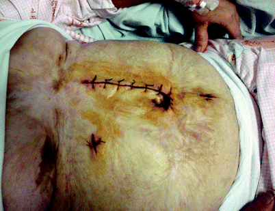
Patients with extensive burns of the abdominal wall (third and fourth degree) represent a significant challenge for laparoscopic access (Fig. 14.1). One must be aware that the omentum will be firmly adherent to the parietal peritoneum of the anterior abdominal wall. A solution here is to use the Hasson technique to carefully introduce the first trocar in the most caudal portion (right or left) because these areas are most likely to be free of adhesions of the greater omentum. The surgeon must then dissect the omentum off the parietal peritoneum to provide access for further trocars.


Fig. 14.1
Burned abdominal wall
Scope Type
There is usually no role for a 0° scope for such complex procedures because, more often than not, the surgeon will need to glance around at adhesions rather than look straight forward. The minimal degree of angulation required is 30°, but sometimes 45° may prove to be more useful. An alternative is a scope with a deflectable tip, and the person holding the camera should be familiar with this device. Our preference is a video laparoscope without an interphase between the scope and the camera as opposed to an optical system; the latter may potentially be associated with “fog” formation and sterility hazards when the hose cable is positioned. Furthermore, the surgeon should be aware that camera chips are located on the tip of the video laparoscope, as opposed to the classical system, where the camera chip lies outside the patient’s abdomen.
Vascular Injuries
Although the Hasson technique will not completely eliminate the risk of major vascular injuries, there is evidence that suggests that this risk is decreased by its more routine use [4]. Although vascular injuries are rare, they must be recognized and repaired with an immediate laparotomy [5]. Before converting, the surgeon may attempt to control the bleeding vessel with a laparoscopic Satinsky clamp (Fig. 14.2). Much of the evidence related to vascular injuries secondary to laparoscopic trocar insertion comes from individual case reports. In a review of 629 reports of trocar incidents, 408 cases were associated with major vascular injuries and resulted in 26 deaths. The vessels most commonly injured were the aorta (23 %) and the vena cava (15 %) [6]. Some surgeons in this analysis used the optical view ports, which indicates that such accessories cannot completely prevent injuries related to trocar insertion.


Fig. 14.2
Laparoscopic Satinsky clamp
Bowel Injuries
Unfortunately, neither the rate of bowel injuries nor their related death rates have been reduced by use of the Hasson technique. It is worth repeating that the Hasson approach should be employed at a site away from preexisting scars [7]. Approximately 40 % of penetrating bowel injuries have been reported to be caused by insertion of a further trocar [8]. Therefore, the insertion of ports must be performed under direct vision and with sufficient intraperitoneal space, which requires the anesthesiologist to provide adequate relaxation of the abdominal wall musculature.
Pneumoperitoneum
There is no question about the need for an adequate pneumoperitoneum in the avoidance of injury. The surgeon should have a thought process that sequentially evaluates all possible causes of insufficient pneumoperitoneum. Once all common causes have been ruled out, the surgeon should communicate with the anesthesiologist to ensure that proper paralysis of the anterior abdominal wall is provided. It is beneficial for patients to be operated upon with the lowest possible intra-abdominal pressure, although there is little difference in the exposure provided with 10 mmHg versus 15 mmHg of intra-abdominal pressure [9]. If the reoperation is performed in the pelvis, it may be sufficient to operate with an intra-abdominal pressure of less than 10 mmHg because the pelvis is relatively nondistendable.
The surgeon also must take into account the patient’s body habitus when inducing and maintaining pneumoperitoneum. An abdominal wall thickness of 10 cm or more will require a 15-cm-long port rather than an ordinary 10-cm-long port. If the trocar is too short to traverse the thickness of the abdominal wall, insufflation likely will lead to significant subcutaneous emphysema with a subsequent increase in tidal volume. Certain medical conditions, including chronic obstructive pulmonary disease and other respiratory diseases as well as congestive heart disease, may be considered contraindications to prolonged pneumoperitoneum. In these circumstances, as the intra-abdominal pressure increases, the splanchnic blood flow, cardiac output, and the renal cortical blood flow concomitantly decrease, resulting in localized or generalized abdominal compartment syndrome [10]. An alternative for patients with these medical conditions may include the use of nitrous oxide or helium for abdominal insufflation, taking into account that these gases are combustible and nonsoluble, respectively.
Operative Procedure
Exposure
Our preference is to position the patient in the lithotomy position on a bean bag with both arms tucked to the side and padded. The lithotomy position also provides an option for intraoperative sigmoidoscopy or colonoscopy as well as for manipulation of the uterus. To avoid any impediment to the surgeon’s elbows or to the use of long laparoscopic instruments, the height of the patient’s knees should never exceed the height of the abdomen. In the obese patient, the surgeon may want to consider strapping the patient’s chest to the operating table because the surgeon must verify that the patient is safely secured to the table before the creation of a sterile field. This is accomplished with remote control of the operating room table and by positioning the patient in a steep Trendelenburg position.
Correctly handling adhesions, the greater omentum, bowel segments, and other organs are necessary steps that permit the surgeon to perform a reoperation safely. It is intuitive that a gentle surgical technique will decrease the risk of unintended organ injury. In this respect, nonlocking bowel graspers should be used to run the length of the bowel. Appropriate exposure may be achieved by pushing adhesions with closed instruments rather than by grasping the bowel; the presence of distended bowel can make exposure difficult and time consuming. Nitrous-based anesthesia should be avoided in this situation because it dilates the small intestine. Other techniques that aid exposure and retraction include a table tilt and intermittent external manual compression of the abdominal wall. Finally, the surgeon should not hesitate to use additional ports to achieve appropriate exposure.
Additional Trocars
The insertion of additional trocars does not require open access but should be performed under direct visualization. The geometry of port placement should be triangular whenever possible. After insertion of the first port, space in the abdominal cavity may be limited because of the presence of dense adhesions. We do not recommend bladeless trocars because they require significant force during insertion, which may result in inward tenting of the abdominal wall; we favor using blade trocars or ultrasonically guided ports [11, 12]. The insertion of multiple trocars has been obviated in colorectal surgery by single port access technology [13, 14].


