70
Triscaphe Degenerative Arthritis
Andrew E. Caputo and H. Kirk Watson
History and Clinical Presentation
A 62-year-old right hand dominant woman who works as a polisher presented with right wrist pain progressively worsening over the last 6 months. She noted significant and progressive difficulty with strength and holding onto small objects. She denied any trauma and noted no specific previous treatment.
Physical Examination
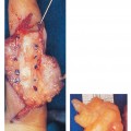
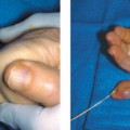
The patient had specific localized tenderness and swelling of the scaphoid-trapezium-trapezoid (STT/triscaphe) joint. This area is located by following the index metacarpal proximally until a recess is palpated. This recess is the dorsal aspect of the triscaphe joint. Passive wrist flexion extension and radial/ulnar deviation were limited with tender end points. Passive forearm rotation was nontender. No other areas of tenderness or swelling were noted. This finding is similar to what is found in most patients with triscaphe degenerative arthritis.
Diagnostic Studies
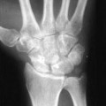
Posteroanterior and lateral views of the right wrist revealed isolated loss of joint space and subchondral sclerosis of the triscaphe joint (Fig. 70–1). No other radiologic signs of degenerative joint disease were noted in the other areas of the hand or wrist.
PEARLS
- Avoid driving the K wires into the radioscaphoid joint. The surgeon should be able to feel the purchase of the K wires into the subchondral bone of the proximal pole of the scaphoid. Intraoperative fluoroscopy can be utilized to facilitate proper placement.
PITFALLS
- Avoid overcorrecting the scaphoid. As noted above, scaphoid position is critical. The surgeon should be holding the scaphoid position as described while an assistant drives the K wires.
Differential Diagnosis
Carpometacarpal arthritis
Nonpantrapezial
Pantrapezial
Radiocarpal arthritis
Scaphocapitate arthritis
Scaphotrapezial trapezoid arthritis
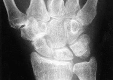
Figure 70–1 Localized degenerative arthritis to the scaphoid-trapezium-trapezoid (STT)/triscaphe joint is noted with sclerosis and complete loss of cartilage space.
Diagnosis
Triscaphe Degenerative Arthritis (Scaphotrapezial, trapezoid)
Triscaphe degenerative arthritis is the second most common type of degenerative arthritis in the wrist. The most common is the pattern of destruction associated with a scapholunate advanced collapse (SLAC) wrist. The diagnosis is relatively easy when radiographs confirm the isolated joint destruction to the STT joint.
Related posts:
Stay updated, free articles. Join our Telegram channel

Full access? Get Clinical Tree


