Triangular and Hatchet Subcutaneous Pedicle Skin Flaps
A. J. J. EMMETT
It is the natural elasticity of skin that enables defects to be closed, and it has been usual to fit the long axis of the defect into the lines of minimal tension (1). Where a rounded defect is to be closed, the shape of the defect may be tapered into the line of closure by adding a triangle at either end of the rounded defect to produce a tapered elliptical shape. These triangles of tissue then are discarded. As the size of this round defect becomes larger, the size of the triangle to be discarded also increases, to the point where these triangles themselves may be used as a flap and slid into the original round defect with a V-Y closure of the base from which they have come (2, 3, 4, 5, 6, 7, 8, 9, 10).
INDICATIONS
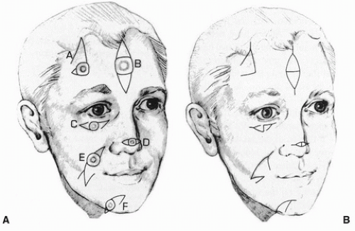
Various triangular flaps are a safe and interesting way to close medium-sized defects in the 1.5- to 3-cm range in all areas of the body. They have been used on the back, foot, hand, face, and chest. They are a versatile design, with a great bulk of pedicle relative to the size of the flap, providing great flap viability. The single triangular flap has an advantage for closure of rounded defects when one side of the defect is closed by a natural boundary. The double-triangular subcutaneous pedicle skin flap has been used for forehead defects above the eyebrow, eyebrow defects, and some hairy scalp defects. I have used the hatchet flap for repair of the chin, eyelid, cheek, and lower lip.
 FIGURE 98.1 A,B: Various forms and modifications of double triangular flaps, single triangular flaps, and hatchet flaps. (From Emmett, ref. 10, with permission.) |
ANATOMY
The pedicle is the subcutaneous tissue that lies beneath triangular flaps, and this can then be extended as deep as necessary. It is all the better if muscle lying beneath is included. The flap is based on a random arterial blood supply, but occasionally, it can be based over a direct nutrient artery.
FLAP DESIGN AND DIMENSIONS
These flaps are all rather free form and have a degree of adjustability; so the surgeon is not rigidly committed to the original cutout flap pattern and can vary the fit of the flap as the operation progresses.
A general principle of these flaps is that the defect is narrowed in around the pedicle. The tail of the flap is closed in a V-Y fashion so that as the donor site is closed, it pushes the tapered tail of the flap forward toward the recipient site. The line of the triangular flap is that of the line of minimal tension closure (Figs. 98.1 and 98.2).
Stay updated, free articles. Join our Telegram channel

Full access? Get Clinical Tree


