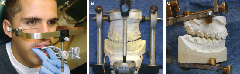
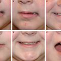
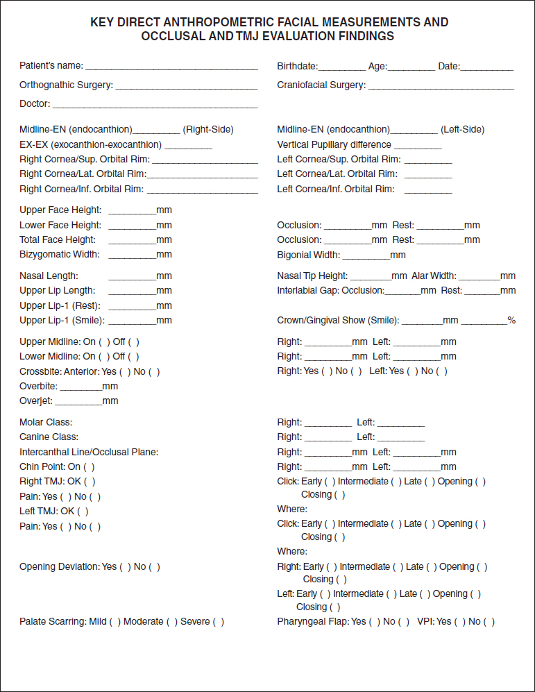
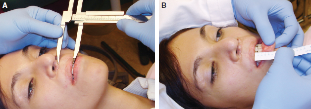
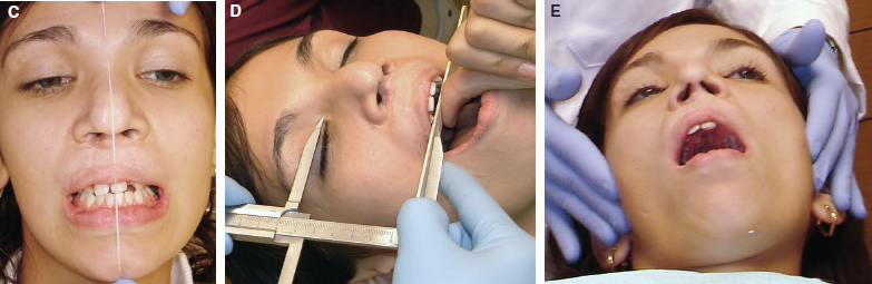
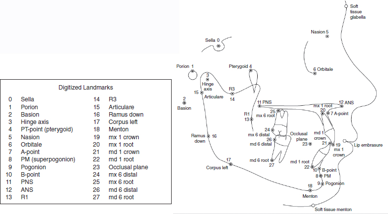
71 ○ Maxillomandibular discrepancies in patients with clefts require a combined surgical and orthodontic treatment approach following established protocols. Close communication between the surgeon and orthodontist is critical. ○ Special considerations should be kept in mind when treating cleft-related maxillomandibular discrepancies, including severity of the skeletal discrepancy, scarring, pharyngeal flap, alveolar or palatal fistulas, missing teeth, and secondary lip and nose deformities. ○ The orthodontist supports the surgeon during planning, arch coordination procedures, preparation of appliances and splints, and finalizing occlusal/dental details and follow-up for patients who require orthognathic surgery and distraction osteogenesis. ○ Important steps during treatment planning include a thorough clinical evaluation, photographic and dental model evaluation, and cephalometric and radiographic analysis. ○ The application of new technologies such as cone beam CT scans and three-dimensional computerized cephalometry improve the diagnostic and planning capabilities available to surgeons and orthodontists as well as reducing the time required for planning. ○ Three-dimensional virtual surgical planning (VSP) allows the clinician a better appreciation of the preoperative condition, including skeletal factors, dental considerations, soft tissues, and airway, and aids selection of the surgical approaches required to treat the maxillomandibular discrepancy. ○ Application of computer-aided design (CAD) and computer-aided manufacturing (CAM) technology and stereolithography allow for preparation of traditional surgical splints as well as the newly introduced surgical guides to assist in accurate repositioning of the maxilla, mandible, and chin. ○ The use of new technologies is likely to improve surgical approaches and outcomes for patients with clefts who require orthognathic surgery and distraction osteogenesis. The care of patients with maxillomandibular skeletal discrepancies requires a protocol that includes the following: Based on these data, a treatment plan is formulated by the various specialists involved in the team management of the patient. These specialists include the maxillofacial surgeon, orthodontist, reconstructive dentist, and speech and language pathologist. On occasion, other team members are included, such as mental health professionals to emotionally support the patients and their families before and during the active phase of treatment. Once the initial diagnostic data are evaluated and all involved professionals understand their role for the particular patient, the preparatory steps for care are initiated. This chapter focuses on treatment planning from the dental, orthodontic, and surgical perspective. Once the overall health and oral health of the patient is determined to be satisfactory, orthodontic treatment can be commenced. The orthodontist is responsible for performing all necessary dental alignment procedures to facilitate surgery and ensure stability of the dentition and occlusion after surgery. The orthodontic appliance will also provide the surgeon with modes of fixation during the intraoperative and postoperative period of surgical care. In addition, the orthodontist must decide on the occlusal relations and specific dental positions required before surgery and the desired postoperative dental occlusion and relations at the completion of treatment. The stability of the dentition is achieved by positioning the teeth with the right inclination relative to their supporting bone bases. This applies to the anterior as well as the posterior dentition. The position of the anterior and posterior teeth relative to their bases is referred to as torque of the crowns and roots. The orthodontist must place the posterior teeth in the correct vertical and transverse position relative to the supporting alveolar bone. The required movements are determined during the clinical examination, cast evaluation, and, to a lesser extent, cephalometric evaluation. The use of a frontal view from a frontal cephalometric radiograph or from a cone beam CT scan might be helpful to evaluate the inclination or torque of the posterior teeth. Also critical to treatment planning are the sagittal positions of the maxillary and mandibular incisors relative to their supporting bone. The position of the incisors is not only important for occlusion but also has a significant effect on lip posture and aesthetics. To best assess incisor position, the cephalometric analysis is of significant value. During the cephalometric evaluation, the orthodontist can not only see the position of the tooth relative to the supporting bone, but also relative to the lips and the opposing incisors. The correction of dental positions before surgery is commonly known as removal of dental compensations, which are abnormal positions that the teeth have attained as a result of the jaw discrepancy, trying to achieve contact despite the skeletal discrepancy. Another critical element during treatment planning is determining which position a tooth will be assigned at the end of treatment. Patients with orofacial clefts are commonly missing teeth, especially the maxillary lateral incisor and second bicuspids. These situations may force the orthodontist to make decisions such as shifting the position of a tooth to replace a missing one (such as a maxillary canine replacing a missing maxillary lateral incisor) or extracting teeth in cases of moderate crowding. Equalizing the number of teeth and achieving symmetry and adequate occlusal relationships are imperative. In cases with missing teeth but minimal crowding, the orthodontist may elect to maintain the space for the missing teeth for future prosthetic replacement. This is particularly important if the existing posterior occlusal relationships are satisfactory. The surgeon and orthodontist must be in close communication to determine what is surgically feasible for the patient’s situation. The orthodontist must rely on the surgeon’s expertise to determine how far dental segments can be moved, or whether the surgery will be done in stages or all in one procedure. The surgeon also should indicate whether the surgery will likely include one or both jaws, segmentalization of the maxilla to close fistulas and eliminate dental gaps, and other issues. However, the orthodontist must be extremely vigilant of orthopedic changes, such as overexpansion of the maxillary arch, during the preparatory phase. If the arch is overexpanded before surgery, the surgeon may be forced to do a segmental procedure rather than a safer, single-piece osteotomy. Once the preparatory phase of orthodontic treatment is completed and the patient is deemed ready for the proposed surgical intervention, the final treatment plan evaluation must be undertaken. The following steps need to be completed to properly and thoroughly evaluate a patient before surgery: The most important part of the presurgical analysis is the actual clinical examination, during which the clinician has the opportunity to directly interact with the patient and casually evaluate overall facial balance, activity of the lips during speech and smiling, head posture, quality and clarity of speech during communication, and the overall attitude of the patient toward the upcoming surgery. Measuring calipers and rulers are used for the clinical evaluation, with the techniques previously published by Farkas.1 The clinician can make innumerable measurements; some of the most helpful measurements are listed in the form on p. 1356. These direct measurements are then used to evaluate the ratios of upper face and lower face; length and width of the nose; length of the upper lip; overall symmetry of the face, nose, upper lip, and chin; and dental midlines. One of the key direct measurements in patients undergoing correction for a maxillomandibular discrepancy includes the exposure of the upper incisors below the upper lip at rest and during a forced smile. In conjunction with the measurement of lip height, these measurements are extremely valuable to determine vertical hypoplasia, hyperplasia, or a short or long lip (Fig. 71-1, A and B). Patients with a cleft may have vertically asymmetrical lips (short on the cleft side); therefore careful decisions must be made to either use the noncleft side as a guideline or plan on postoperative lip revision to improve lip and tooth relations. The position of the chin and the horizontal position or cant of the occlusal plane are critical measurements to assess facial symmetry. The clinician must also determine whether the eyes, nose, and chin are symmetrical. To assess symmetry, a string of dental floss can be used to easily visualize deviations relative to stable structures, such as the eyes (Fig. 71-1, C). A critical aspect of symmetry is the occlusal plane as seen from the frontal view with the lips in repose and during forced smile. A tongue blade is used to determine deviation of the occlusal plane relative to the stable reference, such as the bipupillary distance (Fig. 71-1, D). All the recorded measurements (see form on p. 1356) are integral to the execution of traditional model surgery or VSP for preparation of either traditional or computer-aided design (CAD) and computer-aided manufacturing (CAM)–generated splints; these measurements are also used during the actual surgical intervention. After evaluation of the face, the clinician evaluates the temporomandibular joints to assess the patient’s ability to open the jaw and make lateral excursive movements and determine the presence of abnormal joint signs or symptoms (Fig. 71-1, E). Evaluation during opening movements is performed. Deviations to either side and discrepancies between first occlusal contact (CR) and maximal occlusal intercuspation (CO) are noted. The presence of joint sounds are important to record, because they may indicate internal derangement of the joint. The clinician must determine whether their presence needs special attention before, during, or after the surgical procedure.7,8 In many patients, joint sounds may be in part related to occlusal alterations related to the skeletal discrepancy. Fig. 71-1 A, Direct caliper and, B, ruler measurements of upper lip length and distance between the maxillary incisor and upper lip. Fig. 71-1 C, Use of dental floss to asses position of the nose, maxillary dental midline, and chin. D, Use of calipers and tongue blade to record the cant of the occlusal plane relative to the medial canthi. E, Palpation and evaluation of the temporomandibular joints required to determine presence of abnormal sounds and condylar motion. At this point, the clinician directs attention to the dentition. It is very likely that the patient’s dental health is satisfactory, because the patient has been under the preparatory stages of orthodontic treatment. The clinician must note the presence or absence of crossbites, anterior or posterior open bite, molar and canine relationships, degree of overbite and overjet, any residual crowding, presence or absence of teeth, and dental gaps. If interdental spaces are noted, planning segmentalization of the maxilla is a common approach to consolidate dental spaces, especially in the anterior region. The severity of scarring resulting from previous cleft surgeries and the presence of residual alveolar or palatal fistulas, pharyngeal flap, or a short soft palate should be noted. Based on the clinical examination, the surgeon and orthodontist can perform a preliminary treatment plan that will be eventually confirmed through other studies such as cephalometric prediction tracings, study model analysis, and dental model surgery. An important aspect of the presurgical evaluation to formulate a surgical treatment plan includes the cephalometric analysis of radiographs obtained within 2 months of surgery. With these radiographs, prediction tracings are obtained to evaluate the various desired skeletal movements and outcomes. There are many cephalometric analyses and measurements that have been used by orthodontists and surgeons for many years. Most analyses evaluate dental, skeletal, and soft tissue relations. The selection of a particular analysis is based in great part on the comfort level and experience of the clinician with the analysis. Most practitioners use a combination of measurements derived from various analyses as they select what in their experience yields the most satisfactory overall evaluation for a particular situation. Prediction tracings can be created on acetate paper from the cephalometric radiograph. The clinician will draw first all skeletal and soft tissue structures of interest and subsequently will draw on a separate piece of tracing paper the structures that are planned for movement. In this way, the clinician can evaluate the skeletal and soft tissue change based on the movement of the various skeletal parts over the original tracing.9 Although accurate and inexpensive, they can be time consuming, and the visual effect for patient education may be limited. Most orthodontists and surgeons now have access to computerized programs that allow merging of the cephalometric radiograph with a profile facial photograph. These programs allow the clinician the option for various skeletal manipulations with the approximate concurrent soft tissue changes. These changes are based on published cephalometric studies that have determined, with a great degree of reliability, the changes that occur in the position of the nose and upper lip with advancement of the maxilla. Furthermore, changes in the postion of the lower lip and chin, with anterior or posterior movement of the mandible or its changing position by rotation of the body of the mandible hinged on the temporomandibular joint, can be accurately predicted. Fig. 71-2 indicates skeletal anatomic landmarks commonly used for developing a cephalometric analysis; the cephalometric measurements derived from them can be seen in the subsequent case reports. Most analyses available in the literature provide race-, age-, and sex-specific normative standards. Fig. 71-2 Anatomic cephalometric landmarks used for computerized cephalometric analysis • Adequate nasal tip projection • Proper relationship of the nose to the upper lip (nasolabial angle) • Proper incisor exposure at rest and on smile • Smile fullness • Convexity to the face • Bimaxillary lip projection • Lip fullness • Adequate chin and neck definition It is not uncommon to find vertical and horizontal maxillary hypoplasia in patients with a cleft. In most patients, the mandible is either slightly small or of normal dimensions; however, lower anterior facial height is often increased because of a steep mandibular plane angle and short ramus height.10,11 The clinician must determine whether the deformity can be corrected just with surgery on the maxilla or whether the mandible will also need to be mobilized. To determine the need for mandibular surgery, the first step of the prediction tracing is the counterclockwise autorotation of the mandible, hinged on the center of the mandibular condyle, until proper facial height and upper and lower lip relationships are attained. If, after mandibular autorotation, the mandible is in the right position and the required maxillary movement (to correct the overjet and overbite and obtain adequate upper lip tooth exposure and lip and nose relations) is less than 5 to 6 mm, only maxillary surgery is planned. If the required advancement is less than 5 to 6 mm, conventional orthognathic LeFort I surgery can be performed. However, if the patient has significant palatal scarring, a pharyngeal flap, or hypoplastic bone segments with compromised vascular supply, the surgeon must consider distraction techniques using either an internal or external distraction device. If the proposed maxillary advancement is greater than 8 mm and also requires changes in the infraorbital and paranasal areas, the surgeon must consider maxillary advancement with distraction using an external device.12–18 If the mandibular autorotation produces excessive chin projection or excessive vertical contact with curling of the upper and lower lips, the mandible will likely need to be surgically repositioned. In cases with a vertical facial pattern, an obtuse gonial angle, or reduced posterior ramus height, the occlusal plane will remain steep. In these cases, even if the anteroposterior relationship is favorable, the occlusal plane needs to change and a double-jaw procedure, with counterclockwise rotation of the occlusal plane, is required. After the maxilla and mandible are in the proper position relative to each other and to the face, a critical assessment of chin position is completed. A determination is made for the need of a genioplasty. If the anteroposterior position of the chin is satisfactory but the lower lip and chin definition is deficient because of mandibular incisor forward inclination, a subapical mandibular osteotomy, performed in conjunction with the maxillary advancement and mandibular autorotation, may be considered. Before surgery, and after all necessary orthodontic tooth movements are completed, two sets of maxillary and mandibular dental alginate impressions are obtained. Impressions can also be obtained with a digital scanner. A registration bite is obtained in CO and in CR when a significant discrepancy is noted between CR and CO. The resulting dental models are used to assess arch coordination and occlusal relationships, because the casts are hand- or computer-articulated. The clinician determines the need for maxillary segmentalization with expansion or contraction of the arch and closure of dental gaps. If a double-jaw surgical procedure will be required, and the case will not be planned virtually, a face-bow transfer record needs to be obtained (Fig. 71-3, A). The face-bow will then be used to mount the maxillary and mandibular dental casts in a semi-adjustable articulator to perform model surgery and prepare an intermediate splint (Fig. 71-3, B). If the patient will require only maxillary surgery, in one or more segments, usually a face-bow mounting is not necessary, and the casts are mounted in a hinge articulator to prepare a final splint (Fig. 71-3, C). In situations in which subapical maxillary or mandibular osteotomies will be performed, with or without changes in the posterior aspect of the maxillary and mandibular arches, mounting in a semiarticulator using the face-bow mounting as a guideline is imperative to prepare the necessary splints.
Treatment Planning for Cleft Orthognathic Surgery
Alvaro A. Figueroa, Alexander L. Figueroa, John W. Polley
KEY POINTS
CLINICAL EXAMINATION
TRADITIONAL PLANNING FOR ORTHOGNATHIC SURGERY
Cephalometric Evaluation and Prediction Tracings
Dental Cast Analysis
Plastic Surgery Key
Fastest Plastic Surgery & Dermatology Insight Engine