Rzany B, Mockenhaupt M, Stocker U, Hamouda O, Schöpf E. Arch Dermatol 1993; 129: 1059. HIV testing should be considered for high-risk patients presenting with TEN/SJS.
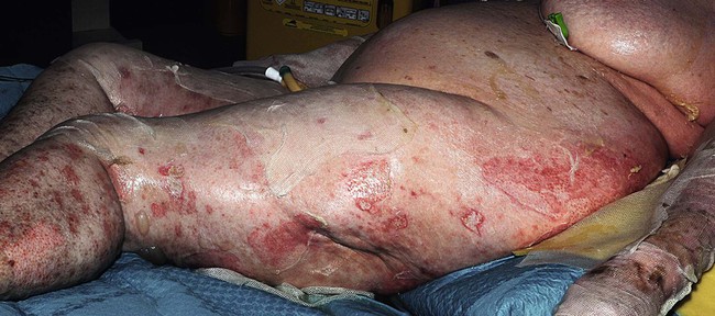
Toxic epidermal necrolysis and Stevens–Johnson syndrome
Specific investigations
Incidence of Stevens–Johnson syndrome and toxic epidermal necrolysis in patients with the acquired immunodeficiency syndrome in Germany.
Toxic epidermal necrolysis and Stevens–Johnson syndrome


 Supportive measures
 Withdrawal of culprit drug
 Analgesia
 Bioengineered skin substitute


