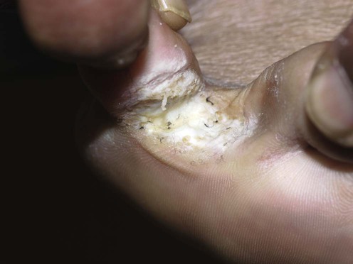
Tinea pedis and skin dermatophytosis


Tinea pedis and skin dermatophytosis


Eirini E. Merika and L. Claire Fuller


Evidence Levels:  A Double-blind study  B Clinical trial ≥ 20 subjects  C Clinical trial < 20 subjects  D Series ≥ 5 subjects  E Anecdotal case reports


image


Superficial fungal infections are the commonest of all mucocutaneous infections and appear to be on the increase. Dermatophytes infect keratinized epithelium, the hair and nails. Tinea pedis (athlete’s foot) describes a dermatophyte infection of the soles of the feet and interdigital spaces; tinea cruris, an infection of the groin; tinea facei, the face; and tinea corporis the rest of the skin.


Management strategy


Skin dermatophytosis rarely causes significant morbidity and certainly not mortality, but there is some evidence that, especially tinea pedis, it acts as a portal of entry for bacteria that produce impetiginization, lymphangitis, and bacterial cellulitis.


Topical antifungal treatment (with topical azoles or allylamines) of tinea pedis is generally adequate, as it is for small areas of tinea corporis and cruris, but for extensive infections, and especially those in immunosuppressed patients, oral therapy may be required. Several antifungal topical therapies are available without prescription, and the vast majority of the disease burden is likely to be managed without intervention from medical personnel. Treatment schedules vary but it is generally accepted that treatment should be applied once to twice daily for 2 to 4 weeks, continuing for at least 1 week after the lesions have cleared. Eradication of tinea elsewhere in the body, such as tinea pedis in the context of tinea cruris or tinea ungium in tinea pedis, is paramount for effective treatment and prevention of relapse.


Extensive forms of infection, failure of local treatment or relapse from inadequate therapy may require oral therapy with systemic antifungals such as terbinafine, fluconazole or itraconazole. Oral therapy remains the most effective agent against extensive or non-responsive forms of tinea. British prescribing guidelines suggest the following regimens: terbinafine 250 mg daily for 14 days, fluconazole 50 mg daily for 2 to 4 weeks (up to 6 weeks in tinea pedis), and itraconazole 100 mg daily for 15 days or 200 mg daily for 7 days (longer for tinea pedis and manuum). Severe macerated forms of tinea pedis may well be superinfected with bacteria justifying concomitant antibiotic therapy, although some topical antifungal agents have in vitro antibacterial activity, e.g., ciclopiroxolamine. Other than general hygiene, there is only anecdotal evidence to support prevention strategies to minimize re-infection, such as using antifungal powder to areas prone to fungal infections after showering, and wearing shower shoes in public bathing facilities.


Specific investigations


The lesions should be scraped carefully, harvesting surface scale with a no. 15 blade or banana-shaped knife. The active edges of large lesions are likely to yield more scale. Avoiding the application of emollients prior to sampling aids sample collection and analysis. Blister and pustule tops may be ruptured and the contents swabbed and placed directly onto agar plates.


Stay updated, free articles. Join our Telegram channel

Aug 7, 2016 | Posted by in Dermatology | Comments Off on Tinea pedis and skin dermatophytosis

Full access? Get Clinical Tree

Get Clinical Tree app for offline access