Tinea
Lawrence Charles Parish
(ICD-9 110.0 CAPITIS; 110.2 MANUUM; 110.3 CRURIS; 110.4 PEDIS; 110.5 CORPORIS)
Symptoms and Signs
Superficial fungal infections may be asymptomatic, pruritic, or burning. They are often referred to as “ringworm” because the characteristic lesion is a round, scaling, red area with central clearing and sharp, elevated borders. Most dermatophyte infections are caused by species of Trichophyton, Epidermophyton, or Microsporon. The infections are described by location.
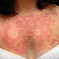
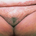
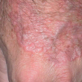
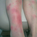
Tinea corporis can involve any part of the body. The lesions are oval to round, red, and scaling patches with a central area of clearing (Fig. 23-1). The patterns can be polycyclic. Tinea cruris, or jock itch, appears as diffuse redness and scaling with sharp borders. Tinea pedis, or athlete’s foot, appears in three forms. The most common, “moccasin foot,” presents as scaling on the soles, sometimes with erythema and crusting. Intertriginous infection manifests as fissuring and scaling between the toes—usually the fourth and fifth toes. Inflammatory and vesicular infection occurs on the instep and sole. Moccasin foot often appears along with tinea manuum, in which the “two-feet–one-hand” pattern is common and is a clue to diagnosis (Fig. 23-2).
Tinea capitis is generally limited to children. Tinea tonsurans causes the so-called black dot type of tinea capitis, in which broken hairs appear as dark spots on the scalp. When the infection is caused by Microsporon canis, there are scaling and well-defined patches of hair loss with broken hairs. With more inflammation and infection, a red boggy area, a kerion, may develop.
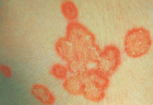 Figure 23-1 Tinea corporis due to Microsporon canis showing red rings with peripheral scale and central clearing.
Related posts:Stay updated, free articles. Join our Telegram channel
Full access? Get Clinical Tree
 Get Clinical Tree app for offline access

|