6
The Traumatized Face
No matter how severe or traumatic the facial injury (Fig. 6–1), these patients still require an appropriate trauma evaluation beginning with the ABCs (airway, breathing, circulation). Facial injuries are rarely life threatening; the patient must be evaluated for all other serious injuries before attempting repair. Usually treatment of any intraabdominal, thoracic, or neurologic injury takes precedence. Coordinate care between the trauma, thoracic, vascular, ENT, orthopedic, ophthalmic, and neurosurgical services.
Any exam should start with a detailed medical, surgical, social, and previous craniofacial injury history. The mechanism of injury should be ascertained to gauge the force of contact and determine where potential fractures or soft tissue injuries may be. Other considerations include loss of consciousness, breathing difficulties, and hearing trouble.
 Airway Establishment
Avoid nasal intubation in patients suspected of having a skull base fracture or excessive midface trauma. Elective oral endotracheal intubation should be considered in patients with severe pan-facial trauma, especially in the midface and mandible. Patients with large posterior base of tongue injuries should also be electively intubated. Any intubation should be done with due consideration of the cervical spine (C-spine): 10% of facial traumas harbor a C-spine injury. Tracheogtomy should be considered in complex cases, particularly when nasal or oral trauma preclude upper airway connulations.
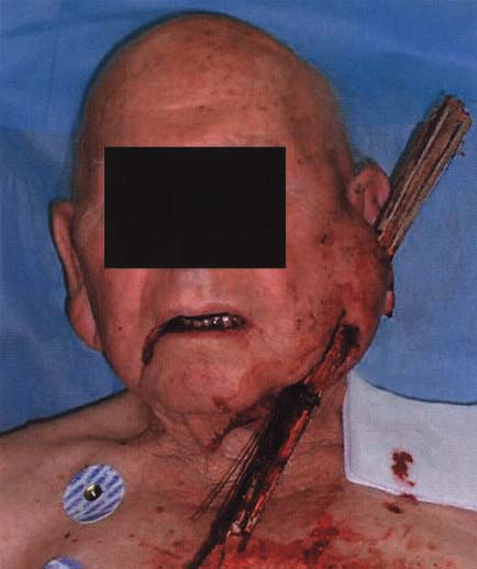
Figure 6–1 Patient impaled with a stick.
 Patient Evaluation
Examination
Remove all necessary articles of clothing and jewelry. Irrigate all dirt, foreign bodies, and dry crusted blood to avoid obscurity of the injury. Note all lacerations, asymmetries, bleeding, bruising, or foreign bodies. An organized systematic approach is recommended to avoid any missed injuries. Check for
• raccoon eyes (periorbital ecchymosis) – skull base fracture
• battle sign (postauricular ecchymosis) – skull base fracture
• otorrhea – skull base fracture, condylar fracture
• hemotympanum – skull base fracture
• perforated tympanic membrane
• epistaxis – nasal fracture
• CSF rhinorrhea – cribriform plate fracture, NOE fracture
• intraoral
 edema
edema
 bleeding
 gingival bleeding
 fractured/loose/displaced teeth
 dental caries
 septal hematoma
Nasal Palpation
• Tenderness
• Crepitus/subcutaneous emphysema
• Bony step-offs
 Scalp – gently palpate to uncover depressions/crepitus
 Forehead – frontal sinus fracture
 Orbital rim
 NOE (naso-orbito ethmoid) – palpate intranasally and inward from medial canthus-bony movement to diagnose NOE fracture
 Nasal bridge
 Zygoma
 Maxilla – gently depress the maxilla with both thumbs to rule out Le Fort fractures. If mobile, grab the central incisors between thumb and index finger with one hand and hold the nasal spine with other hand. Movement of the entire dental alveolus indicates a Le Fort I fracture; movement of the nasal bridge indicates Le Fort II or III.
 Mandible – preauricular pain on palpation can be indicative of a condylar fracture
 Neck exam – performed with caution in relation to the C-spine
Ophthalmic Assessment
• Inspection
 Corrective lens (contacts or eyeglasses)
 Enophthalmos/exophthalmos
 Retrobulbar hematoma
 Interpupillary distance – normally 30 to 32 mm
 Hyphema – blood layering in the inferior aspect of the anterior chamber. An ophthalmologist should be consulted immediately based upon the potential increase in intraocular pressure.
 Cornea abrasion
 Subconjunctival hemorrhage
 Chemosis – scleral edema
 Upper eyelid ptosis
 Fat protrusion
• Visual acuity

Stay updated, free articles. Join our Telegram channel

Full access? Get Clinical Tree


