Reflectance confocal microscopy (RCM) allows the evaluation with superb accuracy of some skin tumors before, during, and after treatment. In clinical trials RCM has been shown to provide useful information for evaluation of efficacy of topical or systemic medication. With the recent introduction of handheld RCM a fast examination of the tumor can be done in minutes. In patients treated with surgery RCM plays a unique role to precisely map margins of the tumor in the skin surface and for the detection of subclinical recurrences. This article reviews the use of RCM in the research of different skin cancer tumor treatments.
Key points
- •
RCM alone or in combination with dermoscopy or OCT has been shown to significantly improve accuracy in detection of tumor margins and recurrences in skin cancer.
- •
This tool can be useful in clinical trials and in clinical practice with this indication in melanocytic and nonmelanocytic tumors using either a handheld device or the standard RCM device to produce vivablocks.
- •
The main advantage of the first technical option is the reduced time of examination to a few minutes.
- •
It is probable that in the future faster scanners with higher resolution will be available in the market with the possibility of anatomic mapping of the tumor.
Introduction
Reflectance confocal microscopy (RCM) allows the evaluation with superb accuracy of some skin tumors before, during, and after treatment. The clinician and the researcher may use RCM for the detection of tumor margins, recurrences, or the investigation of tissue changes during medical treatments. Likewise in clinical trials RCM has been shown to provide useful information for evaluation of efficacy of topical or systemic medication. In this sense noninvasive evaluation of response to treatments substitutes the traditional approach of skin biopsy with the great advantage that the tissue changes can be evaluated in vivo. With the recent introduction of handheld RCM a fast examination of the tumor can be done in minutes. This solves the problem of time-consuming RCM examination using fixed RCM microscope examination. The handheld RCM modality allows a fast examination of tumor margins in melanoma or in patients with multiple tumors.
RCM evaluation is of major relevance in skin cancer assessment when ablative treatments are used or in the case of topical treatments, in particular indications in nonmelanoma skin cancer or in lentigo maligna.
In patients treated with surgery RCM plays a unique role to precisely map margins of the tumor in the skin surface and for the detection of subclinical recurrences. This has been used in melanoma in special situations, such as amelanotic melanoma or in lentigo maligna melanoma (LMM) on the face.
This article reviews evidence in the use of RCM in the research of different skin cancer tumor treatments.
Reflectance confocal microscopy in studies of skin cancer treatment
Reflectance Confocal Microscopy in Actinic Keratosis
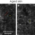
Evaluation of efficacy of treatment of actinic keratosis (AK) and field cancerization in clinical trials traditionally has been done with naked examination and biopsies of lesions. For clinical evaluation, clinical pictures for documentation of the tumor can also be used. However, the clinical naked eye examination cannot assess with accuracy tissue changes or the presence of subclinical lesions, whereas biopsies have the inconvenience of local scar, pain, and the lack of dynamic information in vivo. Response of AKs to topical treatment modalities has been assessed by RCM in several clinical studies used alone or in combination with dermoscopy or different optical coherence tomography (OCT) modalities. The main advantage of RCM is that clinical and subclinical AKs can be monitored and dynamic changes in the tumor can be evaluated in vivo in real time ( Fig. 1 ). This cannot be done with the biopsy of the tumor before, during, or after treatment because the tissue is removed. The main limitation of RCM in AK is in thick tumors with marked hyperkeratosis because of the reduced depth penetration of 200 μm to 300 μm of the microscope. However, in thin AKs or in field cancerization RCM seems to be the best option for monitoring the response to topical treatment; photodynamic therapy (PDT); or ablative options, such as laser or cryotherapy.
There are several studies concerning the efficacy of RCM in monitoring treatments. In 2009 Ulrich and colleagues demonstrated the applicability of RCM for noninvasive monitoring and detection of field cancerization and subclinical AK. The authors in this study described the clinical response of AKs and subclinical AKs to topical treatment in 11 volunteers with imiquimod 5% cream. In nine patients the clinical and RCM evaluation showed clearance of AKs. In one volunteer the clearance was clinical but on RCM examination showed persistence of AK features. The limitation in the evaluation with RCM was in skin areas with intense superficial inflammation or crusting because in these areas the penetration depth was limited, interfering with resolution, and impeding the visualization of cellular detail.
In 2015 Malvehy and colleagues published a study of monitoring treatment with 3% diclofenac sodium plus 2.5% hyaluronic acid and their correlation with histopathology. At RCM the degree of atypical honeycomb decreased and an elevated level of inflammation persisted during the entire period of study. Changes in parakeratosis, inflammation, and dermal collagen remodeling were also observed.
Ulrich and colleagues in 2015 published a case series of eight patients with RCM monitoring for AKs and cancerization field. They suggested the in vivo evaluation with RCM was a promising management approach for subclinical AKs.
A study by Maier and colleagues to evaluate the response of AKs to topical ingenol mebutate using RCM and OCT showed that both tools are noninvasive methods that allow the monitoring of the clinical response to treatment. The combination of RCM and OCT was superior to the naked eye and may help the clinician to decide the therapeutic approach.
Malvehy and colleagues used the combination of RCM and OCT in a clinical trial with 20 patients with diagnosis of AK and subclinical AK treated with a combination of 0.5% 5-FU plus 10% salicylic acid once daily for 6 weeks. The authors evaluated the lesions and field cancerization with RCM and OCT before the treatment and 2 weeks after the end of treatment. The study showed a significant improvement in the imaging scores used to evaluate this lesion (scaling, detached corneocytes, atypical honeycomb, round nucleated cells in the spinosum granulosum layer, round vessels, inflammatory cells).
Recently, Longo and colleagues described the RCM findings of topical treatment response with ingenol mebutate in two patients. At baseline, RCM revealed typical findings seen in AKs, such as the presence of parakeratosis and irregular keratinocytes. Approximately a week after treatment vesicle formation with several inflammatory and necrotic cells was seen. With complete recovery, the RCM shows an epidermis with keratinocytes well-defined and a regular honeycomb pattern.
RCM has also been used in the monitoring of PDT therapy in this indication. In a recent study by Jafari and colleagues efficacy of daylight PDT was assessed in 40 grade I and II AKs in 20 patients. Complete resolution and partial resolution was proved in 80% and 17.5% of lesions, respectively. This study confirmed that vivo RCM examination correlated with clinical findings and that this technology can be used to monitor the efficacy of this treatment modality. The authors in their study proposed a new RCM atypia scoring system based on atypia and cell sizes.
Basal Cell Carcinoma
In the case of superficial basal cell carcinoma (sBCC) topical treatment with imiquimod, PDT, or ablative treatments can be used with excellent results ( Fig. 2 ). However, the precise diagnosis of this subtype needs to be confirmed. For this reason in addition to clinical judgment, dermoscopy, RCM, or OCT have been proposed for an accurate diagnosis.