The use of radiation therapy in the management of skin cancer is variable and often anecdotal. Applied as both primary and adjuvant therapy in patients with both nonmelanoma skin cancer and rarer tumors of the skin, a consensus regarding optimal dosing regimens has not yet been reached. Herein, the authors outline the basic concepts of radiation therapy for tumors of the skin and review its use for high-risk nonmelanoma skin cancer, as well as less common malignancies, including angiosarcoma, Merkel cell carcinoma, and sebaceous carcinoma.
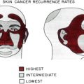
The administration of radiation therapy for the treatment of skin cancers spans several decades. In the context of more effective and efficient surgical methods, such as Mohs micrographic surgery and advances in reconstructive techniques, radiation is less frequently used in the management of skin cancer. However, radiation therapy remains a very useful tool both as a primary treatment and as an adjunctive treatment to enhance cure, specifically for skin cancers of the head and neck. Radiation can also be helpful in palliation for symptomatic relief of incurable skin cancer. This article reviews the current evidence on the use of radiation therapy for the treatment of skin cancer and describes the recommendations of the National Comprehensive Cancer Network (NCCN). Nonmelanoma skin cancers for which radiotherapy can play a role in treatment include basal cell carcinoma, squamous cell carcinoma, Merkel cell carcinoma (MCC), angiosarcoma, dermatofibrosarcoma protuberans, and sebaceous carcinoma.
Techniques of radiation therapy
Radiation therapy can be administered to a cutaneous lesion through a variety of techniques. However, 3 specific methods have been used to deliver radiation to tissue. The first method, orthovoltage radiation, is preferred for more superficial lesions, whereas megavoltage electron beam technology is preferred for reaching deeper tissue. Also, brachytherapy can be used in the interest of delivering radiation directly to the cancer, while sparing noncancerous surrounding tissue.
Orthovoltage X-rays, also known as superficial X-rays, usually range from 75 to 125 kV. Orthovoltage X-rays are the usual choice for radiating lesions thinner than 5 mm, because the X-rays interact maximally with the skin at the surface, before losing strength as they penetrate downward and are attenuated rapidly by interactions with atoms in the tissues traversed. Orthovoltage rays lose 10% of their strength every 0.5 cm that they travel, even when a filter is used. As such, a 100 kV superficial X-ray would diminish to only 90 kV after passing through 0.5 cm of tissue.
Because most cutaneous tumors lie within a few millimeters of the skin surface, superficial X-rays have favorable beam characteristics to more than adequately penetrate such neoplasms. However, some tumors are too thick to be irradiated in this manner. Because of the limited applicability of orthovoltage X-rays for treatment of other cancers, many radiation therapy departments have phased it out in favor of other modes of radiation therapy.
In the place of orthovoltage X-rays, megavoltage electron beam technology has emerged as a more widely available radiotherapy option. This type of radiation is produced by devices known as linear accelerators, which afford clinicians the advantage of being able to generate radiation of variable energy. Electron beams range in strength from 6 to 20 MeV and can reach tissue up to approximately 6 cm deep, radiating the tissue in a relatively even manner. The electron beams provide homogenous tissue coverage to a known depth and then dissipate rapidly. One disadvantage of megavoltage electron beam radiation is the significantly lower than maximum dose delivered at the surface of an air-tissue interface in a low-energy electron beam. The peak energy is delivered below the skin, resulting in a weaker dose being administered to the surface than to underlying tissues. This issue is typically corrected by placing skin-equivalent material on the skin surface to achieve the strongest possible surface dose. In addition, the clinician must take into account the presence of structures, such as the nose or the ears, that have an uneven contour; specific techniques are applied to compensate for such contours when delivering electron beam radiation to preserve the homogeneity of the radiation.
Also, brachytherapy, also known as interstitial therapy, is useful in treating small accessible skin cancers by allowing the delivery of large doses of radiation directly to the lesion. This technique relies on the insertion of radioactive material, usually in the form of iridium 192 seeds, into the tumor to treat the cancer. This insertion is accomplished by placing the seeds in nylon ribbons, which introduce the radioactive seeds manually into the lesion so that they are spaced 1 cm apart. The radiation dose delivered by an interstitial source declines proportionally to the inverse square of the distance from the source. Because of this physical property, very high radiation doses can be delivered close to the source, and the dose declines rapidly with distance. Brachytherapy spares much of the surrounding normal tissue that would otherwise be radiated with electron beam or orthovoltage radiation. The tumor is irradiated continuously for 4 to 6 days, which may be a theoretical advantage because tumor proliferation during the radiation course is limited. Some of the notable disadvantages of brachytherapy include the necessity for the patient to remain hospitalized in isolation during treatment and the insertion of radiation sources under general anesthesia. Thus, external beam therapy is generally more practical.
In treating tumors of the skin with radiotherapy, 2 concepts are fundamental. The first is the half-dose depth, the tissue depth at which half of the surface dose is delivered. Because superficial X-rays penetrate the skin, interaction with atoms in the tissues through which they travel attenuates them, and at a certain depth, only 50% of the surface dose is delivered. If 50% of the surface energy reaches the base of a given tumor, the normal tissues directly deep to the tumor receive 50% of the surface dose, and radiation damage to the normal tissue is limited. Therefore, careful calculation of the radiation dose is imperative to ensure that the amount of radiation delivered to the tumor base and the tissue beneath it is approximately 50% of the dose delivered to the skin surface.
The second concept, known as the 90% isodose concept, relates to electron beam radiation. This type of radiation can be used to treat a tumor homogeneously with the same dose, with less than 10% variation. The base of the tumor receives no less than 90% of its surface radiation dose. As a result, the normal tissue underlying a cancer receives more radiation.
Fractionation of the radiation dose is another important concept in optimizing the cosmetic outcome and minimizing side effects of radiation therapy at the treatment site. By dividing the total radiation dose into fractions that are delivered at intervals over several days or weeks, the side effects of a single large dose are mitigated. The total radiation dose is modified according to the size of each fraction.
Basal cell carcinomas and squamous cell carcinomas
According to the most recent NCCN guidelines of 2010, approximately 1 million cases of basal and squamous cell carcinoma are diagnosed every year, with squamous cell cancers (SCCs) accounting for approximately 300,000 cases and basal cell cancers (BCCs) constituting the remaining cases. According to a recent survey, radiation is used as a first-line treatment for only 8% of BCCs in the United Kingdom and 2% in the Netherlands. Surgical treatments, such as Mohs micrographic surgery or standard surgical excision, are typically the initial treatments for most BCCs and provide histologic evidence of tumor clearance, excellent cure rates, as well as cosmetic and functional preservation. However, because basal and SCCs are relatively radioresponsive, radiotherapy can be used as a primary modality with a curative intent, a preoperative or postoperative adjunctive modality, or as a palliative treatment. NCCN guidelines for treatment of basal and squamous cell carcinomas are helpful in determining when to consider radiation therapy and are described later. Of note, the NCCN guidelines for the use of radiotherapy in management of skin cancers are category 2B, indicating that they are based on a lower level of evidence and nonuniform consensus but without major disagreements among the experts involved in formulating the guidelines. Because of the lack of controlled prospective studies, there are no consensus recommendations for the use of radiation for SCCs of the skin.
Primary Treatment
When surgical excision of a cancer may result in functional or aesthetic compromise, radiation therapy may be the optimal primary treatment, even for superficial lesions such as Bowen disease (cutaneous SCC in situ). Radiotherapy is also suitable as the primary treatment of extensive tumors that are surgically unresectable, such as inaccessible tumors located at the skull base. Deep lesions at the cranial nerve foramina or in the brain are most effectively radiated with electron beam radiation that can deliver radiation dose to a depth of 6 cm. Radiation therapy becomes the primary treatment of choice for patients who are poor surgical candidates; patient preference may also lead to selection of radiation therapy as the primary treatment.
Scouting biopsies around the site of the cutaneous malignancy can help to determine the extent of the cancer and the size of the radiation field when the cancer is ill defined or extensive. The radiation field includes the area involved by the cancer and a margin around it.
Adjuvant Radiotherapy
Adjuvant radiation therapy can be used preoperatively to reduce the extent of a tumor in an effort to make it resectable. In clinical settings where extended surgical excision to obtain negative margins with Mohs micrographic surgery might involve sacrificing major nerve structures and cause significant functional and cosmetic morbidity, adjuvant radiotherapy is a valuable tool. In such a scenario, further excision may be deferred to preserve vital structures, and radiation therapy is added to treat the residual skin cancer. The role of adjuvant radiation in the management of perineurally invasive skin cancer is discussed separately later. Postoperative adjunctive radiation is used when there remains residual cancer after inadequate excision and reexcision is not possible or there is a high likelihood of residual disease after excision and the probability of successful treatment of a recurrence is modest. Tumors excised with positive margins, multiply recurrent tumors, and those with perineural or bony invasion are indications for postsurgical adjunctive radiation.
Management of regional lymph nodes
Elective radiation of clinically uninvolved nodes is performed when there is a high probability (≥20%) of subclinical disease as might be expected in the presence of multiple high-risk features and proximity to the parotid gland. The presence of tumor in palpable regional lymph nodes must be confirmed by fine needle aspiration or lymph node biopsy. Imaging studies help to rule out distant disease in addition to determining the number, size, and location of involved nodes. Lymphadenectomy is sufficient for a solitary cervical lymph node, less than 3 cm, without extracapsular extension. For more advanced disease with larger nodes, multiple nodes, or bilateral nodal involvement, postoperative radiation should be strongly considered and is recommended by some radiation oncologists. Preoperative radiation may be used before lymph node dissection for borderline unresectable clinically positive disease in the neck. Radiation alone is used for unresectable lymph node disease or for patients who are medically inoperable.
Palliative Radiotherapy
Palliative radiotherapy may be helpful in managing symptomatic basal and squamous cell carcinomas in terminally ill patients. The discussion of this subset of patients is beyond the scope of this article.
Perineural Invasion
The histologic finding of perineural invasion (PNI) by cancer cells is associated with aggressive tumor behavior in both basal and SCCs. PNI may be categorized as either microscopic or macroscopic (clinical). It is estimated that approximately 60% to 70% of all PNIs are microscopic, in that they only affect smaller nerves (<1 mm in diameter in the reticular dermis). The remaining 30% to 40% of PNIs manifests with clinical signs or symptoms and involve larger nerves of diameters greater than or equal to 1 mm. Most cases of PNI occur in SCCs, which have a 2.5% to 14% risk of becoming perineurally invasive. However, BCCs may also exhibit such behavior and carry a 1% risk of developing PNI.
A review of the data on the behavior of cutaneous SCC (cSCC) and BCC, described later, demonstrates the role and significance of adjuvant radiotherapy in improving outcomes. PNI carcinomas tend to be larger and have greater subclinical extension and a higher risk of recurrence than those without PNI. In one study, Leibovitch and colleagues reported a 5-year recurrence rate after Mohs surgery for BCCs with PNI of 7.7%, compared with 2.4% for BCCs without PNI. Evidence from many studies suggests that the size of involved nerves in cases of perineurally invasive squamous cell carcinoma can serve as an important prognostic indicator. In a small retrospective cohort study of 48 patients, Ross and colleagues noted rates of local recurrence of 50%, nodal metastases of 38%, distant metastases of 32%, disease-specific death of 32%, and all-cause death of 48% for patients presenting with macroscopic PNI, involving nerves 1 mm or more in diameter. These rates were significantly higher than the 9% local recurrence rate, 0% nodal metastasis rate, 0% distant metastasis rate, 0% disease-specific death rate, and 17% all-cause death rate associated with microscopic PNI, which involved nerves measuring less than 0.1 mm in diameter. In another study, conducted by Mendenhall and colleagues, it was observed that patients presenting with incidental PNI had higher rates of local control over the course of 5 years at 79% when compared with their counterparts who presented with clinical PNI and had a 5-year local control rate of 53%. Hence, adjuvant radiation therapy is recommended for cSCC with extensive perineural disease, even if the margins of resection are tumor free.
The association between PNI and tumor recurrence, as well as the role of adjuvant radiotherapy in the treatment of perineurally invasive disease, has been the subject of several studies. Early detection followed by complete surgical resection provides for the best chance of local and eventual cure. Han and Ratner observed in a 2007 study that the cure rate for cSCCs with microscopic PNI treated with Mohs surgery with or without radiation was 92% to 100%, compared with cure rates of 38% to 100% for those treated with standard excision with or without adjunctive radiotherapy. These findings emphasize the importance of complete excision of the cancer. However, because of the more aggressive biologic behavior and poorer outcomes of cSCC with PNI, adjuvant radiation to the site of the primary cancer may be helpful in achieving better cure rates. Adjuvant radiation may be considered after Mohs surgery in the presence of PNI because of the belief that “skip areas” beyond an excision site may exist, creating a false-negative margin. Many recommendations for radiation therapy are based on retrospective studies of both BCCs and SCCs and do not separately identify recommendations for BCCs. The role of adjuvant radiotherapy for microscopic PNI is not as well defined in the literature as it is for extensive PNI. Total tumor doses of 75 Gy with an altered-fractionation schedule are recommended for tumors with clinical PNI.
Because of the risk of incomplete excision and local recurrence, postoperative radiotherapy is often recommended after standard excision for SCC with PNI. Retrospective data show that radiation therapy to the site of the SCC reduces the risk of local, regional, and distant recurrence, especially for tumors with PNI. Mendenhall and colleagues recommended that most patients with PNI undergo radiation therapy as an adjuvant to surgery in an effort to prevent local and regional recurrence. Local control rates achieved with surgery, with or without adjuvant radiation, in different studies of cSCC with PNI, are summarized in Table 1 . Table 2 compares the local recurrence rate and disease-free survival in patients treated with surgery alone and those treated with surgery plus adjuvant radiation.
| Author | Year | No. of Patients | Treatment | 5-year (Unless Noted Otherwise) Local Control Rate |
|---|---|---|---|---|
| Ampil et al | 1995 | 9 SCC | Surgery ± radiation therapy | 3-y local control rate: 66.7%, |
| Mendenhall et al | 2002 | 46 SCC 16 BCC | Radiation therapy ± surgery | Microscopic PNI: 79% Clinical PNI: 50% |
| Garcia-Serra et al | 2003 | 135 BCC and SCC | Radiation therapy ± surgery | Microscopic PNI: 87% Clinical PNI: 55% |
| Leibovitch et al | 2005 | 70 SCC | MMS ± radiation therapy | Microscopic and clinical PNI: 92% |
| Author | Year | No. of Patients | Treatment | Outcome (5-year) |
|---|---|---|---|---|
| Veness et al | 2007 | 167 | Surgery (21) | Local recurrence rate: 43% Disease-free survival rate: 54% |
| Surgery + ART (146) | Local recurrence rate: 20% Disease-free survival rate: 73% | |||
| Jambusaria-Pahlajani et al | 2009 | 28 | Surgery alone (9) | Local recurrence rate: 0% Disease-free survival rate: 100% |
| Surgery + ART (19) | Local recurrence rate: 0% Disease-free survival rate: 74% |
Elective radiation of clinically normal regional lymph nodes in patients with PNI has been shown to be important in preventing regional recurrences. In a retrospective study of skin cancers (including both BCCs and SCCs) with PNI conducted by Garcia-Serra and colleagues in 2003, the regional (lymph node) recurrence rate in patients with clinical PNI was 5%, compared with 14% in those with microscopic PNI. The difference was attributed to the fact that twice as many patients in the clinical PNI group had been given elective radiation of the regional lymph nodes when compared with those in the microscopic PNI group. This observation underscores the value of elective irradiation of regional lymph nodes in the setting of PNI. These data imply that microscopic metastases may already be present in the lymph nodes and become clinically apparent even though the tumor has been completely excised. Because 15% to 20% of patients with clinically negative nodes are likely to have subclinical disease, Mendenhall and colleagues further recommended that such patients undergo elective radiation therapy to the regional lymph nodes. There is general agreement that adjuvant radiation therapy is warranted in cases of PNI involving larger named nerves. However, adjuvant radiotherapy seldom leads to improvement in cranial nerve deficits, with improvement in symptoms in only 7% of patients without local recurrence.
In a multivariate analysis, Ross and colleagues did not observe involved nerve diameter as a significant risk factor in their small study. Only tumor size and patient age were found to be meaningful predictors of survival. Therefore, they only recommended the use of adjuvant radiation therapy when other high-risk factors associated with poor outcomes are also present in addition to PNI ( Box 1 ). They did, however, acknowledge the necessity of larger prospective studies to confirm this conclusion.
Features:
Diameter greater than 2 cm
Ill-defined margins
Recurrence
Location on areas such as lip and ear
Depth 4 mm or more or infiltration into deep reticular dermis or fat (Clark level IV, V)
Poor differentiation; adenoid, adenosquamous, or desmoplastic histology
Site of chronic inflammation
Rapid growth
Immunosuppression of host
Related posts:
 Mohs Micrographic Surgery Technique
 Mohs Surgery for Squamous Cell Carcinoma
 Flaps and Grafts Reconstruction
 Management of Unusual Cutaneous Malignancies: Atypical Fibroxanthoma, Malignant Fibrous Histiocytoma, Sebaceous Carcinoma, Extramammary Paget Disease
 Special Considerations for Mohs Micrographic Surgery on the Eyelids, Lips, Genitalia, and Nail Unit
Multidisciplinary Approach to Large Cutaneous Tumors of the Head and Neck
Special Considerations for Mohs Micrographic Surgery on the Eyelids, Lips, Genitalia, and Nail Unit
Multidisciplinary Approach to Large Cutaneous Tumors of the Head and Neck
Stay updated, free articles. Join our Telegram channel

Full access? Get Clinical Tree


