Moderate to severe atopic dermatitis (AD) can be debilitating and often requires use of systemic immunosuppressant therapy to achieve adequate disease control. There are currently no US Food and Drug Administration–approved systemic agents for the long-term treatment of AD. Recent insight has identified the T helper 2 cytokines, interleukins 4 and 13, as playing a major role in the pathogenesis of AD. There are multiple novel biologic agents in development that target interleukins 4 and/or 13 for the treatment of moderate to severe AD. The age of targeted biologics for AD has arrived.
Key points
- •
Atopic dermatitis is an inflammatory skin disease mediated by increased T helper 2 inflammation in the skin and blood.
- •
Novel biologics targeting the T helper 2 cytokines, interleukins 4 and 13, show promise for the treatment of moderate to severe atopic dermatitis.
Introduction
Atopic dermatitis (AD) is a chronic, pruritic, inflammatory skin and potentially multisystem disorder that is associated with considerable morbidity. The most common morbidity, severe itch, in AD may result in difficulty falling asleep, staying asleep, more frequent nighttime awakenings, nocturnal scratching, and poor sleep efficiency, ultimately leading to daytime fatigue and impairment of instrumental activities of daily living. AD is also associated with increased symptoms of anxiety and depression and higher rates of diagnosed depression, anxiety, attention-deficit (hyperactivity) disorder, and other mental health disorders in both children and adults. Moreover, chronic itch and AD are associated with impaired productivity at school and work, social and relationship problems, and poor health-related quality of life.
AD is commonly associated with several atopic disorders in children and adults, including asthma, hay fever, and food allergy. The overlap of these disorders suggests potentially overlapping disease mechanisms and/or triggers that extend beyond the skin. Recent studies have identified several previously unrecognized comorbidities of AD, including cardiovascular disease, myocardial infarction, stroke, obesity, osteoporosis, injury and fracture, alopecia areata, and vitiligo. Taken together, the comorbid health conditions occurring in patients with AD suggest that AD is a systemic disease, with widespread harmful effects.
There are currently several unmet needs in the management of AD, particularly pruritus. First, AD is typically managed by treating flares after activity has become full blown. Although this approach is reasonable in patients with only occasional flares, it is inadequate in patients with frequent flares or persistent disease with daily symptoms. In fact, moderate AD may be associated with symptoms 1 out of every 3 days in perpetuity. Second, AD is typically managed using topical therapies, including emollients, corticosteroids (TCS), and calcineurin inhibitors. Topical therapies are typically effective for treating mild disease, but may not be effective in more severe disease and do not address underlying systemic inflammation. Moreover, they are impractical and difficult to apply for patients with extensive disease. Several systemic immunosuppressants have been shown to be effective for treating AD, including intramuscular or oral corticosteroids, cyclosporine, methotrexate, azathioprine, and to a lesser extent, mycophenolate mofetil. Each of these agents has a poor adverse-effect and/or tolerability profile, which limits their use in the clinical dermatology setting and requires laboratory monitoring for adverse effects. For example, systemic corticosteroids can cause glucose intolerance, weight gain, insomnia, depression, psychosis, and adrenal and immune suppression. Cyclosporine can cause, among other side effects, headaches, anemia, nephrotoxicity, hypertension, hirsutism, and electrolyte abnormalities. Methotrexate can cause anemia, leukopenia, gastrointestinal discomfort, weakness, and hepatotoxicity. Even when these agents are successfully used for treating AD, their potential toxicity precludes them from being used long term. The US Food and Drug Administration (FDA) has recommended that cyclosporine not be used in other disorders more than 1 year. Moreover, none of these agents are approved by the FDA for the treatment of AD. Only cyclosporine is approved for the treatment of AD in Europe.
Improved understanding of the immune basis of disease has allowed for the development of multiple novel targeted therapies in the AD pipeline. The principal benefit of more targeted therapy is the combination of improved efficacy and safety. The treatment of moderate to severe psoriasis has been revolutionized by the advent of multiple biologic therapies. This review focuses on the roles of interleukin 4 (IL-4) and/or -13 in the pathophysiology of and development of targeted biologics for AD.
Pathogenesis
AD pathophysiology involves the interaction of epidermal barrier dysfunction with systemic inflammation and immune dysregulation. However, a fundamental debate exists as to whether AD is driven primarily by barrier dysfunction (outside-inside hypothesis) or primarily by an inflammatory response to irritants and environmental allergens (inside-outside hypothesis).
Outside-In Hypothesis
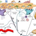
The “outside-in” hypothesis posits that epidermal barrier dysfunction precedes AD and is required for the disease to manifest. The outside-in hypothesis is supported by previous studies demonstrating loss-of-function mutations in the filaggrin gene (FLG). Suboptimal filaggrin proteins may alter epidermal corneocyte shape and change the organization of lamellar bodies, resulting in impaired barrier function of the epidermis. Poor epidermal barrier function leads to increased transepidermal water loss, decreased skin hydration, and vulnerability to exogenous insults. Skin barrier dysfunction might also be acquired secondary to irritants and mechanical disruption. Damaged keratinocytes from the disrupted epidermal barrier may then trigger the recruitment and/or expansion of inflammatory cells via release of thymic stromal lymphopoietin and other cytokines. Epidermal barrier breakdown also permits allergen penetration and binding to Langerhans cells, resulting in increased Th2 inflammation in the skin and systemically; this may also predispose toward atopic diseases, for example, asthma and food allergy.
Inside-Out Hypothesis
The “inside-out” hypothesis posits that inflammation precedes and even causes barrier dysfunction in AD. Recent studies identified multiple polymorphisms of inflammatory genes in patients with AD, including IL-4 receptor-α (IL4Rα), IL-4, IL-13, IL-31, cluster of differentiation 14 (CD14), serine peptidase inhibitor, Kazal type 5, chemokine (C-C motif) ligand 5 (RANTES). These polymorphisms may lead to (a) immune dysregulation and cutaneous inflammation, resulting in (b) impaired keratinocyte differentiation and function, followed by (c) downregulation of filaggrin and antimicrobial peptides (AMPs), thereby (d) allowing penetration of exogenous allergens.
In summary, the outside-in and inside-out hypotheses differ on the sequence of events leading to disease manifestation. It may be that the outside-in hypothesis applies to a subset of patients, such as those with FLG polymorphisms, whereas the inside-out hypothesis applies in patients with polymorphisms of immune-related genes. Regardless of “the chicken or the egg,” both hypotheses agree that all patients have a combination of immune dysregulation, inflammation, and skin-barrier dysfunction.
Role of interleukin 4 and interleukin 13 in atopic dermatitis
Interleukin 4 and Interleukin 13 Activity in Atopic Dermatitis
IL-4 and IL-13 play prominent roles in inflammation, epidermal barrier dysfunction, itch, and susceptibility to infection in AD. In 1994, Hamid and colleagues quantified IL-4 messenger RNA in skin biopsies of both acute and chronic lesions and nonlesional skin in AD patients and normal control skin. IL-4 gene expression was highest in acute AD lesions, but also increased in chronic AD lesions compared with nonlesional AD and normal control skin. In 2001, transgenic murine models expressing epidermal IL-4 produced an AD-like phenotype, including pruritus, xerosis, inflammatory skin lesions, Staphylococcus aureus infection, and histopathology of chronic dermatitis with T cells, eosinophil infiltration, and elevation of total serum immunoglobulin (Ig) E and IgG1. Other studies in both mice and humans substantiated the findings that IL-4 is increased in AD and that it may play a central role in pathogenesis.
IL-13 has also been established as a critical cytokine in AD. Some studies have suggested that IL-13 may even be of greater pathophysiologic importance in AD than IL-4. Increased expression of IL-13 also occurs in acute and chronic AD lesions in human skin, and transgenic mice with cutaneous IL-13 expression also develop an AD-like phenotype. IL-13 is produced by multiple immune cells, including CD4+ and CD8+ T cells in AD lesions as well as mast cells, basophils, and eosinophils. Elevated IL-13 levels in AD lesions may initiate cutaneous inflammation and fibrotic remodeling.
Interleukin 4, Interleukin 13, and Barrier Disruption in Atopic Dermatitis
As reviewed above, epidermal barrier dysfunction and keratinocyte damage are critical to the pathogenesis of AD ( Fig. 1 ). Howell and colleagues found that IL-4 and IL-13 inhibited filaggrin production in vitro during keratinocyte differentiation, suggesting that Th2 inflammation can result in acquired filaggrin deficiency and worsen barrier dysfunction overall. IL-4 and IL-13 were also found to downregulate keratinocyte expression of loricrin and involucrin, 2 important proteins for skin barrier formation and integrity. Exposure to IL-4 in vitro was also found to reduce levels of ceramides, a class of important hydrophobic molecules in the stratum corneum, and desmoglein-3, a key protein in desmosomes. In murine models, IL-4 has an inhibitory effect on barrier recovery, possibly by interfering with IL-1α effects, including DNA and lipid synthesis in keratinocytes.
Interleukin 4, Interleukin 13, Immunoglobulin E, and Allergic Disease
IgE is central to allergic diseases, such as asthma, hay fever, and food allergy. IgE is produced by plasma cells upon stimulation with IL-4 and IL-13. IgE binds to high-affinity receptors on mast cells and basophils and can trigger the release of inflammatory mediators leading to type 1 mediated hypersensitivity reactions in allergic disease. Patients with AD have significantly higher rates of asthma, hay fever, food allergy, and other allergic disorders. Thus, many previously thought that IgE played a critical role in the pathophysiology of AD. However, 20% to 50% of patients with AD may have normal total and allergen-specific IgE levels, suggesting that IgE is not pathogenic in many AD patients. More recent paradigms have placed greater emphasis on the upstream T cells and T helper 2 inflammation, including IL-4 and IL-13, rather than the IgE downstream.
IL-4 and IL-13 also play a critical role in asthma. IL-4 triggers IgE isotype switching, promotes eosinophil migration, increases mucus secretion, induces vascular cell adhesion molecule 1 expression, and causes Th2 differentiation. In murine models, monoclonal antibodies to IL-4 before aerosol challenge neutralized Th2 inflammation that is integral for asthma pathogenesis. Similarly, in murine models, selective blockade of IL-13 followed by antigen challenge failed to produce airway hyperresponsiveness, indicating that IL-13 is necessary for allergic asthma. IL-13 has also been found to promote airway eosinophilia, activate macrophages, and increase airway mucus production.
There is significant overlap in cytokine function as receptors for both IL-4 and IL-13 use the IL4Rα chain for signal transduction, but there are key differences. Both cytokines stimulate plasma cell IgE production, but IL-4 promotes Th2 cell differentiation, whereas IL-13 does not. IL-4 is also involved in eosinophil recruitment. Overall, the scope of function of IL-13 is slightly narrower than that of IL-4 because of a more restricted distribution of the IL-13 receptor, which is most pronounced by the inability of IL-13 to stimulate T-cell differentiation. Some investigators have suggested that IL-4 promotes Th2 cell development, whereas IL-13 promotes Th2 tissue inflammation.
Other Roles of Interleukin 4 and Interleukin 13 in Atopic Dermatitis
Previous studies found that IL-4 and IL-13 downregulate AMP expression in AD skin. Downregulation of AMP in combination with barrier dysfunction and immune dysregulation likely contributes to the increased risk of cutaneous and extracutaneous infections. IL-4 and IL-13 were found to correlate with AD severity and IL-31 levels. Moreover, IL-4 was found to induce IL-31 expression by Th2 cells. IL-31 appears to be an important inflammatory mediator of itch in AD.
Related posts:


Stay updated, free articles. Join our Telegram channel

Full access? Get Clinical Tree


