The Lips


Chapter 17
The Lips


Introduction


‘Eternity was in our lips and eyes’.


William Shakespeare, Antony and Cleopatra (1606–7)1


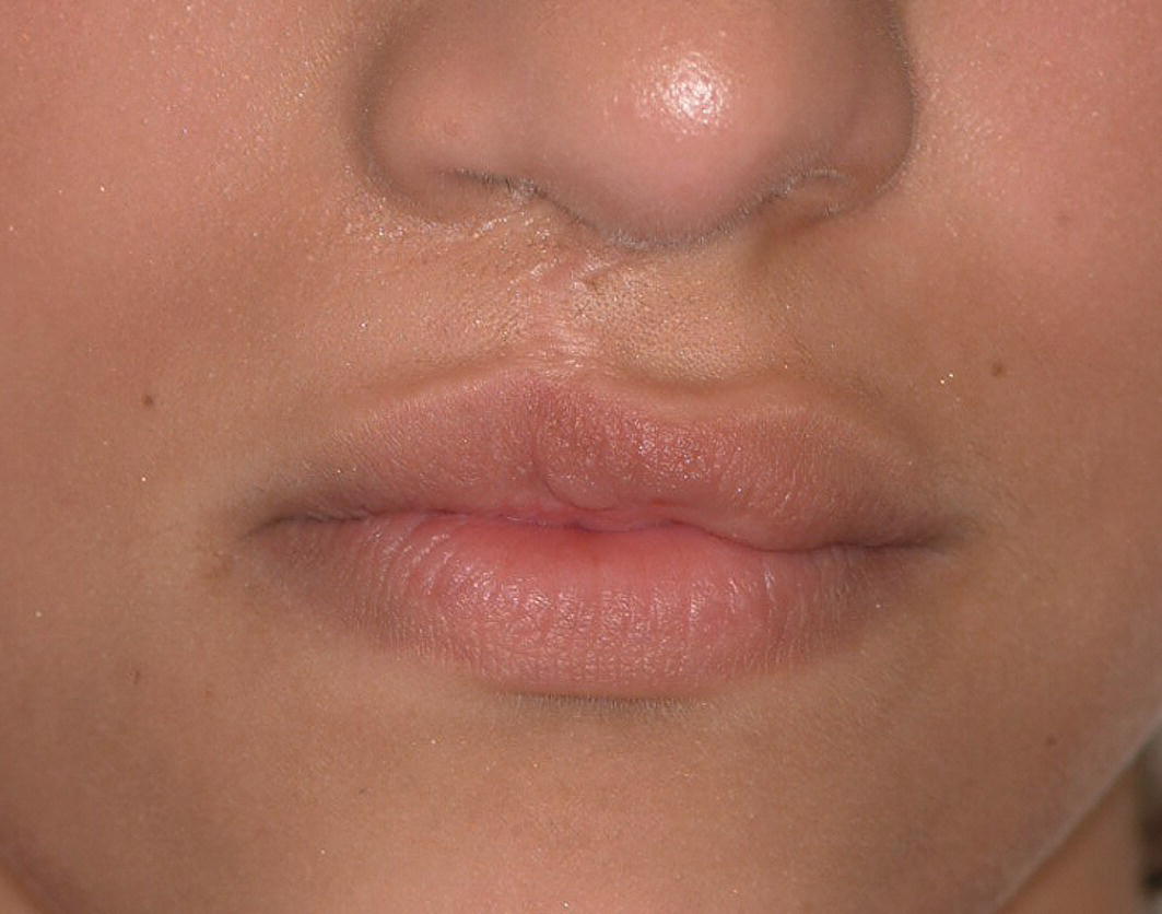
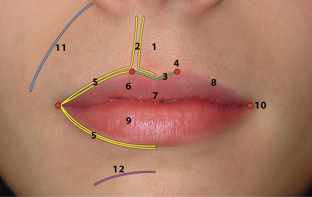
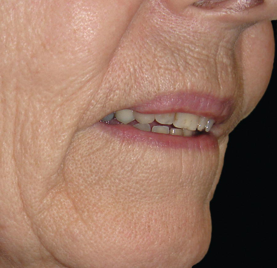
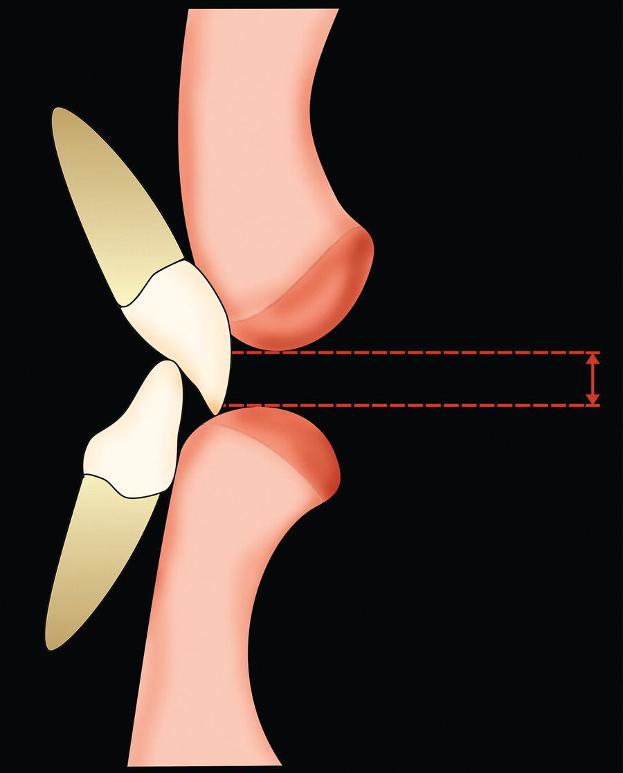
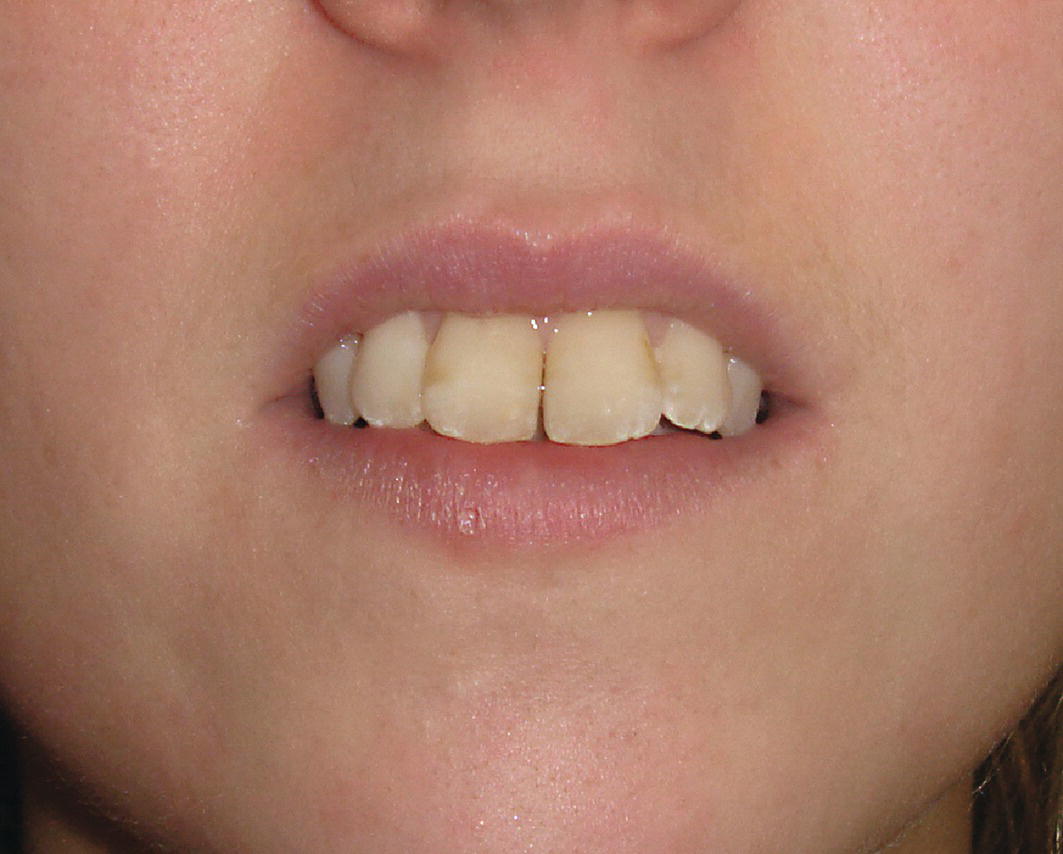
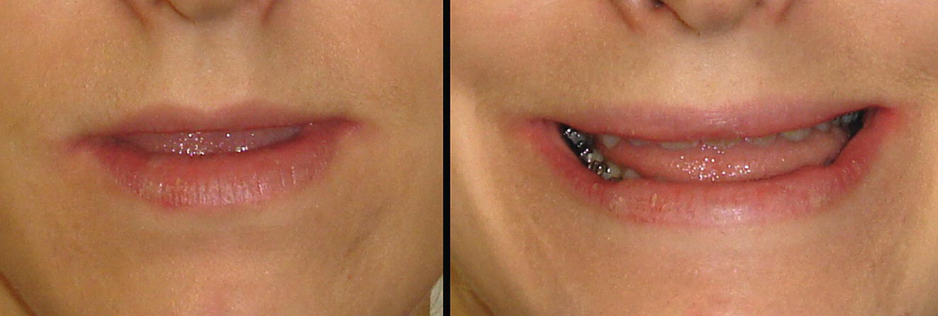
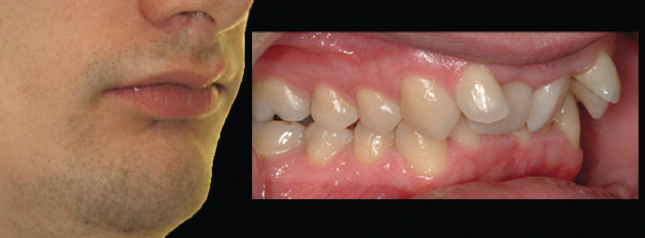
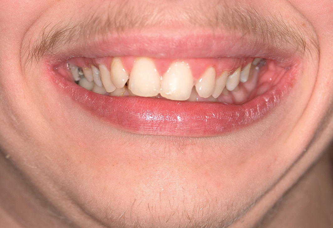
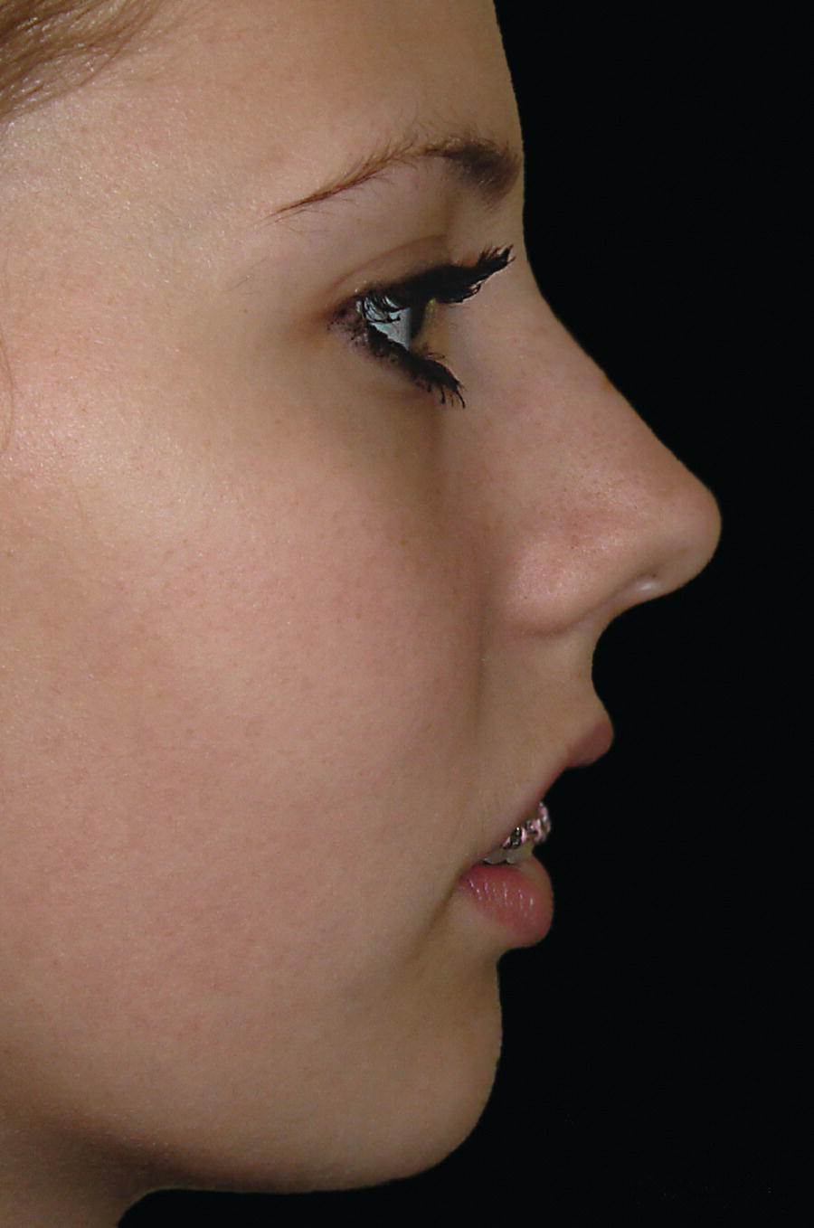
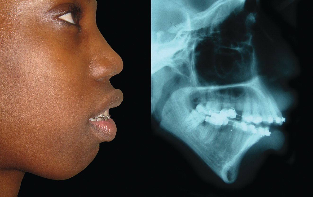
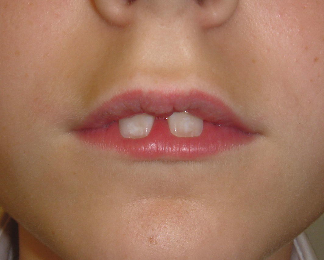
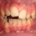
The lips form a transition zone between the facial skin and the oral mucosa and are a significant aesthetic feature of the face (Figure 17.1). They serve important functional roles in phonation and the formation of an anterior oral seal in swallowing. The vermilion of the lips is highly sensitive to light touch sensation and is represented by a large area in the sensory cortex. It is the main exploratory area in babies, before they learn to use their hands for stereognosis.


The lips are perhaps the most movable expressive aesthetic unit of the face. Leonardo da Vinci described the importance of the ‘strongly movable section of the face around the mouth and chin in determining facial expression’, which emphasizes the importance of observing the face in animation as well as in the static state.2

An image of a woman's face indicates vibrant red hair and prominent aesthetically pleasing lips.

Figure 17.1 The lips are a significant aesthetic unit of the face. (Detail, Ginevra de’ Benci, c. 1476–8, Leonardo da Vinci, National Gallery of Art, Washington.)


Anatomy


Embryology


The upper lip forms from fusion of the bilateral maxillary processes with the midline medial nasal process. The characteristic morphology of the philtral ridges and Cupid’s bow of the upper lip are a reflection of this developmental process; a cleft lip is a manifestation of this process going wrong (Figure 17.2).


The lower lip forms from the fusion of the two mandibular processes, resulting in a simpler and less defined structure.


Anatomy


The lips form an aesthetic unit of the face and are anatomically and aesthetically comprised of a number of subunits (Figure 17.3).

An image of a young woman’s face indicates her large nose and repaired cleft lip.

Figure 17.2 Repaired right‐sided cleft lip.

An image of a woman's lips with markings of arrows and numbers indicates a unique artistic expression.

Figure 17.3 Lip anatomy and terminology:


  1. Philtrum
  2. Philtral ridges/columns
  3. Cupid’s bow
  4. High points of the vermilion (bilateral)
  5. White roll
  6. Upper lip vermilion
  7. Upper lip tubercle
  8. Vermilion border (mucocutaneous junction)
  9. Lower lip vermilion
  10. Oral (labial) commissures
  11. Nasolabial groove
  12. Mentolabial groove


  • Philtrum: This is the central region of the upper lip and consists of a shallow vertical groove beneath the nasal columella and between the philtral columns, and the Cupid’s bow, which is comprised of the two high points of the vermilion, corresponding with the inferior end of each philtral ridge, with a ‘V’‐shaped (hence ‘Cupid’s bow’) depression between them.
    An image of a woman indicates wrinkle reduction and changes in lip morphology due to aging.

    Figure 17.4 Changes in lip morphology with ageing.


  • Philtral ridges/columns: These are the normal ridges in the skin of the central region of the upper lip that border the philtrum bilaterally, extending bilaterally from the vermilion border of the upper lip to the nasal columella.
  • White roll/line: This is a raised line of skin, which separates the vermilion from the cutaneous portion of the upper and lower lips. Its function is unknown.
  • Vermilion and vermilion border: The vermilion of the lips is described as the ‘red zone’ due to a lack of keratinization and the rich underlying capillary plexus. It is comprised of a dry and a wet portion. The dry part is essentially the visible vermilion of the lip. Its outer border, or vermilion border, separates the vermilion from the cutaneous portion of the lips. Its inner border is termed the wet line, separating the dry vermilion from the oral mucosa. The size and morphology of the exposed vermilion is subject to considerable age, sex and ethnic variations.
  • Commissures: The oral or labial commissures are the angles (corners) of the mouth. The mouth width is the distance between the oral commissures. The oral commissures are an area where the orbicularis oris muscle decussates and the lip elevators, lip depressors and buccinator muscles join.

Ageing


Lip morphology will tend to change with advancing age (Figure 17.4). However, the ageing process is exacerbated by extrinsic factors, such as excessive sun exposure and smoking. Lip size tends to increase until puberty, due to muscular and glandular hypertrophy, following which it begins to decrease due to changes in the skin and the supporting tissues, including the muscle, adipose tissue and the maxillary and mandibular dentoalveolus. The following changes tend to occur with the ageing process:


  • Lip height: The upper lip height increases due to ‘sagging’, which has a tendency to reduce maxillary incisor exposure; sagging of the lower lip tends to increase mandibular incisor exposure at rest and in animation, particularly in speech.
  • Lip projection: reduces, due to a reduction in volume of the subcutaneous tissues and reduction in muscle tone.
  • White roll: The well‐defined and slightly raised white roll that surrounds the lips flattens.
  • Cupid’s bow: There is loss of definition and blunting of the central defining points and philtrum.
  • Vermilion exposure: This decreases with age.
  • Commissures: Become slightly downturned, giving the impression of a frown.
  • Skin: There is rhytid formation due to loss of volume and loss of skin tone.

Terminology


Labial, labio – (Greek labium: lip).


Oral, oro – (Latin oris: mouth).


Lip protrusion: Refers to an excessively anterior position of the upper or lower lips relative to the facial profile, particularly the nose and chin. Protrusion of both lips is referred to as bilabial protrusion.


Lip retrusion: Refers to an excessively posterior position of the upper or lower lips relative to the facial profile, particularly the nose and chin. Retrusion of both lips is referred to as bilabial retrusion.


Lip procumbence: (Latin procumbere: ‘to fall forwards’) a term used to describe a lower lip that is literally ‘falling forwards’. Procumbence is a description of lip morphology rather than position. Individuals with mandibular deficiency and reduced lower anterior face height (LAFH) will often have a lower lip that is both retrusive and procumbent.


Clinical evaluation


Systematic clinical evaluation of the lips requires analysis of a number of parameters, which may be conveniently remembered with the acronym LAMPP: lip lines, activity (function), morphology (form), posture and prominence.


Lip lines


Lip lines are defined as the vertical position of the upper and lower lips in relation to the anterior dentition. The ‘ideal’ dentolabial relationship results when the upper lip covers approximately the upper two‐thirds of the maxillary incisor crowns at rest, with 2–5 mm maxillary incisor exposure. The interlabial separation is no more than 2–3 mm (Figure 17.5). The upper and lower lips may be brought into contact with little or no circumoral contraction, with the lower lip covering the incisal third of the maxillary central incisors.


  • High upper lip line: (sometimes termed a ‘high lip line’) leads to exposure of the entire clinical crown and associated gingivae of the maxillary incisor teeth, leading to excessive incisor exposure and possibly associated ‘gummy’ appearance at rest (Figure 17.6).
    An image of resting lip lines and 2 to 3 millimeters inter-labial separation.

    Figure 17.5 Resting lip lines: The interlabial separation is no more than 2–3 mm in repose.

    An image of a woman with a high upper lip line indicates her smile transformation after tooth treatment.

    Figure 17.6 High upper lip line.

    An image of a woman's mouth before and after dental work indicates changes in her upper lip and incisor visibility.

    Figure 17.7 Low upper lip line: The maxillary incisor display may be non‐existent at rest and in extreme cases, in speech and in smiling.

    An image of a man's mouth indicates` a missing tooth with the lower lip positioned high on the maxillary incisors.

    Figure 17.8 High lower lip line: The lower lip often rests high on the labial (facial) surface of the maxillary central incisors in Class II division 2 malocclusion, sometimes covering the entire labial surface of the maxillary incisors; pressure from the lower lip is sufficient to retrocline the maxillary central incisors as they erupt.


  • Low upper lip line: (sometimes termed a ‘low lip line’) leads to exposure of less than 2 mm of the maxillary incisor crowns. The maxillary incisor display may be non‐existent at rest and in extreme cases, in speech and in smiling (the ‘no tooth’ smile) (Figure 17.7).
  • High lower lip line: In Class II division 2 malocclusion, the lower lip often rests high on the labial surface of the maxillary central incisors, sometimes covering the entire labial surface, usually as a result of reduced lower anterior face height (LAFH). Pressure from the lower lip is sufficient to retrocline the maxillary incisors as they erupt (Figure 17.8). A hypertonic, strap‐like lower lip may even influence the pre‐eruptive incisal tooth form via pressure on the alveolar process. The maxillary lateral incisors are shorter teeth and erupt later, thereby escaping the influence of the lower lip and assuming a more labial position.
  • Low lower lip line: In patients with a Class II skeletal pattern and increased LAFH, the lower lip is unable to provide coverage of the incisal third of the maxillary incisors, allowing the unopposed resting tongue pressure to procline the maxillary incisors.

Clinical evaluation of lip lines is discussed further in Chapter 23.


Lip activity (function)


Lip tonicity


This refers to the muscular tone of the lips, related to the production of normal muscular contraction and function. The terms hypertonic or hypertonicity refer to a state of abnormally high muscle tone, sometimes described as hyperactivity or overactivity, which is common in Class II division 2 malocclusion, and tends to retrocline the incisor teeth; in Class II division 1 malocclusion, a hypertonic lower lip may retrocline the mandibular incisor teeth (Figure 17.9). The term strap‐like lower lip is sometimes used to describe such overactivity.

An image of a man with a cheerful expression indicates a noticeable gap with a tooth is missing.

Figure 17.9 Hypertonicity of the lower lip.


The terms hypotonic or hypotonicity refer to a state of abnormally low muscle tone, sometimes described as underactivity. Hypotonicity of the upper lip is common in patients with increased lower face height or severe skeletal Class II jaw relationship combined with lip incompetence (Figure 17.10). The lack of function and hypotonicity is due to the anterior oral seal being formed by the tongue and lower lip, without the contraction of the upper lip. Correction of the underlying deformity allows normal lip function and often results in the upper lip developing normal tonicity. In bimaxillary protrusion and/or proclination the lips tend to be soft and rather limp, sometimes described as flaccid (Figure 17.11). As such they offer very little resistance to the labial surface of the teeth and may be involved in the aetiology of the proclination due to the greater tongue pressure on the lingual surface of the incisor teeth.


Dynamic lip evaluation


  • Adaptive anterior oral seal: Ask the patient to swallow and observe the behaviour of the lower lip for signs of excessive contraction of the mentalis muscle. In swallowing, the lips should normally form a seal (anterior oral seal) in front of the anterior teeth. When this is not possible, adaptive patterns of lip and tongue activity occur in order to form an anterior oral seal. In Class II division 1 malocclusion the lower lip will form a seal behind the maxillary incisors (Figure 17.12). The exaggerated muscular contraction of the lower lip may lead to proclination of the maxillary and retroclination of the mandibular incisor teeth.
  • Hypomobile upper lip: A thick, fleshy labial frenum may extend from the inner surface of the upper lip becoming attached to the labial surface of the alveolar process. When this attachment is inferior, it may limit the mobility of the upper lip. Management requires an upper labial frenectomy to reposition the frenum.

Lip morphology (form)


Lip height


Lip height, also referred to as lip length, should be evaluated in terms of absolute linear measurements compared to population norms (Table 17.1) and the proportional relationship between the upper to the lower lips.


  • Upper lip height: This is measured from subnasale (Sn) to stomion superius (Sts). Mean upper lip height in adult Caucasians:

    • Male: 22 ± 2 mm
    • Female: 20 ± 2 mm.
An image of a woman with a nose job indicates noticeable changes and a hypotonic upper lip.

Figure 17.10 Hypotonicity of the upper lip.

An x-ray of a woman's broken jaw with her flaccid lips indicates the severity of her injury.

Figure 17.11 Flaccid lips.


Table 17.1 Upper lip height


Study Caucasian Chinese* Black (African American)*
Male Female Male Female Male Female
Burstone (1967)3 23.8 ± 1.5 (range: 21.5–26.0) 20.1 ± 1.9 (range: 17.0–23.0)


Farkas et al.4,5 (1984; 1994*) 21.8 ± 2.2 19.6 ± 2.4 23.5 ± 2.2 21.6 ± 2.1 26.1 ± 2.5 24.5 ± 3.0
Peck et al.6 (1992) 23.4 ± 2.5 21.2 ± 2.4


Bhatia and Leighton7 (1993) 21.8 ± 1.7 20.9 ± 2.5


Mean adult population norms, measured from subnasale to stomion superius, in mm.

An image of a young girl's mouth with a missing tooth captures her smile and dental malocclusion's effects.

Figure 17.12 In Class II division 1 malocclusion the lower lip will form a seal behind the maxillary incisors; this leads to adaptive patterns of lip and tongue activity in order to form an anterior oral seal in deglutition.


  • Lower lip height: This is measured from stomion (Sto) to sublabiale (Sl):4,5

    • Male: 20 ± 2 mm
    • Female: 18 ± 5 mm.

  • Lower lip/chin height: This is measured from stomion inferius (Sti) to soft tissue menton (Me’):

    • Male: UK7: 47 ± 3 mm (USA:4,5 51 ± 4 mm)
    • Female: UK7: 44 ± 3 mm (USA:4,5 43 ± 3 mm).


  • Ratio of upper lip to lower lip/chin height: Upper lip height may be anywhere between approximately a third (according to Leonardo da Vinci) to a quarter (according to Albrecht Dürer) of lower lip/chin height (see Chapters 2 and 9). Well‐proportioned upper to lower lips are more important than absolute lip heights.
  • Interlabial gap: With the mandible in rest position and the lips in repose, the interlabial gap may be between 0 and 3 mm. An interlabial gap of greater than 4 mm is usually an indication of an incomplete lip seal (lip incompetence).
  • Upper and lower lip vermilion height: Vermilion height and exposure tends to vary quite considerably, both individually and between the sexes and different ethnicities (Figure 17.13).

    • The ratio of upper lip vermilion height (Ls‐Sts) to lower lip vermilion height (Sti‐Li) is approximately 3:4
    • Vermilion height of upper lip:4,5 8–9 ± 1 mm
    • Vermilion height of lower lip:4,5 10 ± 2 mm
    • The ratio of vermilion exposure height to lip height tends to be greater in females, i.e. women have proportionally greater vermilion exposure.
Nov 8, 2025 | Posted by in Aesthetic plastic surgery | Comments Off on The Lips

Full access? Get Clinical Tree

Get Clinical Tree app for offline access