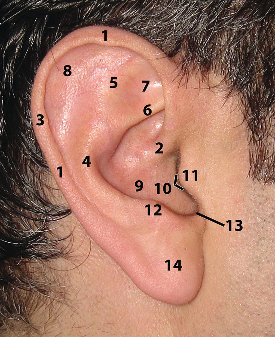
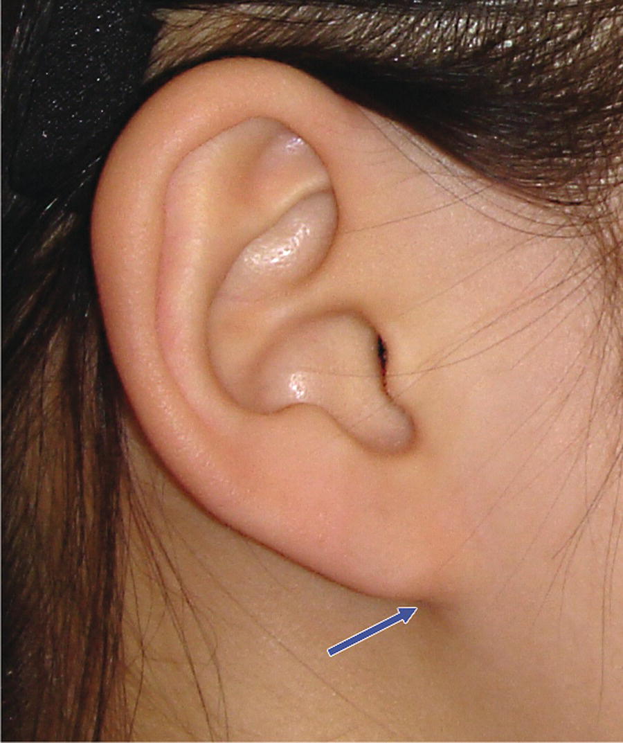
The ear is not strictly speaking part of the face, but is part of the cranial region of the craniofacial complex. However, the ears lie on the same plane as the middle third of the face, and are an important parameter in facial aesthetic evaluation. As such, the clinical evaluation of the ears will be described in this section. The external ear, or auricle, is usually a relatively inconspicuous feature of the craniofacial complex, which may be concealed under long hair in some individuals. However, excessively large, laterally prominent or deformed auricles may be very noticeable. There is considerable individual variation in the size, shape and lateral prominence of the ears. Provided that the proportional relationship of the ears to the rest of the craniofacial complex is within normal limits, and there is relative left–right symmetry, these variations are deemed acceptable. Deformities may be a consequence of considerable deviation from normal morphology and size, and excessive disproportion of the ears. For children in particular, this may well become a source of ridicule by their peers, leading to personal anguish and emotional trauma.1 For such patients, surgical alteration (otoplasty) may have a considerably favourable impact on their self‐esteem.2 The psychosocial experiences of children undergoing otoplasty have been examined, finding that 97% reported an increase in happiness, 92% reported an increase in self‐confidence, 79% noted improved social experience with increased social integration, and 100% reported bullying reduced or stopped; the report concluded that otoplasty is an effective procedure in alleviating psychosocial distress in the vast majority of children that undergo the operation.3 The ear houses the peripheral parts of the auditory and vestibular apparatus and is descriptively divided into the external, middle and internal ear. The anatomy of the external ear is important in facial aesthetic evaluation (Figure 13.1). The external ear consists of the auricle (or pinna) and the external acoustic meatus (or external auditory canal). The auricle projects from the side of the head to collect sound waves, and the meatus leads inwards from the auricle to conduct vibrations to the tympanic membrane. Figure 13.1 Anatomy of the external ear (auricle): The framework of the auricle is the auricular cartilage; this is a single thin plate of resilient elastic fibrocartilage, which is thrown into folds that give its characteristic shape. The cartilage is covered on both surfaces with adherent hairy skin, which continues into the external acoustic meatus. The cartilage of the auricle is prolonged inwards in tubular fashion as the cartilaginous part of the external auditory meatus, whose attachment to bone stabilizes the auricle in position. The lateral surface of the auricle is irregularly concave, faces slightly forwards and displays numerous eminences and depressions; these convolutions may help to locate sound. Its prominent curved rim, or helix, usually bears a small auricular tubercle posterosuperiorly. The antihelix is a curved prominence, parallel and anterior to the posterior part of the helix. The antihelix divides superiorly into two crura (superior and inferior crura of the antihelix), which flank a depressed triangular fossa. The curved depression between the helix and antihelix is the scaphoid fossa. The antihelix encircles the concha of the auricle, which is incompletely divided by the crus or anterior end of the helix. The tragus is a small curved flap below the crus of the helix and in front of the concha; it projects posteriorly, partly covering the meatal orifice. The antitragus is a small tubercle opposite the tragus and separated from it by the intertragic notch. Below this is the lobule (or ear lobe), which is a tag of skin containing fibrous and adipose tissue; the auricular cartilage does not extend into the ear lobe. Figure 13.2 Attached ear lobe.
Chapter 13
The Ears
Introduction
Terminology
Anatomy
Related posts:


Stay updated, free articles. Join our Telegram channel

Full access? Get Clinical Tree


